Development of a surgical guide for minimally invasive corticotomies with a complete digital intraoral and laboratory workflow Entwicklung einer ...
←
→
Transkription von Seiteninhalten
Wenn Ihr Browser die Seite nicht korrekt rendert, bitte, lesen Sie den Inhalt der Seite unten
APPLICATION

Marion Paris, Nathalie Nurdin, Guillermo Manzano, Francesca Caroleo, Yassine Messaoudi, Mark
Bischof, Rabah Nedir, Christian Coachman

Development of a surgical guide for minimally invasive
corticotomies with a complete digital intraoral and laboratory
workflow
Entwicklung einer Operationsschablone mit rein digitalem
intraoralen und labortechnischen Workflow für minimalinvasive
Kortikotomien

 Zusammenfassung                                                  Abstract
 Ziel: Die chirurgisch unterstützte kieferorthopädische           Aim: Surgically facilitated orthodontic treatment is increas-
 Behandlung wird zunehmend vor allem bei Erwachsenen              ingly being used, especially for adults, to facilitate tooth
 eingesetzt, um Zahnbewegungen zu erleichtern und die             movements and reduce the duration of orthodontic treat-
 Dauer der kieferorthopädischen Behandlung zu reduzie-            ment. The present article reports on an innovative, safe, and
 ren. Der vorliegende Artikel beschreibt ein innovatives,         minimally invasive technique to perform flapless corticoto-
 sicheres und minimalinvasives Verfahren zur Durchfüh-            mies using a dedicated surgical guide produced with a com-
 rung lappenloser Kortikotomien mit einer speziellen Ope-         plete digital intraoral and laboratory workflow.
 rationsschablone, die mit einem rein digitalen intraoralen       Materials and methods: A 51-year-old man presented with
 und labortechnischen Workflow angefertigt wird.                  maxillary and mandibular anterior crowding. He required
 Material und Methoden: Ein 51-jähriger Patient stellte           rapid treatment with limited use of braces. Corticotomies
 sich mit einem Engstand im oberen und unteren Front-             were planned for both arches before the use of orthodontic
 zahnbereich vor. Er wünschte eine kurze Behandlung mit           appliances. The matching of the stereolithographic files
 begrenztem Einsatz von Zahnspangen. Vor dem Einset-              obtained from the digital prints of the full arches and the
 zen der kieferorthopädischen Apparaturen wurden an               cone beam computed tomography images allowed for the
 Ober- und Unterkiefer Kortikotomien geplant. Die                 positioning of the cutting planes for corticisions. The guide
 Schnittebenen der kortikalen Inzisionen wurden durch             was printed with a transparent, biocompatible, and pho-
 Abgleich der stereolithografischen Daten beider Kiefer,          topolymerizable resin, and cold sterilized. Minimally inva-
 die bei einer digitalen Abformung erhoben wurden, und            sive corticotomies were performed using a piezoelectric
 der Bilder der digitalen Volumentomografie festgelegt.           instrument. The orthodontic treatment started immediately
 Die Schablone wurde aus durchscheinendem, biokompa-              after surgery.
 tiblem und polymerisierbarem Kunststoff gedruckt und             Results: No adverse events were recorded during surgery.
 kalt sterilisiert. Die minimalinvasiven Kortikotomien wur-       The piezoelectric instrument was guided accurately, and pre-
 den mit einem piezoelektrischen Instrument durchge-              cise application of the corticisions prevented all the anatom-
 führt. Die kieferorthopädische Behandlung begann                 ical elements from being injured. The healing was uneventful
 unmittelbar postoperativ.                                        and the patient experienced no pain.
 Ergebnisse: Während der Operation traten keine uner-             Conclusion: The present report shows that a surgical guide
 wünschten Ereignisse auf. Das piezoelektrische Instrument        specifically and digitally produced for corticotomies allowed
 wurde präzise geführt und die korrekte Durchführung der          for the performance of a minimally invasive flapless tech-
 Kortikotomien verhinderte die Verletzung anatomischer            nique and accurate piezosurgery. The use of such a guide was
 Strukturen. Die Heilung verlief komplikationslos und der         easy to implement, made the procedure safer, and reduced
 Patient war schmerzfrei.                                         postoperative pain.

International Journal of Computerized Dentistry 2020;23(3):1–11                                                               1
APPLICATION

Keywords: guided surgery, corticotomies, surgical guide, com-         Schlussfolgerung: Der vorliegende Bericht zeigt, dass eine
puter-aided design, surgically facilitated orthodontic treatment,     speziell und digital für die Kortikotomien angefertigte
piezoelectric surgery                                                 Operationsschablone die Durchführung des Eingriffs als
                                                                      minimalinvasive, lappenlose Operation mit präziser Piezo-
                                                                      chirurgie ermöglicht. Die Operationsschablone ließ sich
Introduction                                                          gut in das Verfahren einbinden, erhöhte die Sicherheit des
                                                                      Eingriffs und reduzierte die postoperativen Schmerzen.
The repositioning of teeth in adults is a considerable chal-
lenge due to anatomical- and physiologic-related bone con-            Schlüsselwörter: geführte Operation, Kortikotomien, Operati-
straints. The duration of standard orthodontic treatment is 18        onsschablone, Computer-aided Design, chirurgisch unterstützte
to 30 months1,2. Despite the length of treatment, the number          kieferorthopädische Behandlung, piezoelektrische Chirurgie
of adult patients undergoing orthodontic treatment is
increasing significantly3.
      Surgically facilitated orthodontic treatment (SFOT) can         Einleitung
  be used to facilitate tooth movements and can be imple-
  mented from the start of orthodontic treatment. Cortico-            Zahnbewegungen sind bei Erwachsenen aufgrund von
  tomy is one of the techniques available for SFOT and invol-         anatomischen und physiologischen Einschränkungen sehr
  ves the performance of shallow vertical interdental                 schwierig. Normalerweise dauert eine kieferorthopädische
  incisions of the gingiva and cortical bone4,5. The benefits         Behandlung 18–30 Monate1,2. Trotz der Länge der Behand-
  of corticotomy include:                                             lung nimmt die Anzahl der Erwachsenen, die kieferortho-
§ acceleration of tooth movement6-8;                                  pädisch behandelt werden, deutlich zu3.
§ regional acceleratory phenomena for 4 months9;                          Mit der chirurgisch unterstützten kieferorthopädischen
§ 20% to 40% reduction in treatment time4; and                        Behandlung bereits zu Beginn der kieferorthopädischen
§ reduced risk of external apical root resorption, mainly due         Therapie lassen sich Zahnbewegungen vereinfachen. Eine
    to the decreased orthodontic forces applied to the                der dazu verwendeten Techniken ist die Kortikotomie mit
    teeth10,11.                                                       flachen vertikalen interdentalen Inzisionen in Gingiva und
                                                                      Kortikalis4,5. Zu den Vorteilen der Kortikotomie gehören:
Most corticotomies are performed with an invasive flap and            § die Beschleunigung der Zahnbewegung6–8,
require a supply of bone to compensate for bone deepitheliali-        § ein regionales Beschleunigungsphänomen für vier
zation12,13. Some corticotomies are performed flapless; inci-             Monate9,
sions are made between the teeth in a blind manner, without           § die Reduktion der Behandlungsdauer um 20–40 % 4
accurate visualization of the dental roots and their apices or the        und
bone papillae5. Therefore, the indications for corticotomies are      § ein geringeres Risiko für eine externe apikale Wurzel-
limited by the associated risks such as damage of tooth-sup-              resorption überwiegend durch das geringere Einwir-
porting tissue, devitalization of the teeth, injuries to noble ana-       ken kieferorthopädischer Kräfte auf die Zähne10,11.
tomical elements, and postoperative discomfort and pain. The
use of piezoelectric tools has improved the technique5,14.            Meistens werden Kortikotomien mit Lappenabhebung
    Adults often request rapid treatment and the limited              und dem Einbringen von Knochenersatzmaterial zum Aus-
wearing of orthodontic braces; therefore, corticotomies are           gleich der Deepithelialisierung des Knochens durchge-
frequently included in orthodontic treatment. This treatment          führt12,13. Manche der Kortikotomien erfolgen lappenlos,
requires the development of safe and minimally invasive sur-          dabei werden die Inzisionen zwischen den Zähnen blind
gical techniques. The purpose of this report is to present the        und ohne akkurate Darstellung der Zahnwurzeln und ihrer
conception and production of surgical guides for dental cor-          Apices oder des interdentalen Knochens durchgeführt5.
ticotomies with piezosurgery. The guides were developed               Daher sind die Indikationen für Kortikotomien durch die
with a complete digital intraoral and laboratory workflow.            assoziierten Risiken, wie die Schädigung des Zahnhalteap-
With the use of piezoelectric tools, guided surgery enables a         parates, die Devitalisierung der Zähne, die Verletzung
safe, accurate, and minimally invasive technique for corticot-        wichtiger anatomischer Strukturen sowie postoperative
omies as part of SFOT.                                                Beschwerden und Schmerzen, eingeschränkt. Durch den

2                                                                        International Journal of Computerized Dentistry 2020;23(3):1–11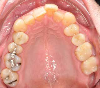
Paris et al

 a                                                                     b

Fig 1 Initial situation: mandible (a) and maxilla (b).
Abb. 1   Ausgangssituation: Oberkiefer (a) und Unterkiefer (b).

 Einsatz piezoelektrischer Instrumente konnte die Technik         Case presentation
 verbessert werden5,14.
                                                                  Surgical planning
     Häufig wünschen Erwachsene eine kurze Behand-
 lungsdauer und möchten nur kurz kieferorthopädische              A 51-year-old man presented in May 2018 for the correction
 Zahnspangen tragen. Daher werden im Rahmen der kie-              of mandibular and maxillary anterior crowding. The clinical
 ferorthopädischen Behandlung oft Kortikotomien durch-            examination revealed a deep bite tendency, convex profile,
 geführt, für die sichere und minimalinvasive chirurgische        retruded mandible, and moderate incisor crowding. There
 Techniken entwickelt werden müssen. Im vorliegenden              was no lip incompetence or gummy smile (Fig 1). Several
 Artikel werden das Konzept und die Produktion von Ope-           treatment options were considered: maxillary second pre-
 rationsschablonen für dentale piezochirurgische Kortiko-         molar extractions, maxillary distalization, and orthognathic
 tomien vorgestellt. Ihre Fertigung erfolgte intraoral und        surgery. The patient refused orthognathic surgery and, for
 labortechnisch rein digital. Mit dem Einsatz piezochirurgi-      professional reasons, requested rapid orthodontic treatment
 scher Instrumente ist die geführte Chirurgie ein sicheres,       and to wear braces for a short duration. To facilitate tooth
 präzises und minimalinvasives Verfahren zur Durchfüh-            movement and speed up the orthodontic treatment, cortico-
 rung von Kortikotomien im Rahmen der chirurgisch unter-          tomies were planned for both arches before the installation
 stützten kieferorthopädischem Behandlung.                        of orthodontic appliances and the extraction of the maxillary
                                                                  second premolars. Braces were to be installed on the same
                                                                  day, immediately after the corticotomies. The patient under-
 Fallbericht                                                      stood and accepted the treatment.
                                                                      A digital impression of the full arches was taken (Trios 3
 Operationsplanung
                                                                  Move; 3Shape, Copenhagen, Denmark). A recording of the
 Ein 51-jähriger Patient stellte sich zur Korrektur eines         occlusal forces was used to match cone beam computed
 Engstands im oberen und unteren Frontzahnbereich vor.            tomography (CBCT) images with the digital print, whereas a
 Die klinische Untersuchung ergab eine Tendenz zum tie-           mucosal approach was used to adjust the surgical guide.
 fen Biss, ein konvexes Profil, eine Retrusion des Unterkie-          Radiography was performed using CBCT (Model CS 9300;
 fers und einen mäßigen Engstand der Schneidezähne.               Carestream Health, Rochester, NY, USA). Images of the full
 Eine Lippeninkompetenz oder ein Zahnfleischlächeln               arches were acquired with a 0.3-mm pixel size, 90 kV tube
 lagen nicht vor (Abb. 1). Es wurden mehrere Behand-              voltage, 4 mA beam current, and 8 s exposure time. CBCT had

International Journal of Computerized Dentistry 2020;23(3):1–11                                                               3
APPLICATION
lungsoptionen erwogen: die Extraktion der oberen zwei-
ten Prämolaren, die Distalisierung im Oberkiefer und die
orthognathe Chirurgie. Der Patient verweigerte die
orthognathe Chirurgie und wünschte aus beruflichen
Gründen eine kurze kieferorthopädische Behandlung mit
kurzer Tragezeit einer Zahnspange. Zur Erleichterung der
Zahnbewegung und Beschleunigung der kieferorthopä-
a
dischen Behandlung wurden an Ober- und Unterkiefer
vor dem Einsetzen der kieferorthopädischen Apparatu-
ren und der Extraktion der oberen zweiten Prämolaren
Kortikotomien geplant. Die Zahnspangen wurden noch
am Operationstag, unmittelbar nach den Kortikotomien
eingesetzt. Der Patient verstand die Behandlung und
stimmte ihr zu.
b Von beiden Kiefern wurde eine digitale Abformung
angefertigt (Trios 3 Move; Fa. 3Shape, Kopenhagen,
Fig 2 Cutting plan (a) and guide (b) simulation for corticoto- Dänemark). Die Bilder der digitalen Volumentomografie
mies.
(DVT) und des digitalen Abdrucks wurden anhand der
Abb. 2 Simulation der Schnittebenen (a) und der Schablone (b) Bestimmung der Okklusalkräfte abgeglichen; die Opera-
für die Kortikotomien. tionsschablone wurde mit einem mukosalen Ansatz
angepasst.
Die radiologischen Aufnahmen erfolgten mittels DVT
(Model CS 9300; Fa. Carestream Health, Rochester, NY,
USA). Die Bilder von Ober- und Unterkiefer wurden mit
to include the teeth and vestibular and lingual cortices as well einer Pixelgröße von 0,3 mm, einer Röhrenspannung von
as the alveolar nerve for the mandible and floor of the nasal 90 kV, einer Stromstärke von 4 mA und einer Belichtungs-
cavities, and the maxillary sinuses for the maxilla. Files of the zeit von 8 s aufgenommen. Die DVT sollte die Zähne, die
scans were acquired in DICOM (Digital Imaging and Commu- vestibuläre und linguale Kortikalis sowie im Unterkiefer
nications in Medicine) format. den N. alveolaris und am Oberkiefer den Sinusboden und
die Sinus maxillares erfassen. Die Bilddaten wurden im
DICOM-Format (Digital Imaging and Communications in
Guide conception
Medicine) gespeichert.
The digital print images were saved as standard tessellation
language (STL) and CBCT files in DICOM format. These digital
Entwerfen der Schablone
files were sent to the DSD Planning Center (Madrid, Spain)
and integrated through computer-aided design (CAD) soft- Die digitalen Druckdaten wurden im STL-Format (Standard
ware (NemoStudio, version 12.12.0; Nemotec, Spain). Data Tessellation Language) und die DVT-Daten im DICOM-For-
were then processed with the dedicated software (NemoStu- mat gespeichert. Diese digitalen Dateien wurden zum DSD
dio) and built into three-dimensional (3D) models, which Planning Center (Madrid, Spanien) gesendet und mithilfe
were superimposed by matching the surfaces. Each tooth eines CAD-Programms (NemoStudio, Version 12.12.0;
was individualized on the 3D reconstruction; the bone sup- Fa. Nemotec, Spanien) integriert. Dann wurden die Daten
porting the teeth and the inferior alveolar nerve were distin- mit dieser speziellen Software (NemoStudio) zu dreidi-
guishable. mensionalen Modellen weiterverarbeitet, die anhand
The guides were modelled by taking the space between eines Abgleichs der Oberflächen übereinandergelagert
the guide and the teeth stabilizing the guide to be 0.15 mm, wurden. Jeder Zahn wurde in der dreidimensionalen
and the thickness of the guides to not exceed 3 mm. To Rekonstruktion einzeln dargestellt. Der Knochen, der die
ensure the correct and complete insertion of the guides at Zähne stützt, und der N. alveolaris inferior waren gut von-
the time of surgery, positioning windows were modelled as einander abgrenzbar.
4 International Journal of Computerized Dentistry 2020;23(3):1–11Paris et al
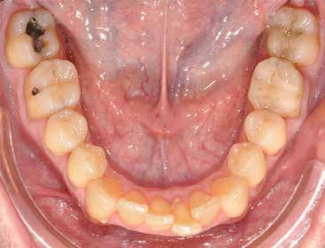
Fig 3 Front view of mandibular guide (a)
and occlusal view of maxillary guide
showing positioning control openings (b).
Abb. 3 Frontalansicht der Unterkiefer-
schablone (a) und okklusale Ansicht der
Oberkieferschablone mit Ausrichtung der
a b
Kontrollöffnungen (b).
Für die Schablonen wurde davon ausgegangen, dass part of the guides. These openings were located on the
der Abstand zwischen der Schablone und den sie tragen- occlusal surfaces of the teeth. Cutting plans simulating the
den Zähnen 0,15 mm beträgt und die Schablone selbst insertion of piezoelectric tools (Fig 2) were placed according
nicht dicker als 3 mm ist. Damit die Schablonen bei der Ope- to the following criteria:
ration korrekt und komplett eingesetzt werden konnten, § Corticotomies were realized between each tooth from the
wurden Positionierfenster eingearbeitet. Diese Öffnungen distal part of the right second premolar to the distal part
lagen auf der Okklusalseite der Zähne. Die Schnittebenen of the left second premolar.
für das Einführen der piezoelektrischen Instrumente (Abb. 2) § The upper limit of the cutting plan was 2 mm under the
wurden nach folgenden Kriterien platziert: bone papilla; the lower limit corresponded to the apex of
§ Die Kortikotomien erfolgten zwischen allen Zähnen in the roots.
dem Bereich zwischen dem distalen Anteil des rechten § The thickness of the grooves for the corticision was
zweiten Prämolars und dem distalen Anteil des linken 0.8 mm.
zweiten Prämolars. § The incision path was as parallel as possible to the central
§ Die Schnittebene reichte oben bis 2 mm unter den axes of the teeth and located equidistantly between two
interdentalen Knochen und unten bis zur Wurzelspitze. teeth.
§ Die Breite der Schneidrillen für die Kortikotomien
wurde mit 0,8 mm festgelegt. The modelling of the guides with the cutting plans and
§ Die Inzision sollte möglichst parallel zur senkrechten grooves in place (Fig 2) was controlled by the surgeon, par-
Zahnachse und genau mittig zwischen den beiden ticularly the upper and lower limits of the section plans;
Zähnen verlaufen. access to the entire thickness of the vestibular bone; and dis-
tances between the section plans and the alveolar nerve,
Die Schnittebenen und Schnittbreiten der Schablonen sinus, and teeth. After approval by the surgeon, the models
(Abb. 2) wurden vom Operateur kontrolliert, vor allem die were converted to STL files and printed (Fig 3) with a desktop
oberen und unteren Grenzen der Schnittebenen, das stereolithography 3D printer (3D Printer Form 2; Formlabs,
Erfassen der gesamten Dicke des vestibulären Knochens Somerville, MA, USA). A transparent, biocompatible, pho-
und die Abstände der Schnittebenen zu N. alveolaris, Sinus topolymerizable resin was used to improve the visual control
und Zähnen. Nach der Freigabe durch den Chirurgen wur- of the surgery (Photopolymer Resin, Clear, FLGPCL02; Form-
den die Modelle in STL-Dateien umformatiert und mit labs). Before surgery, the guide was subjected to cold chem-
einem stereolithografischen 3-D-Desktop-Drucker (3D ical sterilization.
Printer Form 2; Fa. Formlabs, Somerville, MA, USA)
gedruckt (Abb. 3). Um die visuelle Kontrolle der Operation
Surgery
zu verbessern, wurde ein durchscheinender, biokompatib-
ler, lichtpolymerisierbarer Kunstharz (Photopolymer Resin, The surgical procedure was performed under antibiotic proph-
Clear, FLGPCL02; Fa. Formlabs) verwendet. Vor der Opera- ylaxis initiated two hours before surgery (amoxicillin 2 g;
tion wurde die Schablone chemisch kalt sterilisiert. Amoxi-Mepha; Mepha Pharma, Aesch, Switzerland). After local
International Journal of Computerized Dentistry 2020;23(3):1–11 5APPLICATION
anesthesia (articaine 68 mg per carpule; adrenaline 1/200 000, Operation
8.5 μg/carpule), the surgical guide was positioned on the teeth
(Fig 4). The position of the guide was adjusted using specially Die Operation wurde unter Antibiotikaprophylaxe durch-
created windows in the guide to allow for visual inspection, geführt. Dazu erhielt der Patient zwei Stunden präopera-
and its stability and fit were carefully controlled. tiv Amoxicillin 2 g (Amoxi-Mepha; Fa. Mepha Pharma,
Full thickness mucogingival incisions in the cutting Aesch, Schweiz). Nach der Lokalanästhesie (Articain
grooves were performed using a 15C blade (Schreiber 68 mg per Karpule, Adrenalin 1/200 000, 8,5 μg/Karpule)
Instrumente, Fridingen/Donau, Germany). The guide was wurde die Operationsschablone auf die Zähne gelegt
then removed. The bone had to be free of periosteum and (Abb. 4). Ihre Position wurde mit speziell angelegten
gingival mucosa before further incisions were made with a Fenstern ausgerichtet, die eine visuelle Inspektion
piezosurgical device; therefore, access to the bone through ermöglichen, und ihre Stabilität und Passung sorgfältig
the mucosal incision path was controlled using a small strip- kontrolliert.
per. The guide was re-placed, and corticotomies were car- Mit einer 15C-Klinge (Fa. Schreiber Instrumente, Fri-
ried out through the grooves in the guide with abundant dingen/Donau) wurden in den Schneidrillen mukogingi-
irrigation (Figs 5 and 6). A 0.5-mm–thick scalpel insert (CS3 vale Vollschichtinzisionen durchgeführt. Anschließend
Crest Splitting Kit; Satelec Acteon Group, Mérignac, France) wurde die Schablone entfernt. Da der Knochen für weite-
was used for the corticotomies. Piezoelectric osseous cuts re Inzisionen mit dem piezochirurgischen Instrument frei
(Piezotome Solo; Satelec Acteon) were carefully made in von Periost und gingivaler Mukosa sein musste, wurde
low-frequency cutting mode. Each incision was closed with der Zugang zum Knochen durch die mukosalen Inzisio-
one or two non-absorbable interrupted sutures (Supramid; nen mit einem kleinen Stripper überprüft. Anschließend
B. Braun Vet Care, Tuttlingen, Germany; Fig 7). The same sur- wurde die Schablone wieder eingelegt und die Kortikoto-
gical procedure was used for the corticotomies of the other mien wurden unter reichlicher Spülung durch die
arch. Schneidrillen der Schablone ausgeführt (Abb. 5 und 6).
Ibuprofen (400 mg) was administered for pain relief. The Für die Kortikotomien wurde ein 0,5 mm dicker Skalpell-
patient was instructed not to change his dental hygiene einsatz (CS3 Crest Splitting Kit; Fa. Satelec Acteon Group,
habits, to eat cold and soft food for the first day following Mérignac, Frankreich) verwendet. Die piezolektrischen
the procedure, and to begin mouth washing 24 h after sur- Knochenschnitte (Piezotome Solo; Satelec Acteon) wur-
gery. He was requested to assess his pain on a digital scale den vorsichtig im niederfrequenten Schneidmodus
from 0 to 10 during surgery, two hours after surgery, and durchgeführt. Jede Inzision wurde mit ein oder zwei
one day after surgery; the respective assessments were 0, 2, nicht resorbierbaren Einzelknopfnähten verschlossen
and 0. The orthodontic treatment started immediately after (Supramid; Fa. B. Braun Vet Care, Tuttlingen; Abb. 7). Die
surgery with the placement of the appliances. Healing was Kortikotomien in Ober- und Unterkiefer wurden mit dem
assessed 1 week after surgery and the sutures were gleichen Operationsverfahren durchgeführt.
removed. The clinical examination took place one month Zur Schmerzlinderung erhielt der Patient Ibuprofen
later. (400 mg). Er wurde angewiesen, seine Maßnahmen zur
Mundhygiene unverändert durchzuführen, am ersten
postoperativen Tag nur weiche, kalte Speisen zu sich zu
Results nehmen und 24 h nach der Operation mit Mundspülun-
gen zu beginnen. Die Schmerzen bei der Operation,
No adverse events were recorded during or after the surgery. zwei Stunden sowie einen Tag danach sollte er auf einer
No injury occurred to the noble anatomical elements such as digitalen Skala von 0 bis10 einstufen; er gab die
the teeth and the inferior alveolar nerve. The patient report- Schmerzstärke mit 0, 2 und 0 an. Die kieferorthopädi-
ed no pain during surgery or one day, one week and sche Behandlung begann sofort postoperativ mit dem
one month after surgery, but he complained about moderate Einsetzen der Apparaturen. Eine Woche nach der Opera-
pain two hours after surgery. The healing was uneventful. No tion wurde der Heilungsverlauf überprüft und wurden
bone or mucosal infection or inflammation was observed. die Fäden gezogen. Die klinische Untersuchung erfolgte
One month after the surgery, the sites were completely einen Monat später.
healed (Fig 8).
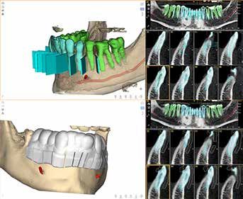
6 International Journal of Computerized Dentistry 2020;23(3):1–11Paris et al Fig 4 Guide positioned on the mandibular teeth. Abb. 4 Auf die Unterkieferzähne gesetzte Schablone. Fig 5 Abundant irrigation (100 ml/min) during bone incisions. Fig 6 Mucoperiosteal incisions and corticotomies on the mandible. Abb. 5 Reichliche Spülung (100 ml/min) während der Knochen- Abb. 6 Mukoperiostale Inzisionen und Kortikotomien am inzisionen. Unterkiefer. a b Fig 7 Sutures and orthodontic appliances: mandible (a) and maxilla (b). Appliances were placed on the same day as the surgery. Abb. 7 Nähte und kieferorthopädische Apparaturen: Unterkiefer (a) und Oberkiefer (b). Die Apparaturen wurden noch am Operations- tag eingesetzt. Ergebnisse Während und nach der Operation traten keine uner- wünschten Ereignisse auf. Es wurden keine wichtigen ana- tomischen Strukturen, wie die Zähne oder der N. alveolaris inferior, verletzt. Der Patient gab weder während der Ope- ration, noch einen Tag, eine Woche oder ein Jahr später Schmerzen an; allerdings bestanden zwei Stunden post- operativ mäßige Schmerzen. Die Heilung verlief ohne Komplikationen. Es fand sich keine Infektion oder Entzün- dung von Knochen oder Mukosa. Einen Monat postopera- Fig 8 Uneventful healing one month after surgery. tiv waren alle Bereiche vollständig abgeheilt (Abb. 8). Abb. 8 Komplikationslose Heilung einen Monat postoperativ. International Journal of Computerized Dentistry 2020;23(3):1–11 7
APPLICATION

Discussion                                                         Diskussion
The aim of the present case report was to present the digital      Ziel des hier geschilderten Fallberichts war die Vorstellung
conception and production of a surgical guide for corticoto-       des digitalen Entwurfs und der digitalen Produktion einer
mies undertaken within a SFOT. The guide was tailored to           Operationsschablone zur Durchführung von Kortikotomien
perform a safe, flapless piezosurgery before the commence-         im Rahmen der chirurgisch unterstützten kieferorthopädi-
ment of orthodontic treatment.                                     schen Behandlung. Die Schablone wurde so gestaltet, dass
    Digital technology is rapidly becoming increasingly useful     sie vor Beginn der kieferorthopädischen Behandlung eine
and essential at every stage of the treatment of patients          sichere, lappenlose Piezochirurgie ermöglichte.
requiring dental care, particularly for diagnosis, treatment           Der Nutzen der digitalen Technologie nimmt rasant zu
planning, and implementation15. Guided surgery has become          und sie wird in allen Stadien der Behandlung von Patienten,
widely available in the field of implantology. Orthodontic sur-    bei denen eine zahnärztliche Behandlung erforderlich ist,
gery refers to different procedures that include surgery of        immer entscheidender – insbesondere für Diagnostik,
impacted teeth (disimpaction and extraction), placement of         Behandlungsplanung und -durchführung15. Die geführte
orthodontic anchors, and SFOT. To the authors’ knowledge,          Chirurgie steht inzwischen allgemein in der Implantologie
however, and unlike in the case of implantology, few studies       zur Verfügung. Als orthodontische Chirurgie werden ver-
have described orthodontically guided surgical procedures.         schiedene Verfahren bezeichnet, zu denen die Operation
Cassetta et al used guided surgery for the placement of            impaktierter Zähne (Aufrichtung und Extraktion), das Set-
orthodontic miniscrews in implant surgery16 . Wang et al           zen orthodontischer Anker und die chirurgisch unterstützte
reported on the navigation-guided extraction of impacted           kieferorthopädische Behandlung gehören. Soweit die Auto-
supernumerary teeth17. Only two studies have reported on           ren wissen und anders als in der Implantologie, gibt es
the use of surgical guides obtained by 3D printing for cortico-    jedoch nur wenige Studien zu orthodontisch geführten
tomies11,18. To decrease the risk of intraoperative damage,        Operationsverfahren. In der Implantatchirurgie setzten Cas-
Cassetta et al planned the location and depth of the corticot-     setta et al. mithilfe der geführten Chirurgie orthodontische
omies using preoperative CBCT, and used a surgical guide           Minischrauben16. Wang et al. beschrieben die navigations-
obtained by 3D printing11. Hou et al designed a translucent,       geführte Extraktion impaktierter überzähliger Zähne17. Nur
rigid, porous guide to improve visibility, guidance, and irriga-   zwei Studien befassten sich mit dem Einsatz von Operati-
tion during surgery18.                                             onsschablonen, die mittels 3-D-Druck für Kortikotomien
    Most software used for modelling has been developed for        angefertigt wurden11,18. Um die Gefahr einer intraoperati-
research purposes11,18. However, some apps and software            ven Verletzung zu reduzieren, planten Cassetta et al. Lage
were designed to assist esthetic dentistry practitioners (treat-   und Tiefe der Kortikotomien anhand der präoperativen DVT
ment planning and clinical evaluation) as well as implant sur-     und verwendeten eine mittels 3-D-Druck angefertigte Ope-
geons (guided implant surgery) 19-22. The use of a complete        rationsschablone11. Hou et al. entwarfen eine durchschei-
digital intraoral and laboratory workflow and versatile soft-      nende, starre, poröse Schablone zur perioperativen Verbes-
ware specially developed for dentistry might render guided         serung von Sicht, Führung und Spülung18.
corticotomies accessible to a large number of practitioners.           Die meisten Programme zur Modellbildung wurden zu
Furthermore, the development of such safer procedures              Forschungszwecken entwickelt11,18 . Manche Apps und
should also increase acceptance of the treatment by patients.      Programme wurden jedoch so entworfen, dass sie die auf
    In the present report, the information collected from the      dem Gebiet der ästhetischen Zahnheilkunde tätigen Ärzte
preoperative CBCT images and digital print allowed for the         (bei Behandlungsplanung und klinischer Evaluation) sowie
precise analysis of the anatomical situation, including the        die Implantatchirurgen (durch geführte Implantationen)
position, shapes, and axes of the roots as well as their dis-      unterstützen können19–22. Durch komplett digitale intra-
tance from each other and the lower alveolar nerve. The            orale und labortechnische Arbeitsabläufe und vielseitige
thickness of the vestibular cortex was measured to ensure          Programme, die speziell für die Zahnheilkunde entwickelt
that the piezosurgical instrument crossed the entire thick-        wurden, dürften Kortikotomien für noch mehr Ärzte als
ness, not only of the guide and the gums but also of the cor-      Behandlungsoption infrage kommen. Außerdem sollte die
tex. The cutting planes for carrying out the corticotomies         Entwicklung derartiger sicherer Verfahren die Akzeptanz
were positioned precisely according to the anatomy of the          seitens der Patienten erhöhen.

8                                                                     International Journal of Computerized Dentistry 2020;23(3):1–11
Paris et al
Im vorliegenden Bericht ermöglichten die mit den prä- patient. This step was carefully controlled by the guide
operativen DVT-Aufnahmen und dem digitalen Abdruck designer and the surgeon. In this way, the guide rendered the
gewonnenen Informationen eine präzise Analyse der ana- surgical procedure safer and more accurate.
tomischen Situation, einschließlich Position, Form und The guide was easy to use, effective, and compatible with
Ausrichtung der Zahnwurzeln sowie deren Abstand von- flapless piezosurgery. As a result of its rigidity, the guide
einander und zum N. alveolaris inferior. Um sicherzustel- resisted the mechanical stresses on the teeth during insertion
len, dass das piezochirurgische Instrument nicht nur die and disinsertion, and was not deformed on the depressible
Schablone und Zahnfleisch, sondern auch die Kortikalis mucous areas during the use of surgical instruments. The cor-
auf voller Dicke durchtrennt, wurde die Dicke der vestibu- rect position of the guide was controlled by its transparency
lären Kortikalis ermittelt. Die Schnittebenen der Kortikoto- and through its openings. The distance of 0.15 mm between
mien wurden gemäß der Anatomie des Patienten präzise the guide and the teeth stabilizing it was similar to that of
festgelegt. Dieser Schritt wurde sorgfältig vom Planer der dental-supported guides for implantology. If this space is too
Schablone und dem durchführenden Chirurgen überprüft. small, the friction on the teeth is high and the insertion of the
Auf diese Weise erhöhte die Schablone Sicherheit und Prä- guide is a delicate procedure; if the space is too wide, the
zision des operativen Eingriffs. guide lacks stability.
Die Schablone war leicht in der Handhabung, effektiv The guide allowed for the performance of piezoelectric
und mit der lappenlosen Piezochirurgie kompatibel. Auf- surgery. To control the depth of the corticotomies, the thick-
grund ihrer Starrheit widerstand sie den mechanischen ness of the guide did not exceed 3 mm, such that the working
Belastungen der Zähne beim Einsetzen und Herausneh- part of the 10-mm–long drill of the piezoelectric tool passed
men und wurde während des Einsatzes der chirurgischen through the guide, gum, and vestibular cortex. The drill pro-
Instrumente nicht auf den eindrückbaren mukosalen truded a few millimeters into the cancellous bone without
Bereichen verformt. Die korrekte Lage der Schablone any additional risk. Furthermore, the use of low piezoelectric
konnte aufgrund ihrer Durchsichtigkeit und anhand ihrer frequencies needed to cut the mineralized structures might
Öffnungen überprüft werden. Der Abstand von 0,15 mm have protected the soft tissue from injury23. There was no
zwischen der Schablone und den sie stützenden Zähnen sign of bone warming because of the abundant irrigation uti-
ähnelte demjenigen bei zahngelagerten Schablonen in lized and the regular pauses taken during surgery. The need
der dentalen Implantologie. Wird dieser Abstand zu klein for sufficient irrigation was confirmed by Schlee et al and
gewählt, kommt es zur starken Reibung an den Zähnen Lajolo et al24,25.
und das Einsetzen der Schablone ist sehr schwierig. Wird Unlike the technique reported by Dibart et al, the present
er zu groß gewählt, sitzt die Schablone nicht stabil. procedure involved no flaps, yet it was not blind5. Preopera-
Durch die Verwendung der Schablone war eine piezo- tive CBCT permitted radiographic visualization of the differ-
elektrische Operation möglich. Um die Tiefe der Kortikoto- ent anatomical elements, and the guide enabled the optimal
mien zu kontrollieren, war die Schablone nicht dicker als positioning of the surgical instruments. Therefore, the risk of
3 mm, sodass das Arbeitsteil des 10 mm langen Bohrers des damaging a noble anatomical element such as a root or the
piezoelektrischen Instruments Schablone, Zahnfleisch und lower alveolar nerve was considerably decreased. Flapless
vestibuläre Kortikalis erfasste. Der Bohrer reichte ein paar Mil- surgery has been shown to reduce postoperative pain and
limeter in die Spongiosa, ohne dass dies mit einem zusätzli- the need for analgesics26.
chen Risiko assoziiert gewesen wäre. Außerdem dürfte der Since the present report was limited to a short follow-up,
Einsatz niederfrequenter piezoelektrischer Frequenzen zum conclusions could not be drawn about the orthodontic treat-
Schneiden der mineralisierten Strukturen die Weichgewebe ment. Further research with a larger sample of patients is
vor Verletzungen geschützt haben23. Aufgrund der reichli- needed to bring the protocol described in this article into
chen Spülung und der regelmäßigen Pausen während der general use. The follow-up of patients to the end of the
Operation gab es keine Anzeichen für eine Knochenerwär- orthodontic treatment is necessary to evaluate the benefit of
mung. Die Bedeutung einer ausreichenden Spülung wurde corticotomies in terms of treatment duration.
von Schlee et al. und Lajolo et al bestätigt24,25.
Im Gegensatz zu der von Dibart et al. beschriebenen
Technik wurden bei dem hier beschriebenen Verfahren keine
Lappen mobilisiert – trotzdem wurde es aber nicht blind
International Journal of Computerized Dentistry 2020;23(3):1–11 9APPLICATION
Conclusion durchgeführt5. Die präoperative DVT stellte die verschiede-
nen anatomischen Elemente radiologisch dar, und die Scha-
In the search for a SFOT technique that is safe, easy to imple- blone ermöglichte die optimale Ausrichtung der chirurgi-
ment, minimally invasive, and painless, the surgical guide for schen Instrumente. Dadurch wurde das Risiko für eine
corticotomies developed with a complete digital intraoral Verletzung wichtiger anatomischer Strukturen, wie Zahnwur-
and laboratory workflow might be a tool of choice. The guide zeln oder N. alveolaris inferior, deutlich reduziert. Lappenlose
was inserted easily, placed on the teeth accurately, and Eingriffe erzeugen postoperativ nachweislich weniger
remained stable. It allowed for the performance of a minimal- Schmerzen, sodass weniger Analgetika erforderlich sind26.
ly invasive flapless technique and accurate piezosurgery with Aufgrund der kurzen Beobachtungszeit des vorliegenden
reduced risk of root and lower alveolar nerve damage. How- Berichts ist keine abschließende Bewertung der kieferortho-
ever, further studies on a larger number of patients are need- pädischen Behandlung möglich. Es sind weitere Studien an
ed to establish whether the use of a surgical guide for cortico- größeren Patientenpopulationen erforderlich, damit das in
tomies is reproducible and reliable. diesem Artikel beschriebene Protokoll breitflächiger einge-
setzt werden kann. Außerdem müssen die Patienten nach
Abschluss der kieferorthopädischen Behandlung weiter
Acknowledgment
beobachtet werden, um den Nutzen von Kortikotomien hin-
The authors thank the team at LDC Laboratoire Dentaire de sichtlich der Behandlungsdauer bewerten zu können.
Chauderon (Lausanne, Switzerland) for the production of the
surgical guide.
Schlussfolgerung
Disclaimer
Bei der Suche nach einem sicheren, leicht durchführbaren,
The authors have no conflicts of interest in relation to this minimalinvasiven und schmerzlosen Verfahren zur chirur-
study. gisch unterstützten kieferorthopädischen Behandlung könn-
te die Operationsschablone für Kortikotomien, die mit einem
komplett digitalisierten intraoralen und labortechnischen
References
Workflow angefertigt wird, ein Instrument der Wahl sein. Die
1. Robb SI, Sadowsky C, Schneider BJ, BeGole EA. Effectiveness and dur- Schablone ließ sich leicht einsetzen, präzise auf den Zähnen
ation of orthodontic treatment in adults and adolescents. Am J Orth- platzieren und blieb stabil. Sie ermöglichte die Durchführung
od Dentofacial Orthop 1998;114:383–386.
einer minimalinvasiven lappenlosen Operation mit präziser
2. Skidmore KJ, Brook KJ, Thomson WM, Harding WJ. Factors influenc-
Piezochirurgie und einem reduzierten Risiko für Verletzun-
ing treatment time in orthodontic patients. Am J Orthod Dentofacial
Orthop 2006;129:230–238. gen der Zahnwurzeln und des N. alveolaris inferior. Allerdings
3. Khan RS, Horrocks EN. A study of adult orthodontic patients and their
sind weitere Studien an größeren Patientenpopulationen
treatment. Br J Orthod 1991;18:183–194. erforderlich, um zu klären, ob der Einsatz einer Operations-
4. Wilcko WM, Wilcko T, Bouquot JE, Ferguson DJ. Rapid orthodontics schablone für Kortikotomien reproduzierbar und reliabel ist.
with alveolar reshaping: two case reports of decrowding. Int J Perio-
dontics Restorative Dent 2001;21:9–19.
5. Dibart S, Sebaoun JD, Surmenian J. Piezocision: a minimally invasive, Danksagung
periodontally accelerated orthodontic tooth movement procedure.
Compend Contin Educ Dent 2009;30:342–344, 346, 348–350.
Die Autoren danken dem Team von LDC Laboratoire Den-
6. Alikhani M, Raptis M, Zoldan B, et al. Effect of micro-osteoperfora-
tions on the rate of tooth movement. Am J Orthod Dentofacial
taire de Chauderon (Lausanne, Schweiz) für die Anferti-
Orthop 2013;144:639–648. gung der Operationsschablone.
7. Al-Naoum F, Hajeer MY, Al-Jundi A. Does alveolar corticotomy accel-
erate orthodontic tooth movement when retracting upper canines?
A split-mouth design randomized controlled trial. J Oral Maxillofac Interessenkonflikt
Surg 2014;72:1880–1889.
8. Fleming PS, Fedorowicz Z, Johal A, El-Angbawi A, Pandis N. Surgical Die Autoren geben bezogen auf diese Studie keine Interes-
adjunctive procedures for accelerating orthodontic treatment.
senkonflikte an.
Cochrane Database Syst Rev 2015;6:CD010572.
10 International Journal of Computerized Dentistry 2020;23(3):1–11Paris et al
9. Abbas NH, Sabet NE, Hassan IT. Evaluation of corticotomy-facilitated 18. Hou HY, Li CH, Chen MC, et al. A novel 3D-printed computer-assisted
orthodontics and piezocision in rapid canine retraction. Am J Orthod piezocision guide for surgically facilitated orthodontics. Am J Orthod
Dentofacial Orthop 2016;149:473–480. Dentofacial Orthop 2019;155:584–591.
10. Shoreibah EA, Ibrahim SA, Attia MS, Diab MM. Clinical and radio- 19. Vercruyssen M, Cox C, Coucke W, Naert I, Jacobs R, Quirynen M. A
graphic evaluation of bone grafting in corticotomy-facilitated ortho- randomized clinical trial comparing guided implant surgery (bone-
dontics in adults. J Int Acad Periodontol 2012;14:105–113. or mucosa-supported) with mental navigation or the use of a pilot-
11. Cassetta M, Pandolfi S, Giansanti M. Minimally invasive corticotomy drill template. J Clin Periodontol 2014;41:717–723.
in orthodontics: a new technique using a CAD/CAM surgical tem- 20. Somogyi-Ganss E, Holmes HI, Jokstad A. Accuracy of a novel proto-
plate. Int J Oral Maxillofac Surg 2015;44:830–833. type dynamic computer-assisted surgery system. Clin Oral Implants
12. Gonen ZB, Alkan A, Ekizer A, Kutuk N, Tasdemir Z. Evaluation of ves- Res 2015;26:882–890.
tibular bone thickness in Class I malocclusion treatment with cortico- 21. Coachman C, Calamita MA, Coachman FG, Coachman RG, Sesma N.
tomy-assisted rapid orthodontics. J Craniofac Surg 2019;30: Facially generated and cephalometric guided 3D digital design for
e727–e733. complete mouth implant rehabilitation: a clinical report. J Prosthet
13. Shoreibah EA, Salama AE, Attia MS, Abu-Seida SM. Corticotomy-facil- Dent 2017;117:577–586.
itated orthodontics in adults using a further modified technique. J Int 22. Coachman C, Georg R, Bohner L, Rigo LC, Sesma N. Chairside 3D digi-
Acad Periodontol 2012;14:97–104. tal design and trial restoration workflow [epub ahead of print 7 Jan
14. Chandra S, Vaidya M, Avinash BS, Jyothikiran H, Raghunath N. An 2020]. J Prosthet Dent 2020. doi:10.1016/j.prosdent.2019.10.015.
innovative approach for faster orthodontic tooth movement – A case 23. Brugnami F, Caiazzo A, Mehra P. Piezosurgery-assisted, flapless split
report. Int J Med Dent Case Rep 2018;5:1–4. crest surgery for implant site preparation. J Maxillofac Oral Surg
15. van der Meer WJ, Vissink A, Ren Y. Full 3-dimensional digital workflow 2014;13:67–72.
for multicomponent dental appliances: a proof of concept. J Am 24. Schlee M, Steigmann M, Bratu E, Garg AK. Piezosurgery: basics and
Dent Assoc 2016;147:288–291. possibilities. Implant Dent 2006;15:334–340.
16. Cassetta M, Altieri F, Di Giorgio R, Barbato E. Palatal orthodontic 25. Lajolo C, Valente NA, Romandini WG, Petruzzi M, Verdugo F, D’Addo-
miniscrew insertion using a CAD-CAM surgical guide: description of na A. Bone heat generated using conventional implant drills versus
a technique. Int J Oral Maxillofac Surg 2018;47:1195–1198. piezosurgery unit during apical cortical plate perforation. J Periodon-
17. Wang J, Cui NH, Guo YJ, Zhang W. Navigation-guided extraction of tol 2018;89:661–668.
impacted supernumerary teeth: a case report. J Oral Maxillofac Surg 26. Fortin T, Bosson JL, Isidori M, Blanchet E. Effect of flapless surgery on
2017;75:1136.e1–1136.e1–1136.e5. pain experienced in implant placement using an image-guided sys-
tem. Int J Oral Maxillofac Implants 2006;21:298–304.
Marion Paris, DDS, MS Yassine Messaoudi, DSD, MS
Ardentis Clinique Dentaire Vevey, Swiss Dental Ardentis Clinique Dentaire Vevey, Swiss Dental
Clinics Group, Vevey, Switzerland Clinics Group, Vevey, Switzerland
Nathalie Nurdin, PhD Mark Bischof, DMD, MS
Ardentis Clinique Dentaire Vevey, Swiss Dental Ardentis Clinique Dentaire Vevey, Swiss Dental
Clinics Group, Vevey, Switzerland Clinics Group, Vevey, Switzerland
Guillermo Manzano, DDS, MS Rabah Nedir, DMD, MS
DSD Planning Center, Madrid, Spain Ardentis Clinique Dentaire Vevey, Swiss Dental
Clinics Group, Vevey, Switzerland
Francesca Caroleo, DMD, MS
Clinique Dentaire Ardentis EPFL, Swiss Dental Christian Coachman, DDS, CDT
Marion Paris Clinics Group, Ecublens, Switzerland DSD Planning Center, Madrid, Spain
Address Dr Marion Paris, Ardentis Clinique Dentaire Vevey, Swiss Dental Clinics Group, Rue du Collège 3, 1800 Vevey, Switzerland; Email:
publications@ardentis.ch
International Journal of Computerized Dentistry 2020;23(3):1–11 11Sie können auch lesen