Systematische Identifizierung von Krankheitsgenen für intestinale und urogenitale Fehlbildungen mittels "Whole Exome Sequencing" (WES) - bonndoc
←
→
Transkription von Seiteninhalten
Wenn Ihr Browser die Seite nicht korrekt rendert, bitte, lesen Sie den Inhalt der Seite unten
Systematische Identifizierung von Krankheitsgenen
für intestinale und urogenitale Fehlbildungen mittels
         „Whole Exome Sequencing“ (WES)

                     Inaugural-Dissertation

                zur Erlangung des Doktorgrades

               der Hohen Medizinischen Fakultät

         der Rheinischen Friedrich-Wilhelms-Universität

                             Bonn

        Franziska Kause-Zriouil, geb. Kause
                         aus Lüneburg
                             2021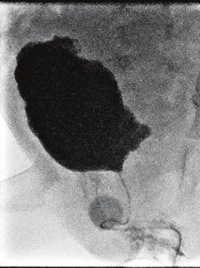
Angefertigt mit der Genehmigung
der Medizinischen Fakultät der Universität Bonn

1. Gutachter*in: Prof. Dr. med. Heiko M. Reutter

2. Gutachter*in: PD Dr. Alfredo Estanislao Ramirez Zuniga

Tag der Mündlichen Prüfung: 10.05.2021

Aus dem Institut für Humangenetik
Direktor: Prof. Dr. med. Markus M. Nöthen
3

Inhaltsverzeichnis

      Abkürzungsverzeichnis                                                 5
1.    Deutsche Zusammenfassung                                              6
1.1   Einleitung                                                            6
1.2   Material und Methoden                                                 8
1.3   Ergebnisse                                                           10
1.4   Diskussion                                                           14
1.5   Zusammenfassung                                                      16
1.6   Literaturverzeichnis der deutschen Zusammenfassung                   17
2.    Publikation A: HSPA6: A new autosomal recessive candidate gene for
      the VATER/VACTERL malformation spectrum
      Abstract                                                             22
      Introduction                                                         23
      Materials and Methods                                                23
      Results                                                              24
      Discussion                                                           26
      References                                                           27
3.    Publikation B: Whole exome sequencing identifies a mutation in
      EYA1 and GLI3 in a patient with branchiootic syndrome and
      esophageal atresia: coincidence or a digenic mode of inheritance?
      Abstract                                                             29
      Introduction                                                         29
      Materials and Methods                                                30
      Results                                                              30
      Discussion                                                           32
      References                                                           33
4.    Publikation C: CAKUT and autonomic dysfunction caused by
      acetylcholine receptor mutations
      Abstract                                                             35
      Report                                                               35
      References                                                           41
4 5. Danksagung 43
5

Abkürzungsverzeichnis

ARM           Anorektale Malformation(en)
Abb.          Abbildung
BO-Syndrom    Branchio-otisches Syndrom
BOR-Syndrom   Branchio-oto-renales Syndrom
CAKUT         congenital anomalies of the kidney and urinary tract
              (kongenitale Anomalien der Nieren und ableitenden Harnwege)
cDNA          complementary deoxyribonucleic acid
              (komplementäre Desoxyribonukleinsäure)
CNV           copy number variation (Kopienzahl-Veränderung)
DNA           Desoxyribonukleinsäure
EA/TEF        esophageal atresia/tracheoesophageal fistula
              (Öophagusatresie/Tracheoösophageale Fistel)
LUTO          lower urinary tract obstruction
              (Verengung der unteren ableitenden Harnwege)
qPCR          quantitative polymerase chain reaction
              (quantitative Polymerase-Kettenreaktion)
VACTERL       Vertebral defects (Wirbelkörperfehlbildungen)
              Anorectal malformations (anorektale Fehlbildungen)
              Cardiac defects (kardiale Fehlbildungen)
              Tracheoesophageal fistula (tracheo-ösophageale Fistel)
              Esophageal atresia (Ösophagusatresie)
              Renal malformations (renale Fehlbildungen)
              Limb defects (Extremitätenfehlbildungen)
WES           whole exome sequencing (Exom-Sequenzierung)
6 1. Deutsche Zusammenfassung 1.1 Einleitung Anorektale Malformationen (ARM) und Ösophagusatresien, mit oder ohne tracheoösophagealer Fistel (EA/TEF), sind seltene Fehlbildungen des embryonalen Vorder- und Hinterdarms. Sie kommen mit einer Prävalenz von 1 pro 3.000-4.000 Lebendgeburten vor (Cuschieri, 2001; Depaepe et al., 1993). Ein gemeinsames Auftreten mit anderen Fehlbildungen ist in 50-60% der Fälle zu beobachten (De Jong et al., 2010; Cuschieri et al., 2001; Balanescu et al., 2013). Hierbei ist am häufigsten zusätzlich der Urogenitaltrakt mit etwa 80% betroffen (Stoll et al., 2007). Eine alleinige Kombination von ARM und Fehlbildungen der oberen Gliedmaßen tritt in etwa 6% der Fälle auf (van den Hondel et al., 2016). ARM und EA/TEF treten ebenfalls als Teil von genetischen Syndromen und Assoziationen auf, z.B. im Rahmen der VATER/VACTERL-Assoziation. Diese beschreibt das gleichzeitige Auftreten von mindestens drei Fehlbildungen in folgenden Organsystemen: Wirbelsäule (V), anorektale Malformation (A), kardiale Defekte (C), tracheoösophageale Fistel mit oder ohne Ösophagusatresie (TE), renale Fehlbildungen (R), Fehlbildungen der Extremitäten (L). Liegen nur in zwei dieser Organsysteme Fehlbildungen vor, wird dieses als VATER/VACTERL-ähnliche Assoziation bezeichnet. Über die genetischen Ursachen von ARM und EA/TEF ist bisher wenig bekannt. Saisawat et al. konnten ein rezessives Krankheitsgen, TRAP1, in Zusammenhang mit ARM bzw. der VATER/VACTERL-Assoziation und kongenitalen Anomalien der Nieren und der abführenden Harnwege (congenital anomalies of the kidney and urinary tract, CAKUT) beschreiben (Saisawat et a., 2014). In Mausmodellen wurden Gli2 und Gli3 als essenziell für die embryologische Entwicklung der Speiseröhre identifiziert (Motoyoma et al, 1998). Beide sind Teil des Hedgehog-Signalwegs. CAKUT sind der häufigste Grund für eine chronische Niereninsuffizienz in den ersten drei Lebensdekaden und treten mit einer Prävalenz von 3-6 pro 1.000 Lebendgeburten auf. Eine Verengung der unteren ableitenden Harnwege (Lower Urinary Tract Obstruction, LUTO) ist hierbei durch sekundäre Folgen eines Harnaufstaus, wie zum Beispiel bei vesikoureteralem Reflux (VUR) oder Hydronephrose, eine wichtige Ursache für die chronische Niereninsuffizienz. Monoallelische und biallelische Varianten in über 40 Genen sind als verursachend für CAKUT identifiziert worden, können jedoch nur bei ca.
7

5%-20% der Patienten die Fehlbildungen erklären (van der Ven et al., 2018).
Vorherrschend sind hierbei Gene, die in die Regulation wichtiger embryologischer
Entwicklungsprozesse des Urogenitaltraktes eingebunden sind (van der Ven et al., 2018;
Vivante et al., 2014). Zu beachten sind jedoch auch Gene, die verantwortlich für die
neuronale   Regulation   der   Blasenentleerung    sind.   Bisher   wurden   in   diesem
Zusammenhang biallelische Varianten in dem Gen CHRM3 gefunden, welche zu einer
Verengung der unteren ableitenden Harnwege führen (Weber et al., 2011).

Das Ziel der drei Studien zu ARM, EA/TEF und CAKUT war, Kandidatengene mittels
„whole exome sequencing“ (WES) zu identifizieren und zu re-sequenzieren.

In Publikation A lag der Schwerpunkt auf der Suche nach autosomal-rezessiven
(biallelischen) und X-chromosomal-rezessiven Kandidatengenen in drei Familien mit ARM
oder VATER/VACTERL-ähnlichem Phänotyp.

Publikation B beschreibt einen Indexpatienten, bei dem eine EA/TEF mit dem seltenen
autosomal-dominant vererbten branchio-otischen Syndrom (BOS1: OMIM #602588)
zusammenfällt. Dieses hat eine Geburtenprävalenz von 1 pro 40.000 Lebendgeburten
(Fraser et al., 1980) und ist gekennzeichnet durch Fehlbildungen der embryonalen
Kiemenbogen und der Ohren. Das BO-Syndrom ist allelisch zum branchio-oto-renalen
Syndrom (BOR1: OMIM #113650) (Vincent et al., 1997), bei welchem zusätzlich
Fehlbildungen der Nieren auftreten. Ursächlich für beide Krankheitsbilder, BOS1 und
BOR1, sind monoallelische Varianten in EYA1 und SIX1 (Abdelhak et al., 1997, Vincent
et al., 1997; Ruf et al., 2003; Ruf et al., 2004). Für das BOR-Syndrom sind zusätzlich
ursächliche Varianten in SIX5 (Hoskins et al., 2006) bekannt. Hier sollte ein möglicher
Zusammenhang zwischen der EA/TEF und dem BO-Syndrom im Indexpatienten
genetisch untersucht werden.

Ziel der in Publikation C besprochenen Studie war ebenfalls, autosomal-rezessive
(biallelische) Kandidatengene bei einem Patienten mit CAKUT und neurogener
Blasenstörung zu identifizieren. Für diese Studie führte die Arbeitsgruppe von Prof. Dr.
Friedhelm Hildebrandt am Boston Children’s Hospital, Department of Pediatrics/Boston
8

(USA) ein „Homozygosity mapping“ (Homozygotie-Kartierung) durch (Hildebrandt et al.,
2009). Die aus dem „Homozygosity mapping“ generierten Daten wurden auf Anhäufung
homozygoter Regionen im Genom hin untersucht. Die durch WES gewonnenen Sequenz-
Daten filterte ich systematisch auf rezessive (biallelische) Varianten in diesen
homozygoten Regionen. Zur funktionellen Untersuchung des Kandidatengens führte ich
unter anderem zellbiologische Methoden an HEK293 Zellen durch (HEK=human
embryonic kidney).

1.2 Material und Methoden

In den drei Studien kamen bei den Indexpatienten und -familien grundlegend
molekulargenetische Methoden zum Einsatz: Für Publikation A und B bereitete ich die
„DNA libraries“ der einzelnen Patienten vor. Die WES-Daten generierte dann unser
Kooperationspartner Cologne Center for Genomics/Universität Köln mit einem
IlluminaHiSeq2500 Sequencer. Ich wertete diese mit dem Exom- und Genom-Analyse-
Programm „Varbank“ aus (https://varbank.ccg.uni-koeln.de). Für Publikation C wurden
mittels einer Illumina™ Plattform WES-Daten generiert, die ich mit dem Exom-und-
Genom-Analyse-Programm CLC Genomics Workbench (version 6.5.1) (CLC bio)
auswertete. Der Schwerpunkt lag hier auf Regionen, die sich im „Homozygosity mapping“
als vererbt homozygot zeigten. Die Priorisierung der Varianten erfolgte mithilfe einer
Begrenzung der Allelfrequenz auf
9

In Publikation A wurden drei Geschwisterpaare mit ARM oder VATER/VACTERL-
ähnlichem Phänotyp und deren gesunde Eltern untersucht (Familie 1098, Familie 971,
Familie 1346). In Familie 1346 zeigte das betroffene Geschwisterpaar einen
VATER/VACTERL-ähnlichen Phänotyp mit ARM, Hypoplasie und Ankylose beider kleinen
Finger bei der Schwester und isolierter Radiushypoplasie bei dem Bruder (Abb. 1). Mittels
WES und Sanger-Sequenzierung suchte ich in den drei Familien nach rezessiven
homozygoten, compound-heterozygoten und X-chromosomalen Varianten. Bei Familie
1346 wandte ich zusätzlich eine auf SYBR® Green basierte qPCR (quantitative
polymerase chain reaction) zur Identifizierung von copy number variations (CNVs) an. Zur
weiteren Untersuchung des in der Folge beschriebenen Kandidatengens re-sequenzierte
ich dieses nach Sanger in 167 Patienten mit VATER/VACTERL- und VATER/VACTERL-
ähnlicher Assoziation, die mit ARM und Fehlbildungen der Gliedmaßen einen ähnlichen
Phänotyp wie die betroffenen Kinder dieser Familie hatten.

In Publikation B wies der Indexpatient eine EA/TEF vom Typ Vogt IIIb (Vogt, EC, 1929)
und ein BO-Syndrom mit beidseitigen Hals- und Ohrfisteln auf. Seine Mutter und seine
Schwester zeigten ebenfalls Merkmale des BO-Syndrom mit einseitigem Hörverlust und
unilateraler Ohrfistel bei der Mutter und bilateralen Ohrfisteln sowie unilateraler Halsfistel
bei der Schwester. Der Bruder wies lediglich ein unspezifisches präaurikuläres Grübchen
auf (Abb.2). Initial untersuchte ich den Indexpatienten auf Varianten in EYA1, SIX1 und in
Genen, die mit beiden im Hedgehog-Signalweg interagieren. Für alle identifizierten
Varianten führte ich dann eine Segregationsanalyse bei Geschwistern und Eltern durch.
Zusätzlich untersuchte ich mit Sequenzierung nach Sanger 18 weitere Patienten mit
EA/TEF und BO-Syndrom assoziierten Anomalien (Hörverlust oder Fehlbildungen der
Ohren).

In Publikation C zeigte der Indexpatient eine neurogene Blasenstörung mit
vesikoureteralem     Reflux    (VUR)    Grad       5,   Hydronephrose    und    progressiver
Niereninsuffizienz. Zusätzlich fielen in der augenärztlichen Untersuchung weite und
fixierte Pupillen, entsprechend einer Mydriasis, auf (Abb. 3). Entsprechend der familiären
Vererbung der Fehlbildungen (gesunde Eltern und betroffenes Kind) filterte ich die WES-
Daten nach rezessiven (biallelischen bzw. X-chromosomalen) Varianten. Über das online-
10

Netzwerk   Genematcher      (https://genematcher.org/statistics)   und   Screening   einer
hauseigenen Kohorte von 380 Familien mit CAKUT suchte ich nach weiteren Patienten
mit rezessiven Varianten in dem durch WES identifizierten Krankheitsgen. Die über
Genematcher einbezogenen Patienten wurden durch unsere Kooperationspartner an den
Universitäten von Bristol (Vereinigtes Königreich) und Exeter (Vereinigtes Königreich)
klinisch untersucht, die auch ihre Sequenzdaten generierten. Unser Kooperationspartner
an der Yale Universität, Department of Genomics/New Haven (USA) generierte die WES-
Daten des Indexpatienten und der hauseigenen Kohorte. Für die funktionelle
Untersuchung der Mutationen führte ich Immunofluoreszenz-Experimente in HEK293
Zellen durch. In cDNA (complementary deoxyribonucleid acid) fügte ich hierfür die
identifizierten Mutationen mittels zielgerichteter Mutagenese ein und brachte sie als C-
terminale GFP-markierte Konstrukte in die HEK293 Zellen ein. Wildtyp- und mutierte
Proteine lokalisierte ich mit konfokaler Fluoreszenzmikroskopie in den Zellen. Zusätzlich
wurden durch unsere Kooperationspartner an der University of Texas, Southwestern
Medical Center/Texas (USA) ektrophsyiologische Studien mit patch-clamp-Technik
ebenfalls in HEK293 Zellen durchgeführt.

1.3 Ergebnisse

Publikation A: Die hier untersuchten Daten der drei Familien wurden auf rezessive
homozygote, compound-heterozygote und X-chromosomale Varianten hin gefiltert. In
Familie 1346 identifizierte ich bei dem betroffenen Geschwisterpaar eine zunächst
scheinbar homozygote Variante in HSPA6 (c.1340G>T, gemäß ENSEMBL GRCh37/hg19
Transkript ENST00000309758). Die Segregationsanalyse zeigte jedoch, dass nur die
Mutter heterozygote Trägerin der Variante war und der Vater einen Wildtyp aufwies.
Mittels qPCR konnte ich folgend nachweisen, dass der Vater eine Deletion von HSPA6
trägt. Beide Kinder erbten diese Deletion vom Vater. Somit sind beide Kinder compound-
heterozygot für die Deletion ihres Vaters und die Punktmutation ihrer Mutter (Abb. 1). Als
Regulator von Zellzyklen und Teil apoptischer Signalkaskaden ist HSPA6 ein wichtiger
protektiver Faktor während der embryonalen Entwicklung (Luft & Dix, 1999). Um weitere
Familien mit Varianten in HSPA6 zu identifizieren, sequenzierte ich 167 Patienten mit
11

ARM und VATER/VACTERL-ähnlichem Phänotyp. Dabei ließen sich keine weiteren
biallelischen Varianten in HSPA6 identifizieren.
In Familie 971 zeigten sich compound-heterozygote Varianten in ITSN2 (c.1442T>A und
c.4367A>G, entsprechend ENSEMBL GRCh37/hg19 Transkript ENST00000355123).
Variante c.1442T>A (p.Ile481Asn) stufte ich mithilfe der Vorhersage-tools als schädigend
ein. Variante c.4367A>G (single nucleotide polymorphism [SNP]: rs139986826;
p.Asn1456Ser) erwies sich jedoch als benigne, da ein geringer Einfluss auf die
Proteinfunktion und -konformation vorlag und die Aminosäure Alanin an dieser Stelle nur
schwach konserviert ist. Hiermit konnten beide Varianten als Ursache der Fehlbildungen
in einem compound-heterozygoten Erbgang ausgeschlossen werden.
In Familie 1098 wurden keine rezessiven Varianten identifiziert.

                      I.1                  I.2

 HSPA6 wt/del                            HSPA6 wt/G447V

                      II.1                 II.2

HSPA6 del/G447V                           HSPA6 del/G447V

Abb. 1: Stammbaum der in Publikation A untersuchten Familie 1346.
1346_II.1: isolierte radiale Hypoplasie beidseits.
1346_II.2: anorektale Malformation in Form einer perinealen Fistel, Hypoplasie und
Ankylose beider kleinen Finger.
Durch WES und qPCR identifizierte Mutationen und Wildtyp sind angezeigt. HSPA6,
heat shock 70kD protein 6; wt, Wildtyp; del, Deletion; G, Glycin; V, Valin.

Publikation B: Meine WES-Analyse ergab bei dem Indexpatienten eine vorbeschriebene
EYA1-Mutation in der Donor-Spleißstelle von Exon 10 (c.966+5G>A, entsprechend
ENSEMBL Transkript ENST00000340726) (Stockley et al., 2009; Krug et al., 2011; Song
et al., 2013; Berkheineria et al., 2017). Diese konnte ebenfalls bei den vom BO-Syndrom
betroffenen Familienmitgliedern, Schwester und Mutter des Indexpatienten, identifiziert
12
werden. Das weitere Filtern der WES-Daten zeigte in den mit EYA1 und SIX1
interagierenden Genen folgende Varianten: GLI1 (p.Thr176Met), GLI3 (Spleiß-Variante
c.1028+3A>G, rs368499795, entsprechend ENSEMBL Transkript ENST00000395025),
NRP1 (p.Asp601Asn) und SMO (p.Arg168His) (Abb. 2). Von den Varianten in GLI1, GLI3,
NRP1 und SMO berücksichtigte ich in der Folge nur die Varianten, die der gesunde Vater
ausschließlich auf den Indexpatienten übertrug, da hier möglicherweise durch das
Zusammenspiel mit der EYA1-Mutation im Sonic-hedgehog pathway das Auftreten von
BO-Syndrom und EA/TEF im Indexpatienten verursacht wurde. Alleine die heterozygote
GLI3 Spleiß-Variante c.1028+3A>G erfüllte dieses Kriterium. Laut „Human Splicing Finder
3.0“ (Desmet et al., 2009) sowie nach Shapiro und Senapathy (Shapiro und Senapathy,
1987) kann diese den korrekten Spleiß-Vorgang behindern. Die Varianten in GLI1, NRP1
und SMO wurden ausgeschlossen, da diese entweder von der Mutter, welche keine
EA/TEF aufwies, an die Kinder vererbt wurden oder, im Fall der NRP1- und SMO-
Variante, gering konserviert bzw. zu häufig waren. Das Screening einer Kohorte von 18
Patienten mit EA/TEF und Merkmalen des BO-Syndroms konnte keine weiteren potentiell
krankheitsverursachenden Varianten in EYA1 aufdecken.
I.1 I.2
EYA1 wt/wt EYA1 wt/Spleiß
GLI3 wt/Spleiß GLI3 wt/wt
SMO wt/wt SMO wt/R168H
GLI1 wt/wt GLI1 wt/T176M
NRP1 wt/wt NRP1 wt/wt
II.1 II.2 II.3
EYA1 wt/wt EYA1 wt/Spleiß EYA1 wt/Spleiß
GLI3 wt/wt GLI3 wt/wt GLI3 wt/Spleiß
SMO wt/R168H SMO wt/R168H SMO wt/R168H
GLI1 wt/wt GLI1 wt/wt GLI1 wt/T176M
NRP1 wt/wt NRP1 wt/wt NRP1 wt/D601N
Abb. 2: Stammbaum der in Publikation B untersuchten Familie.
I.2: BO-Syndrom (einseitiger Hörverlust, unilaterale Ohrfistel)
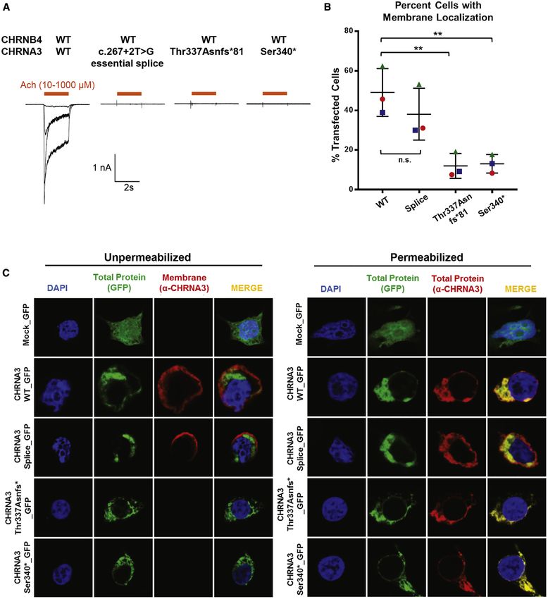
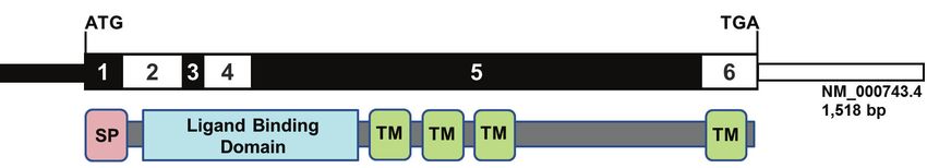
II.2: BO-Syndrom (bilaterale Ohrfisteln, unilaterale Halsfistel)13 II.3: Öophagusatresie/Tracheoösophageale Fistel Typ Vogt IIIb, BO-Syndrom (beidseitige Hals- und Ohrfisteln). Der Indexpatient wird durch einen Pfeil markiert. Durch WES und Sanger Sequenzierung identifizierte Mutationen und Wildtyp sind angezeigt. BO-Syndrom, Branchio-otisches Syndrom; wt, Wildtyp; Spleiß, Spleiß-Variante; EYA1, Drosophila eyes absent; GLI, GLI family zinc finger; SMO, smoothened, frizzled class receptor; NRP1, neuropilin 1; R, Arginin; H, Histidin; T, Threonin; D, Asparaginsäure; M, Methionin; N, Asparagin. Publikation C: Bei dem Indexpatienten und seinem betroffenen Bruder identifizierte ich rezessiv-homozygote Varianten in CHRNA3 (entsprechend ENSEMBL Transkript ENST00000326828.5; c.1010_1011delCA, p.T337Nfs*81) (Abb. 3). CHRNA3 kodiert die α-Untereinheit des α3β4-nikotinischen Rezeptors (α3β4-nAchR). Über Genematcher fand sich dann ein betroffenes Geschwisterpaar von konsanguinen Eltern, welches eine homozygote Variante aufwies (c.1019C>G, p.S340*). Beide Varianten, p.T337Nfs*81 und p.Ser340*, sind potentiell trunkierend und können möglicherweise zu einer Verkürzung des Proteins vor der vierten Transmembranhelix führen. Nach Screening von 666 WES- Datensätzen (380 Familien) der eigenen CAKUT-Kohorte konnte eine Spleiß-Variante in einer Patientin identifiziert werden (c.267+2T>G), welche vermutlich zum Aussetzen von Exon 3 und einer in-frame Deletion von 15 Aminosäuren in der extrazellulären Ligandenbindungsstelle führt. Alle drei genannten Patienten wiesen einen ähnlichen urogenitalen und ophtalmologischen Phänotyp auf wie der Indexpatient. CHRNA3 ist ein Membranprotein, welches als Teil des α3β4-nAchR fungiert. In den Lokalisationsstudien in HEK293 Zellen zeigte sich, dass nur der Wildtyp vollständig an der Zellmembran vorhanden war. Die den Mutationen p.T337Nfs*81 und p.S340* entsprechenden trunkierten Proteinen wanderten nur zu einem geringfügigen Anteil bis zu der Zellmembran und verblieben zu einem großen Teil im zellulären Plasma. In den elektrophysiologischen Studien an transfizierten HEK293 Zellen konnte bei beiden trunkierten Proteinen und bei dem durch die Spleiß-Variante veränderten Protein kein Natrium-Einwärtsstrom, ausgelöst durch Bindung von Acetylcholin, nachgewiesen werden. Hier zeigte sich somit ein vollkommener Funktionsverlust des α3β4-nAchR.
14

             I.1                     I.2

             II.1                    II.2

 CHRNA3                     CHRNA3
 p.T337Nfs*81/              p.T337Nfs*81/
 p.T337Nfs*81               p.T337Nfs*81

Abb. 3: Stammbaum der in Publikation C untersuchten Indexfamilie.
II.1: nicht-neurogene neurogene Blasenstörung, vesikoureteraler Reflux Grad 5,
Hydronephrose, progressive Niereninsuffizienz, Mydriasis.
II.2: rezidivierende Harnwegsinfekte, verminderter Lichtreflex der Pupillen.
Der Indexpatient wird durch einen Pfeil markiert. Durch WES und Sanger-Sequenzierung
identifizierte Mutationen sind angezeigt. CHRNA3, cholinergic receptor nicotinic alpha 3
subunit; T, Threonin; N, Asparagin; fs, frameshift.

1.4 Diskussion

Bei den in Publikation A-C untersuchten Familien war das Ziel, monogene Ursachen für
die jeweiligen Fehlbildungen zu identifizieren.

Thema von Publikation A ist die Identifikation von HSPA6 als ein rezessives
Kandidatengen für Fehlbildungen im Zusammenhang mit der VATER/VACTERL
Assoziation. Nicht nur seine embryonale Funktion, sondern auch das vorbeschriebene
TRAP1, welches wie HSPA6 für ein Hitzeschockprotein kodiert, legen seine ursächliche
Rolle bei der Entstehung der beschriebenen Fehlbildungen nahe. Es fanden sich jedoch
keine weiteren biallelischen Varianten in einer Kohorte von 167 Patienten mit ARM und
VATER/VACTERL ähnlichem Phänotyp. Erstrebenswert wären eine Re-Sequenzierung
in   einer      größeren   Kohorte     und   tiefergehende   Studien   zur   Beteiligung   von
15 Hitzeschockproteinen an der Entstehung von ARM und Komponenten der VATER/VACTERL-Assoziation. Die Mikrodeletion von HSPA6 in Familie 1346 zeigt auf, dass auch CNVs zu biallelischen Krankheitsbildern als ursächliche Variante beitragen können. Darüber hinaus wäre es wünschenswert, die Beteiligung von CNVs bei Patienten zu untersuchen, bei denen keine monogenetische Ursache identifiziert werden kann. Publikation B beschreibt das gleichzeitige Auftreten von EA/TEF und BO-Syndrom bei einem Patienten. Hier kann die EYA1-Mutation als alleinige Ursache gewertet werden, da EYA1 im Hedgehog-Signalweg als upstream-Koordinator mit FGF10 interagiert, welches im Tiermodell in die Entwicklung der Speiseröhre involviert ist (Korzh et al., 2011; Hajduk et al, 2010). Die EYA1-Mutation kann jedoch auch im Zusammenspiel mit Mutationen in weiteren Genen gesehen werden, welche im Hedgehog-Signalweg mit EYA1 interagieren. Die GLI3-Mutation liegt hier in Kombination mit der EYA1-Mutation nur im Indexpatienten vor und suggeriert eine digenische Ursache für die EA/TEF beim Indexpatienten. Es könnte daher sein, dass die Kombination beider Varianten die Signalkaskade des Sonic hedgehog Signalwegs stört. Auch hier ist eine größere Kohorte mit EA/TEF und BO-Syndrom assoziierten Merkmalen wünschenswert, um einer möglichen Involvierung von EYA1 in die Entstehung von EA/TEF nachzugehen. Einschränkend können ursächliche de novo Mutationen nicht ausgeschlossen werden. Diese hätten nur in einer Trio-basierten WES-Analyse identifiziert werden können. In Publikation C werden rezessive (biallelische) Varianten in CHRNA3 in drei nicht verwandten Familien beschrieben. CHRNA3 greift als Teil des α3β4-nAchR in die neuronale Regulation der Blase ein. Es wird sowohl in autonomen Ganglien als auch im Urothel exprimiert (Beckel et al., 2006; Fowler et al., 2008). Die Expression in autonomen Ganglien erklärt die dysautonomen Merkmale der betroffenen Patienten: diese reichen von der Fehlregulation der Blasenkontraktion und -relaxation bis zu einer Mydriasis. Die Übereinstimmung mit dem Mausmodell, in dem Chrna-/- Mäuse eine Megacystis, rezidivierende Harnwegsinfekte und Mydriasis zeigen, ist hier unübersehbar (Xu et al., 1999). Für LUTO als Unterform von CAKUT sind bisher nur wenige Gene beschrieben worden, welche hauptsächlich in die Kontraktion von glatten Muskelzellen mittels Aktin oder in
16

synaptische neuronale Signalweiterleitung eingreifen (Weber et al., 2011; Wangler et al.,
2014; Milewicz et al., 2010; Daly et al., 2010; Stuart et al., 2013). Einzig für das Gen
CHRM3, welches wie CHRNA3 in die neuronale Regulation der Blase involviert ist, sind
ebenfalls Patienten mit persistierender Mydriasis beschrieben worden (Weber et al.,
2011). Das unabhängige Auftreten der Mutationen in unterschiedlichen Familien und die
funktionale Untersuchung der mutierten Proteine unterstützen CHRNA3 als gutes
Kandidatengen. Das Augenmerk sollte so nicht nur auf Gene gerichtet werden, die in die
embryonale Entwicklung des Harntraktes oder in mechanische Vorgänge eingreifen,
sondern auch auf Gene, die in die neuronale Regulation von beteiligten Strukturen
involviert sind. Die hier untersuchten Patienten zeigen eine phänotypische Variabilität mit
zum Teil nur versteckten oder fehlenden dysautonomen Merkmalen.
Voraussetzung für ein besseres Verständnis der Genotyp-Phänotyp-Korrelation ist die
Identifikation weiterer Patienten mit CHRNA3-Mutationen.

1.5 Zusammenfassung

In den vorliegenden Studien (Publikationen A-C) untersuchte ich die genetischen
Ursachen intestinaler Atresien und von CAKUT mittels Exom-Sequenzierung. Dabei
standen monogene Erklärungsansätze im Mittelpunkt. HSPA6 als Kandidatengen für
ARM als Teil der VATER/VACTERL-Assoziation konnte nur in einer betroffenen Familie
identifiziert werden. Im Fall des Patienten mit BO-Syndrom und EA/TEF suggeriert das
Ergebnis ein digenisches Modell durch die Kombination von monoallelischen Varianten in
EYA1 und GLI3. In CHRNA3 konnten wir für mehrere Familien mit gleichem Phänotyp
ursächlich biallelische Varianten nachweisen. Funktionelle Studien unterstützen diese
genetischen Ergebnisse. Im Kontext der neuronalen autonomen Regulation der Blase
durch den α3β4-nAchR entsteht so eine pathophysiologische Sequenz, in deren Rahmen
die   neurogene   Blasenstörung     sekundär     zu   CAKUT   führt.   Die   systematische
Identifizierung weiterer genetischer Ursachen und weiterer Patienten mit ursächlichen
Varianten in den beschriebenen Genen wird zu einem umfassenderen Verständnis der
molekularen Vorgänge und der Krankheitsbilder führen. Im Fall der Patienten, die
17

ursächliche Varianten in CHRNA3 tragen, ergeben sich gegebenenfalls, anders als bei
Patienten mit mechanischen LUTOs, medikamentös therapeutische Optionen.
Darüber hinaus ist ein besseres Verständnis der genetischen Ursachen unerlässlich für
die genetische Beratung von Patienten und deren Familien.

1.6 Literaturverzeichnis

Abdelhak S, Kalatzis V, Heilig R, Compain S, Samson D, Vincent C, Weil D, Cruaud C,
Sahly I, Leibovici M, Bitner-Glindzicz M, Francis M, Lacombe D, Vigneron J, Charachon
R, Boven K, Bedbeder P, van Regemorter N, Weissenbach J, Petit C. A human
homologue of the Drosophila eyes absent gene underlies Branchio-Oto-Renal (BOR)
syndrome and identifies a novel gene family. Nature Genetics. 1997; 15: 157-164

Balanescu    RN,   Topor    R,   Moga     A.     Anomalies   associated   with   anorectal
malformations.Chirurgia. 2013; 108: 38-42

Beckel JM, Kanai A, Lee SJ, de Groat WC, Birder LA. Expression of functional nicotinic
acetylcholine receptors in rat urinary bladder epithelial cells. American journal of
physiology-Renal physiology. 2006; 290: F103-110

Bekheirnia MR, Bekheirnia N, Bainbridge MN, Gu S, Akdemir ZHC, Gambin T, Janzen
NK, Jhangiani SN, Muzny DM, Michael M, Brewer ED, Elenberg E, Kale AS, Riley AA,
Swartz SJ, Scott DA, Yang Y, Srivaths PR, Wenderfer SE, Bodurtha J, Applegate CD,
Velinov M, Myers A, Borovik L, Craigen WJ, Hanchard NA, Rosenfeld JA, Lewis RA,
Gonzales ET, Gibbs RA, Belmont JW, Roth DR, Eng C, Braun MC, Lupski JR, Lamb DJ.
Whole-exome sequencing in the molecular diagnosis of individuals with congenital
anomalies of the kidney and urinary tract and identification of a new causative gene.
Genetics in Medicine. 2017; 19: 412-420

Cuschieri A. Descriptive epidemiology of isolated anal anomalies: A survey of 4.6 million
births in Europe. American Journal of Medical Genetics. 2001; 103: 207–215

Daly SB, Urquhart JE, Hilton E, McKenzie EA, Kammerer RA, Lewis M, Kerr B, Stuart H,
Donnai D, Long DA, Burgu B, Aydogdu O, Derbent M, Garcia-Minaur S, Reardon W,
18

Gener B, Shalev S, Smith R, Woolf AS, Black GC, Newman WG. Mutations in HPSE2
cause urofacial syndrome. American Journal of Human Genetics. 2010; 86: 963-969

De Jong EM, Douben H, Eussen BH, Felix JF, Wessels MW, Poddighe PJ, Nikkels PG,
de Krijer RR, Tibboel D and de Klein A. 5q11.2 deletion in a patient with tracheal agenesis.
European Journal of Human Genetics. 2010; 18: 1265-1268

Depaepe A, Dolk H and Lechat MF. The epidemiology of tracheo-esophageal fistula and
oesophageal atresia in Europe. EUROCAT Working Group. Archives of Disease in
Childhood. 1993; 68: 743-748

Desmet FO, Hamroun D, Lalande M and Collod-Béroud G, Claustres M and Béroud C.
Human splicing finder: An online bioinformatics tool to predict splicing signals. Nucleic
Acids Research. 2009; 37: e67

Fowler CJ, Griffiths D, de Groat WC. The neural control of micturition. Nature Reviews
Neuroscience. 2008; 9: 453-466

Fraser FC, Sproude JR and Halal F. Frequency of the branchio-oto-renal (BOR) syndrome
in children with profound hearing loss. American Journal of Medical Genetics. 1980; 7:
341-349

Hajduk P, Murphy P and Puri P. Fgf10 gene expression is delayed in the embryonic lung
mesenchyme in the adriamycin mouse model. Pediatric Surgery International. 2010; 26:
23-27

Hildebrandt F, Heeringa SF, Rüschendorf F, Attansio M, Nürnberg G, Becker C, Seelow
D, Huebner N, Chernin G, Vlangos CN, Zhou W, O’Toole JF, Hoskins BE, Wolf MTF,
Hinkes BG, Chaib H, Ashraf S, Schoeb DS, Ovunc B, Allen SJ, Vega-Warner V, Wise E,
Harville HM, Lyons RH, Washburn J, MacDonald J, Nürnberg P, Otto EA. A systematic
approach to mapping recessive disease genes in individuals from outbred populations.
Plos Genetics. 2009; 5: e1000353

Hoskins BE, Kramer CH, Silvius D, Zou D, Raymond RM, Orten DJ, Kimberling WJ, Smith
RJ, Weil D, Petit C, Otto EA, Xu PX, Hildebrandt F. Transcription factor SIX5 is mutated
19

in patients with branchio-oto-renal syndrome. American Journal of Human Genetics. 2007;
80: 800-804

Korzh S, Winata CL, Zheng W, Yang S, Yin A, Ingham P, Korzh V and Gong Z. The
interaction of epithelial Ihha and mesenchymal Fgf10 in zebrafish esophageal and
swimbladder development. Developmental Biology. 2011; 359: 262-276

Krug P, Moriniére V, Marlin S, Koubi V, Gabriel HD, Colin E, Bonneau D, Salomon R,
Antignac C and Heidet L. Mutation screening of the EYA1, SIX1 and SIX5 genes in a large
cohort of patients harboring branchio-oto-renal syndrome calls into question the
pathogenic role of SIX5 mutations. Human Mutation. 2011; 32: 183-190

Milewicz DM, Ostergaard JR, Ala-Kokko LM, Khan N, Grange DK, Mendoza-Londono R,
Bradley TJ, Olney AH, Adès L, Maher JF, Guo D, Buja LM, Kim D, Hyland JC, Regalado
ES. De novo ACTA2 mutation causes a novel syndrome of multisystemic smooth muscle
dysfunction. American Journal of Medical Genetics Part A. 2010; 152A: 2437-2443.

Motoyama J, Liu J, Mo R, Ding Q, Post M and Hui CC. Essential function of Gli2 and Gli3
in the formation of lung, trachea and oesophagus. Nature Genetics. 1998; 20: 54-57

Ruf RG, Xu PX, Silvius D, Otto EA, Beekmann F, Muerb UT, Kumar S, Neuhaus TJ,
Kemper MJ, Raymond RM Jr, Brophy PD, Berkman J, Gattas M, Hyland V, Ruf EM,
Schwartz C, Chang EH, Smith RJ, Stratakis CA, Weil D, Petit C, Hildebrandt F. SIX1
mutations cause branchio-oto-renal syndrome by disruption of EYA1-SIX1-DNA
complexes. Proceedings of the National Academy of Sciences USA. 2004; 101:
8090-8095

Shapiro MB and Senapathy P: RNA splice junctions of different classes of eukaryotes:
Sequence statistics and functional implications in gene expression. Nucleic Acids
Research.1987; 15: 7155-7174

Saisawat P, Kohl S, Hilger AC, Hwang D-Y, Gee HY, Dworschak GC, Tasic V,
Pennimpede T, Natarajan S, Sperry E, Matassa DS, Stajić N, Bogdanovic R, de Blaauw
I, Marcelis CL, Wijers CH, Bartels E, Schmiedeke E, Schmidt D, Märzheuser S, Grasshoff-
Derr S, Holland-Cunz S, Ludwig M, Nöthen MM, Draaken M, Brosens E, Heij H, Tibboel
20

D, Herrmann BG, Solomon BD, de Klein A, van Rooij IA, Esposito F, Reutter HM,
Hildebrandt F. Whole exome resequencing reveals recessive mutations in TRAP1 in
individuals with CAKUT and VACTERL association. Kidney International. 2014; 85: 1310–
1317

Song MH, Kwon TJ, Kim HR, Jeon JH, Baek JI, Lee WS, Kim UK, Choi JY. Mutational
analysis of EYA1, SIX1 and SIX5 genes and strategies for management of hearing loss
in patients with BOR/BO syndrome. PLoS One. 2013; 8: e67236

Stockley TL, Mendoza-Londono R, Propst EJ, Sodhi S, Dupuis L and Papsin BC. A
recurrent EYA1 mutation causing alternative RNA splicing in branchio-oto-renal
syndrome: Implications for molecular diagnostics and disease mechanism. American
Journal of Medical Genetics Part A. 2009; 149A: 322-327

Stoll C, Alembik Y, Dott B, Roth MP. Associated malformations in patients with anorectal
anomalies. European Journal of Medical Genetics. 2007; 50: 281-290

Stuart HM, Roberts NA, Burgu B, Daly SB, Urquhart JE, Bhaskar S, Dickerson JE,
Mermerkaya M, Silay MS, Lewis MA, Olondriz MB, Gener B, Beetz C, Varga RE, Gülpınar
O, Süer E, Soygür T, Ozçakar ZB, Yalçınkaya F, Kavaz A, Bulum B, Gücük A, Yue WW,
Erdogan F, Berry A, Hanley NA, McKenzie EA, Hilton EN, Woolf AS, Newman WG. LRIG2
mutations cause urofacial syndrome. American Journal of Human Genetics. 2013; 92:
259-264

van den Hondel D, Wijers CHW, van Bever Y, de Klein A, Marcelis CLM, de Blaauw I,
Sloots CEJ, Ijsselstijn H. Patients with anorectal malformation and upper limb anomalies:
Genetic evaluation is warranted. European Journal of Pediatrics. 2016; 175: 489–497

van der Ven AT, Vivante A, Hildebrandt F. Novel Insights into the Pathogenesis of
Monogenic Congenital Anomalies of the Kidney and Urinary Tract. Journal of the
American Society of Nephrology: JASN. 2018; 29: 36-50

Vincent C, Kalatzis V, Abdelhak S, Chaib H, Compain S, Helias J, Vaneecloo FM and
Petit C. BOR and BO syndromes are allelic defects of EYA1. European Journal of Human
Genetics. 1997; 5: 242-246
21

Vivante A, Kohl S, Hwang DY, Dworschak GC, Hildebrandt F. Single-gene causes of
congenital anomalies of the kidney and urinary tract (CAKUT) in humans. Pediatric
Nephrolology. 2014; 29: 695-704

Vogt EC: Congenital esophageal atresia. American Journal of Roentgenology.1929; 22:
463-465

Wangler MF, Gonzaga-Jauregui C, Gambin T, Penney S, Moss T, Chopra A, Probst FJ,
Xia F, Yang Y, Werlin S, Eglite I, Kornejeva L, Bacino CA, Baldridge D, Neul J, Lehman
EL, Larson A, Beuten J, Muzny DM, Jhangiani S; Baylor-Hopkins Center for Mendelian
Genomics, Gibbs RA, Lupski JR, Beaudet A. Heterozygous de novo and inherited
mutations in the smooth muscle actin (ACTG2) gene underlie megacystis-microcolon-
intestinal hypoperistalsis syndrome. PLoS genetics. 2014; 10: e1004258

Weber S, Thiele H, Mir S, Toliat MR, Sozeri B, Reutter H, Draaken M, Ludwig M, Altmüller
J, Frommolt P, Stuart HM, Ranjzad P, Hanley NA, Jennings R, Newman WG, Wilcox DT,
Thiel U, Schlingmann KP, Beetz R, Hoyer PF, Konrad M, Schaefer F, Nürnberg P, Woolf
AS. Muscarinic Acetylcholine Receptor M3 Mutation Causes Urinary Bladder Disease and
a Prune-Belly-like Syndrome. American Journal of Human Genetics. 2011; 89: 668-674
22

Received: 30 December 2018          Revised: 21 February 2019    Accepted: 3 March 2019
DOI: 10.1002/bdr2.1493

ORIGINAL ARTICLE

HSPA6: A new autosomal recessive candidate gene
for the VATER/VACTERL malformation spectrum
Franziska Kause1                    | Rong Zhang1,2 | Michael Ludwig3 | Eberhard Schmiedeke4 |
Anke Rissmann5 | Holger Thiele6 | Janine Altmueller6,7 | Stefan Herms2,8,9 | Alina C. Hilger1,10 |
Friedhelm Hildebrandt11 | Heiko Reutter1,12

1
 Institute of Human Genetics, University of Bonn,
Bonn, Germany                                            Background: The VATER/VACTERL association refers to the nonrandom co-
2
 Department of Genomics, Life & Brain Center,            occurrence of at least three of the following component features (CFs): vertebral
Bonn, Germany                                            defects (V), anorectal malformations (ARM) (A), cardiac defects (C), tracheoesophageal
3
 Department of Clinical Chemistry and Clinical           fistula with or without esophageal atresia (TE), renal malformations (R), and limb defects
Pharmacology, University of Bonn, Bonn,
                                                         (L). Patients presenting with two CFs have been termed VATER/VACTERL-like
Germany
4
                                                         phenotypes.
 Clinic for Paediatric Surgery and Paediatric
Urology, Klinikum Bremen-Mitte, Bremen,                  Methods: We surveyed the exome for recessive disease variants in three affected sib-
Germany                                                  pairs. Sib-pair 971 consisted of two brothers with ARM and additional hydronephrosis
5
 Malformation Monitoring Centre Saxony-Anhalt,           in one brother. Sib-pair 1098 consisted of two sisters with ARM. In family 1346, the
Medical Faculty, Otto-von-Guericke University,           daughter presented with ARM and additional hypoplasia of both small fingers and
Magdeburg, Germany
6
                                                         ankyloses. Her brother presented with unilateral isolated radial hypoplasia. Sib-pairs
 Cologne Center for Genomics, University of
Cologne, Cologne, Germany
                                                         971 and 1346 resembled a VATER/VACTERL-like phenotype.
7
 Center for Molecular Medicine Cologne
                                                         Results: We detected a novel maternally inherited missense variant (c.1340G > T) and
(CMMC), University of Cologne, Cologne,                  a rare paternally inherited deletion of the trans-allele in HSPA6 in both siblings of family
Germany                                                  1346. HSPA6 belongs to the heat shock protein (HSP) 70 family. Re-sequencing of
8
 Institute of Medical Genetics and Pathology,            HSPA6 in 167 patients with VATER/VACTERL and VATER/VACTERL-like pheno-
University Hospital Basel, Basel, Switzerland
9
                                                         types did not reveal any additional bi-allelic variants.
 Department of Biomedicine, Human Genomics
                                                         Conclusions: Until now, only TNF-receptor associated protein 1 (TRAP1) had
Research Group, University of Basel, Basel,
Switzerland                                              been reported as an autosomal recessive disease-gene for the VATER/VACTERL
10
 Children's Hospital, University of Bonn, Bonn,          association. TRAP1 belongs to the heat shock protein 90 family (HSP90). Both
Germany                                                  Hsp70 and Hsp90 genes have been shown to be important embryonic drivers in the
11
 Division of Nephrology, Boston Children's               formation of mouse embryonic forelimb tissue. Our results suggest HSPA6 as a
Hospital, HMS, Boston, Massachusetts
                                                         new candidate gene in VATER/VACTERL-like phenotypes.
12
  Department of Neonatology and Pediatric
Intensive Care, Children's Hospital, University
                                                         KEYWORDS
of Bonn, Bonn, Germany
Correspondence                                           anorectal malformations, autosomal recessive inheritance, HSPA6,
Franziska Kause, Institute of Human Genetics,            VATER/VACTERL association, whole-exome sequencing
University of Bonn, Bonn, Germany.
Email: s4frkaus@uni-bonn.de
Funding information
BONFOR/ University of Bonn, Grant/Award
Numbers: O-149.0096, O.149.0123; Deutsche
Forschungsgemeinschaft, Grant/Award Number:
RE 1723/4-1; Else-Kröner-Fresenius-Stiftung,
Grant/Award Number: 2014_A14; National
Institutes of Health, Grant/Award Number:
DK076683

Birth Defects Research. 2019;1–7.                               wileyonlinelibrary.com/journal/bdr2                   © 2019 Wiley Periodicals, Inc.    1
23
2 KAUSE ET AL.
1 | INTRODUCTION esophageal atresia, www.great-konsortium.de). Written in-
formed consent was obtained from every family. The study
Anorectal malformations (ARMs) are rare disorders of the was approved by the Ethics Committee of the Medical Faculty
embryonic distal hindgut. They occur in about 1 in 4.000 of the University of Bonn (Lfd. Nr. 073/12 & 146/12). Geno-
live births (Cuschieri, 2001; Bartels et al., 2012). They are mic DNA was isolated from whole blood using the Chemagic
caused by defective differentiation of the primitive hindgut DNA Blood Kit special (Chemagen, Baesweiler, Germany).
(Matsumaru et al., 2015). ARM present with a broad pheno- Genomic DNA from saliva samples was isolated with the
typic spectrum ranging from perineal fistula to severe cloacal Oragene DNA Kit (DNA Genotek Inc., Kanata, Canada).
malformations. For phenotypic classification of ARM, Our study included three affected sib-pairs and their healthy
the Krickenbeck classification system is usually applied parents. In Family 971, two affected brothers (971_II.1,
(Holschneider et al., 2005). Around 60% of ARM patients 971_II.2) presented with ARM and additional hydronephrosis
present with co-occurring congenital malformations, most of in 971_II.1 and ARM in form of a perineal fistula in 971_II.2.
which belong to the congenital anomaly spectrum of the Sib-pair 1098 consisted of two affected sisters (1098_II.1,
VATER/VACTERL association. The VATER/VACTERL 1098_II.2) presenting with ARM in form of a perineal fistula.
association refers to the nonrandom co-occurrence of at least In Family 1346, the affected daughter (1346_II.1) presented
three of the following component features (CFs): vertebral with ARM in form of a perineal fistula and additional hypo-
defects (V), ARMs (A), cardiac defects (C), tracheoesophageal plasia of both small fingers and ankyloses. Her brother
fistula with or without esophageal atresia (TE), renal mal- (1346_II.2) presented with unilateral isolated radial hypopla-
formations (R), and limb defects (L) (Belloni et al., 2000; sia. Hence, Sib-pairs 971 and 1346 resemble the phenotypic
Kohlhase, Wischermann, Reichenbach, Froster, & Engel, spectrum of a VATER/VACTERL-like phenotype (Table 1).
1998; Solomon et al., 2014). Patients presenting with two CFs For re-sequencing of the top candidate gene, we
have been termed VATER/VACTERL-like phenotypes. ARM chose 167 patients presenting with VATER/VACTERL
in combination with major upper limb malformations as and VATER/VACTERL-like phenotypes.
defined by van den Hondel et al. occurs in about 6% of ARM
patients (van den Hondel et al., 2016). 2.2 | Whole-exome sequencing (WES) and data
After the detection of causative copy number variations analysis
in patients with ARM and ARM as part of their VATER/
VACTERL association (Bartels et al., 2011; Dworschak et al., WES was performed at the Next Generation Sequencing
2013; Schramm, Draaken, Bartels, Boemers, Aretz, et al., Laboratory of the Institute of Human Genetics of the Univer-
2011; Schramm, Draaken, Bartels, Boemers, Schmiedeke, sity of Bonn. For the enrichment of exonic and adjacent
et al., 2011; Schramm, Draaken, Tewes, et al., 2011) Saisawat intronic sequences from genomic DNA, the NimbleGen
et al. (2014) were the first who identified a recessive disease- SeqCap EZ Human Exome Library v2.0 enrichment kit was
gene, namely TNF-receptor associated protein 1 (TRAP1), in used. WES was performed on an Illumina HiSeq2500
patients with VATER/VACTERL association and congenital sequencer using a 100 bp paired-end read protocol, follow-
anomalies of the kidney and urinary tract (CAKUT) (Saisawat ing the manufacturer's recommendations. Data analysis and
et al., 2014). TRAP1, located on 16p13.3, belongs to the heat filtering of mapped target sequences was accomplished
shock protein 90 family (HSP90) (Chen, Piel, Gui, Bruford, & with “Varbank” exome and genome analysis pipeline v.2.1
Monteiro, 2005). (https://varbank.ccg.uni-koeln.de). We especially filtered
Here, we aimed to identify rare autosomal recessive for high-quality (coverage of more than six reads, a mini-
disease-genes for isolated ARM or ARM as part of VATER/ mum quality score of 10, VQSLOD greater then −8) and
VACTERL-like phenotypes. For this purpose, we performed rare (allele frequency < 0.5%) autosomal variants. In Sib-
whole-exome sequencing (WES) in three affected sib-pairs pair 971 with two affected brothers, we also filtered for
and their healthy parents and re-sequenced the top candidate X-chromosomal recessive variants. A detailed description of
gene in 167 patients with VATER/VACTERL and VATER/ filter criteria has been described elsewhere (Zhang et al., 2016).
VACTERL-like phenotypes.
2.3 | Confirmation of variants detected by WES
Variants identified by WES and called to be possibly causa-
2 | M AT E R IA L S A ND M E T H O DS
tive were amplified from the DNA by polymerase chain
reaction (PCR). Automated sequence analysis was carried
2.1 | Patients, controls, and DNA isolation out using standard procedures. From the UCSC Human
Patients and families were recruited through the German net- Genome Browser (https://genome.ucsc.edu/) and Ensembl
work for congenital uro-rectal malformations (CURE-Net, (http://www.ensembl.org/index.html) genomic and cDNA
www.cure-net.de) and the great-consortium (genetic risk of sequences were obtained to compile primers. PCR products24
KAUSE ET AL. 3
TABLE 1 Phenotypic features of the patients of Family 1098, org.uk/], PROVEAN [http://provean.jcvi.org/index.php], Muta-
971, and 1346
tion Taster [http://www.mutationtaster.org/], PolyPhen-2 [http://
Family genetics.bwh.harvard.edu/pph2/], and the CADD-PHRED-score
number Patient Anorectal malformation Other features
[http://cadd.gs.washington.edu/home]).
1098 II.1 Anorectal malformation in form
of a perineal fistula
II.2 Anorectal malformation in form
of a perineal fistula 3 | RESULTS
971 II.1 Anorectal malformation Hydronephrosis, left
II.2 Anorectal malformation in form Using WES, we investigated three affected sib-pairs and
of a perineal fistula their healthy parents for autosomal recessive disease vari-
1346 II.1 Anorectal malformation in form Hypoplasia and
of a perineal fistula ankyloses of both
ants. In the case of affected male brothers, we also surveyed
small fingers the exome for X-chromosomal disease variants (Table 1).
II.2 Radial hypoplasia Analysis of the WES-data revealed two candidate genes in
Family 971 and Family 1346. No X-chromosomal disease
variant was found in Sib-pair 971. No autosomal recessive
were subjected to direct automated BigDye Terminator se- disease variants were found in Sib-pair 1098.
quencing (3130XL Genetic Analyzer, Applied Biosystems,
Foster City, CA). Both strands from each amplicon were
3.1 | Sib-pair #971
sequenced and presence of the variants in each family was
confirmed by sequencing the respective PCR product (primer We detected two possible disease-causing variants in ITSN2,
sequences are available upon request). suggesting a compound heterozygous background in the
affected brothers 971_II.1 and 971_II.2 (c.1442 T > A and
2.4 | Re-sequencing of candidate gene HSPA6 c.4367A > G, according to ENSEMBL GRCh37/hg19
transcript ENST00000355123). The variant c.1442 T > A
For the amplification of DNA, PCR primers were designed
(p.Ile481Asn, ENSP00000347244) was predicted to be dele-
for exon 1 of HSPA6. The resultant PCR products were
terious by five out of seven in silico prediction tools (delete-
referred to direct automated sequencing with the 3130XL
rious in SIFT [score: 0], medium functional impact by
Genetic Analyzer (Applied Biosystems).
Mutation Assessor [FI-score: 2.175], tolerated in fathmm
[score: 0.08], deleterious in PROVEAN [score: −5.80],
2.5 | Quantitative polymerase chain reaction
disease-causing in Mutation Taster [p value: 1], probably
Quantitative PCR (qPCR) was performed on a Roche Light damaging by PolyPhen-2 [humvar score 0.996], CADD
Cycler 480 II (Roche, Basel, Switzerland) with Roche Light score of 25.4). In contrast, for the second variant c.4367A >
Cycler Sybr Green I Master Mix. Each assay included the G (single nucleotide polymorphism [SNP]: rs139986826; p.
respective DNA specimen at a final concentration of 10 ng/μL Asn1456Ser [ENSP00000347244]), the amino acid change
in triplicate. Reaction mixtures (10 μL) contained 0.2 mmol of was predicted to be deleterious by two out of the seven in
each primer and 5 μL of SYBR Green Master Mix (Roche) silico prediction tools (tolerated in SIFT [score 0.64], low
with cycling conditions as follows: initiation 50! C for 2 min, functional impact in Mutation Assessor [score: 1.01], toler-
denaturation 95! C for 10 min, followed by 40 cycles at 95! C ated in fathmm [score: −0.26], deleterious in PROVEAN
for 15 s, and a combined annealing and extension step at 64! C [score: −3.67], disease-causing in Mutation Taster [p value:
for 60 s. The threshold cycle (Ct) values were normalized using 0.926], benign in PolyPhen-2 [humvar score 0.009],
the Ct value of three reference genes (BNC1, CFTR, and CADD-PHRED-score of 17.3). The zebrafish (Danio rero)
RNAseP subunit p38) for each DNA sample. Relative quantifi- and the green spotted pufferfish (Tetraodon nigroviridis)
cation was done using the comparative Ct method normalizing present with a Serine at the homologous position. Previ-
to the mother as a control sample, since she was found to be ously, high-throughput sequencing revealed recessive muta-
heterozygous for variant c.1340G > T and therefore could not
tions of ITSN2 as causing nephrotic syndrome (NS) in humans
carry a deletion of this region (primer sequences are available
(Ashraf et al., 2018). Neither of the two brothers (971_II.1 and
upon request).
971_II.2) presented with symptoms of NS. Of course, we can-
not exclude that the two brothers will manifest NS in the future.
2.6 | In silico prediction of variants However, none of the initially reported patients with NS and
To predict the effect of the variants on the function and structure recessive disease variants in ITSN2 exhibited any CF of the
of the encoded proteins, we used seven online prediction VATER/VACTERL association spectrum. Hence, we assume
tools (SIFT [http://sift.jcvi.org/], Mutation Assessor 3 [http:// that the variants in ITSN2 are probably not disease causing for
mutationassessor.org/r3/], fathmm [http://fathmm.biocompute. the development of ARM in an autosomal recessive context in25
4 KAUSE ET AL.
our patients. Screening for X-linked variants did not reveal any
Note. CADD v1.3: combined annotation dependent depletion v1.3 PHRED like scaled C-score; Ce: Caenorhabditis elegans; Ci: Ciona intestinalis; Cp: Cavia porcellus; D: deleterious; D.C.: disease causing; Dm: Drosophila melano-
gaster; Dr: Danio rerio; FATHMM: functional analysis through Hidden Markov models (v2.3); G: Glycine; Gg: Gallus gallus; h: heterozygous; MutA: Mutation Assessor 3 functional impact score; MutT: Mutation Taster prediction
Sc
G
–
disease variant for the two brothers.
Dm
G
–
3.2 | Sib-pair #1346
Ce
G
–
In Sib-pair 1346, WES revealed an apparent homozygous
class; PROVEAN: protein variation effect analyzer; PP2: PolyPhen2 humvar score; Sc: Saccharomyces cervisiae; SIFT: sorting intolerant from tolerant prediction class; T: tolerated; V: valine; Xt: Xenopus tropicalis.
mutation in heat shock protein family A (HSP70) member
Ci
G
–
6 (HSPA6 or HSP70B', here referred to as HSPA6)
Dr
(c.1340G > T, according to ENSEMBL GRCh37/hg19 tran-
G
–
script ENST00000309758) in exon 1. Polyphen2, SIFT,
Xt
G
Mutation Taster, Mutation Assessor and PROVEAN
–
predicted the mutation to be damaging (Table 2). With a
Gg
G
–
CADD-score of 25.5, it is among 1% of the most deleterious
variants in the human genome. The HSPA6 p.Gly447Val
CP
G
–
mutation (SNP: rs56366425) alters a highly conserved
amino acid. It is conserved throughout the species to
Maternal inheritance
Paternal inheritance
Segregation/State
Saccharomyces cerevisiae with no orthologue in mice
(Leung, Hall, Rajendran, Spurr, & Lim, 1992) (Table 2).
of alleles
Segregation analysis revealed a heterozygous variant in the
mother. The father presented with a wild type allele and
qPCR revealed a deletion of HSPA6 on the trans-allele. Both
CADD v1.3
Mutations in HSPA6 (NM_002155.3) in patients 1346_II.1 and 1346_II.2 with anorectal malformations and limb deformities
children inherited variant c.1340G > T from their mother.
They also inherited a microdeletion harboring HSPA6 from
25.5
their father on the trans-allele (Figure 1). In our in-house
–
control cohort, 1 out of 1.320 healthy individuals carried a
PROVEAN
deletion at the HSPA6 locus (data not shown). Copy number
variations (CNVs) in our in-house controls were identified
D
via the human Infinium Omni 2.5 BeadChip (Illumina, San
–
Diego, CA) and evaluated with the Illumina Genome Viewer
FATHMM
program. The frequency of 1 in 1.320 (≈0.0008) of the
observed deletion in our population-based control cohort is
T
–
clearly below the population-based frequency of ARM and
ARM as part of the VATER/VACTERL association in a
MutA
4.085
recessive disease-model (1 in 4.000 and 1 in 40.000, respec-
–
tively; Cuschieri, 2001; Bartels et al., 2012). HSPA6 is a
SIFT
member of the HSP70 protein family coding for HSPs
D
–
throughout the human organism (Leung, Rajendran, Monfries,
Hall, & Lim, 1990). It contains only one exon of 2.731
1.000
PP2
base pairs (according to ENSEMBL GRCh37/hg19 transcript
–
ENST00000309758), a conserved N-terminal nucleotide-
MutT
D.C.
binding domain as well as a C-terminal peptide substrate-
–
binding domain (Wisniewska et al., 2010).
acid change
To follow up on this finding, we re-sequenced HSPA6 in
G447 V
167 patients with VATER/VACTERL or VATER/VACTERL-
Amino
like phenotypes resembling the phenotypes of patients
–
1346_II.1 and 1346_II.2 (ARM and limb malformation).
Exon
Sanger sequencing revealed four exonic heterozygous mis-
1
1
sense variants with minor allele frequencies (MAF) > 0.08.
Additionally, we found three synonymous rare exonic variants
Nucleotide alteration/
(MAF < 0.08) and two exonic synonymous common SNPs
deletion (zygosity)
c.1340G > T (h)
were detected. In the untranslated region, three common SNPs
Deletion (h)
as well as one rare variant (MAF unknown) were detected.
TABLE 2
Overall, we did not detect any further recessive disease variants
in the investigated cohort.26
KAUSE ET AL. 5
(a) I.1 I.2 (b) I.1 I.2
ITSN2 wt/N1456S ITSN2 wt/I481N
II.2 II.1
II.2 II.1
I481N/N1456S I481N/N1456S
(c) I.1 I.2
HSPA6 wt/del HSPA6 wt/G447V
II.2 II.1
HSPA6 del/G447V HSPA6 del/G447V
FIGURE 1 Pedigrees of the families. (a) 1098_II.1 and 1098_II.2 presented both with ARM in form of a perineal fistula. (b) 971_II.1 presented with ARM
and left hydronephrosis. 971_II.2 presented with ARM in form of a perineal fistula. (c) 1346_II.1 presented with ARM in form of a perineal fistula,
hypoplasia and ankylosis of both small fingers. 1346_II.2 presented with isolated radial hypoplasia. The presence or absence of genetic variants detected by
whole-exome sequencing and (quantitative) polymerase chain reaction are indicated. Members affected with anorectal or skeletal malformations are shown in
black, while unaffected members are shown in white. Males and females are represented in squares and circles, respectively. del: deletion; HSPA6: heat shock
70kD protein 6; ITSN2: intersection 2; wt: wild type
4 | DISCUSSION live births, respectively; Cuschieri, 2001; Bartels et al., 2012).
Screening 167 patients with VATER/VACTERL or VATER/
Our WES analysis identified compound heterozygous vari- VACTERL-like phenotypes resembling the phenotypes of
ants in an affected sib-pair with congenital anomalies of the patients 1346_II.1 and 1346_II.2 (ARM and limb malforma-
VATER/VACTERL association spectrum. Both carried a tion) did not reveal any recessive disease variants.
novel missense variant c.1340G > T in HSPA6, inherited The highly conserved HSPA6 gene (Daugaard, Rohde, &
from their healthy mother and a deletion of HSPA6 on the Jäättelä, 2007; Gupta & Golding, 1993) belongs to the multi-
trans-allele inherited from their healthy father. The variant gene HSP family A (HSP70). Little is known about the func-
was absent in the gnomAD browser beta (http://gnomad. tion of HSPA6, a gene present in the human genome but
broadinstitute.org/ [Lek et al., 2016] or in the exome variant absent from mouse and rat (Deane & Brown, 2018). To our
server database (Exome Variant Server, NHLBI GO Exome knowledge, there are no animal models for functional
Sequencing Project (ESP), Seattle, WA (URL: http://evs.gs. screening of the orthologous gene although it is present in
washington.edu/EVS/) [12/2018]). In the ExAC Browser other vertebrates and species such as frog, fruit fly, or yeast
(Beta) (http://exac.broadinstitute.org/ [Lek et al., 2016]) the (Table 2). Emerging research suggests the HSPA6 protein to
allele count for this variant was 36 in 121098 alleles without be involved in apoptotic response and cellular differentiation
any homozygous individuals listed and with an allele fre- and to contribute to protection of differentiated human neu-
quency of about 0.0003. Fifteen studies listed in the database ronal cells from cellular stress (Deane & Brown, 2018).
of genomic variants describe the loss of genome sequences Only when induced after severe stress events (Parsian et al.,
in the region of HSPA6 (http://dgv.tcag.ca/dgv/app/home? 2000), it functions as a secondary responder to proteotoxic
ref=; (MacDonald, Ziman, Yuen, Feuk, & Scherer, 2014)) stress and maintains cellular survival by directly binding to
without mentioning population-based frequencies. Here, we Apaf-1 and inhibiting the assembly of a functional apoptosome
investigated a healthy control cohort of 1.320 individuals (Beere et al., 2000; Noonan, Place, Giardina, & Hightower,
and found one of these 1.320 healthy individuals to carry a 2007). The involvement of HSP70s in regulation of the cell
deletion at the HSPA6 locus revealing a population-based cycle and apoptotic pathways is essential for human embryonal
frequency clearly below the population-based frequency of development as they protect embryonal cells from cell stress
ARM and ARM as part of the VATER/VACTERL associa- such as hyperthermia or chemical teratogens (Luft & Dix,
tion in a recessive disease-model (1 in 4.000 and 1 in 40.000 1999). This is supported by the identification of HSPA6 inSie können auch lesen