Medizinerkolleg Münster Abschlusskolloquium der Kohorte 2020_1 und Auftaktveranstaltung der Kohorte 2021_1 - am 26. und 27. Juli 2021
←
→
Transkription von Seiteninhalten
Wenn Ihr Browser die Seite nicht korrekt rendert, bitte, lesen Sie den Inhalt der Seite unten
Medizinerkolleg Münster

Abschlusskolloquium der Kohorte 2020_1
                   und

Auftaktveranstaltung der Kohorte 2021_1

          am 26. und 27. Juli 2021
Liebe Teilnehmer*innen,
wir begrüßen Sie herzlich zum Abschlusskolloquium des Medizinerkollegs 2020_1. In das
Kolloquium ist die Auftaktveranstaltung für die neue Kohorte 2021_1 integriert, die wir vorab
ebenfalls ganz herzlich willkommen heißen.
Da wir das Kolloquium leider nicht als Präsenzveranstaltung, sondern als Zoom-Konferenz abhalten
müssen, möchten wir Ihnen vorab ein paar Informationen zu diesem Format zukommen lassen:
Bei den Vorträgen ist eine Redezeit von 15min und Diskussionszeit von 5min vorgesehen.
Sie sind alle herzlich eingeladen, sich aktiv an den Diskussionen zu beteiligen und gerne viele Fragen
zu stellen. Wenn Sie gerade keinen Redebeitrag leisten möchten, bitten wir Sie das Mikrofon
auszustellen.
Im Rahmen eines wertschätzenden Miteinanders während Zoom-Konferenzen möchten wir Sie
bitten ihre Kamera einzuschalten, sofern dies für Sie technisch möglich ist.
Neben den Vorträgen wird es ein weiteres Präsentationsformat geben, welches die in den
vergangenen Jahren durchgeführten Postersessions ersetzt. Diese alternativen Postersessions
werden wie folgt ablaufen:
- Die Präsentierenden bekommen jeweils einen Breakoutraum zugeteilt und präsentieren Ihr
Thema in einer 5minütigen Kurzpräsentation. Anschließend kann 10min diskutiert werden.
Abstracts zu den Kurzpräsentationen finden Sie ab Seite 5 des Programmheftes
- Alle anderen Teilnehmer*innen können sich aussuchen, welchen Raum Sie besuchen und
werden gebeten sich möglichst gut auf die 5 angebotenen Räume zu verteilen.
- Nach 15min werden die Breakouträume geschlossen und für eine weitere Runde erneut
geöffnet. Sie haben die Möglichkeit 4 der 5 angebotenen Räume zu besuchen.
Wir wünschen allen Teilnehmer*innen ein spannendes und erfolgreiches Kolloquium,
Das Organisations-Team
Maurice Dellin (Kohortensprecher 2020_1)
Aliska Brugmans (Kohortenprecherin 2020_1)
Prof. Dr. Rupert Hallmann (Sprecher des MedK)
Melanie Wilbers (Studienkoordinatorin MedK)
1Programm 26.07.2021:
8:15 Begrüßung Prof. Dr. Rupert Hallmann
Vorträge
8:30 “Sonic Hedgehog N-terminal peptide characterization in signaling”, Sophia Ehlers
Prof. Dr. Grobe, Institut für Physiologische Chemie und
Pathobiochemie
8:50 „Impact of Standardized Endurance Exercise on Experimental Daniel Schiffmann
Autoimmune Encephalomyelitis in NOD Mice“, Prof. Dr. Klotz, Klinik
für Neurologie mit Institut für Translationale Neurologie
9:10 „The Regulatory Role of Smarcb1 in the Proliferation of Neuronal Aliska Brugmans
Stem Cells“ – PD Dr. Kerl, Klinik für Kinder- und Jugendmedizin -
Pädiatrische Hämatologie und Onkologie
9:30 Pause 10 min
Postersession I
9:40 PSBI-BR01: “Cross-validation of arterial input functions by Florian Gierse
simultaneous recordings of MR contrast agents and their radioactive
analogs in mice”, Univ.-Prof. Dr. Schäfers, Klinik für Nuklearmedizin
PSBI-BR02: “In vitro effects of the RNA-binding protein Musashi-1 on Isabel Falke
radiation and chemotherapy in endometrial carcinoma”, Prof. Dr.
Greve, Klinik für Strahlentherapie - Radioonkologie
PSBI-BR03: “Effect of fluid shear stress on the membrane integrity of Ann Marleen Starke
endothelial cells”, Univ.-Prof. Dr. Gerke, Institut für Medizinische
Biochemie
PSBI-BR04: “The role of PATJ in cilia maintenance”, Univ.-Prof. Dr. Thomas Mönnig
Dr. Krahn, Medizinische Klinik D
PSBI-BR05: “Non-genomic Action of Steroids on the Slo3 Potassium- Johannes Lorenz
Channel”, Univ.-Prof. Dr. Strünker, Centrum für
Reproduktionsmedizin und Andrologie
10:40 Pause 20 min
2Vorträge
11:00 IGF-IR/PI3K/AKT-dependent regulation of beta-catenin in synovial Hanna Wattendorff
sarcoma, Univ. Prof. Dr. Hartmann, Gerhard-Domagk-Institut –
Sektion für Translationale Pathologie
11:20 “Uptake of the Cell-Penetrating Effector Protein IpaH9.8 of Shigella Franziska Laing
flexneri in Tissue Culture Models”, PD Dr. Rüter, Institut für
Infektiologie
11:40 “The Impact of Intermittent Fasting on Autoimmune Victoria Lampkemeyer
Encephalomyelitis in NOD Mice”, Univ.-Prof. Dr. Klotz, Klinik für
Neurologie mit Institut für Translationale Neurologie
12:00 Mittagspause 60 min
Vorträge
13:00 “In vitro studies on the function of cell surface heparan sulfate in Stefan Krautschneider
radiation resistance of triple negative breast carcinoma”, Prof. Dr.
Greve, Klinik für Strahlentherapie - Radioonkologie
13:20 „Alterations of spermatogonia in testicular tissues from men with Lena Schülke
normal and impaired spermatogenesis“, PD Dr. Neuhaus, Centrum
für Reproduktionsmedizin und Andrologie
13:40 Pause 10 min
Postersession II
13:50 PSBII-BR01: “KCNQ1 and PI(3,5)P2 - a functional and structural Maurice Dellin
analysis of potential binding pockets”, Univ.-Prof. Seebohm, Institut
für Genetik von Herzerkrankungen, Abtl. Zelluläre Elektrophysiologie
PSBII-BR02: “Characterization of TREK1 ion channel activators”, Lucas Spohler
Univ.-Prof. Dr. Dr. Dr. h. c. Meuth, Klinik für Neurologie mit Institut für
Translationale Neurologie
PSBII-BR03: “Characterization of radial spoke defects in patients with Alina Biegemeier
Primary Ciliary Dyskinesia“, Univ.-Prof. Dr. Omran, Klinik für Kinder-
und Jugendmedizin - Allgemeine Pädiatrie
PSBII-BR04: “Characterizing Inner Dynein Arm defects in motile cilia Greta Zweigart
and flagella”, Univ.-Prof. Dr. Omran, Klinik für Kinder- und
Jugendmedizin - Allgemeine Pädiatrie
PSBII-BR05: „Characterization of ciliogenesis defects and the impact Claus Seelmann-Eggebert
of HAS3-Mutations on congenital hydrocephalus“, PD Dr.
Schlingmann, Klinik für Kinder- und Jugendmedizin - Allgemeine
Pädiatrie
314:50 Pause 10 min
Vorträge
15:00 “Validation of potential compounds to promote remyelination using Aurelia Seitz
human oligodendrocytes”, Univ.-Prof. Dr. Kuhlmann, Institut für
Neuropathologie
15:20 “Studies on the influence of Fcy receptor stimulation on macrophage Annika Schwacha
polarization”, Univ.-Prof. Dr. Kiefer, European Institute of Molecular
Imaging (EIMI)
15:40 „Influence of the metabolic state on neuronal signal processing“, Hannes Schmidt
Univ.-Prof. Dannlowski, Institut für Translationale Psychiatrie
16:00 Wrapup Tag 1 (10 min) Prof. Dr. Rupert Hallmann
Programm 27.07.2021:
Vorträge
8:15 “Epigenetic pathways of resistance to BRD4-inhibitor therapy in ETP- Lisa Hüchtker
ALL”, Univ.-Prof. Dr. Rössig, Klinik für Kinder- und Jugendmedizin -
Pädiatrische Hämatologie und Onkologie
8:35 “Biofilm formation and phenotypical characterization of Carolin Gawin
Staphylococcus aures isolates from urine cultures”, Prof. Dr. Kahl,
Institut für Medizinische Mikrobiologie
8:55 „The Interaction of JAM-A with RhoGDI, a Regulator of the Rho Niklas Beckmann
Family of GTPases“, Prof. Dr. Ebnet, Institut für Medizinische
Biochemie
9:15 Pause 10 min
Postersession III
9:25 PSIII-BR01: “Membrane potential dynamics during chemotaxis and Stina Becker
respiratory burst”, Univ.-Prof. Dr. Schwab, Institut für Physiologie II -
Vegetative Physiologie
PSIII-BR02: „Function of Dead End Protein in controlling cell fate of Solveig Reinecke
Primordial Germ Cells“, Univ.-Prof. Dr. Raz, Institut für Zellbiologie
4PSIII-BR03: “Role of Neuregulin1 dependend circRNA´s in the Helen Haupt
functional network of the brain of the mouse”, Univ.-Prof. Dr. Zhang,
Klinik für Psychische Gesundheit
PSBIII-BR04: “Longitudinal rs-fMRI and graph theoretical analysis Leo Hebbelmann
reveal brain network changes in the GAERS rat model of absence
epilepsy”, Univ.-Prof. Dr. Faber, Klinik für Radiologie
PSBIII-BR05: “ Studies on colibactin polyketide expression in E. Ann-Kathrin Alraun
coli”,Univ.-Prof. Dr. Dobrindt, Institut für Hygiene
10:25 Pause 20 min
Vorträge
10:45 “Stress-induced sex-specific changes in brain structures of ZDHHC7 Sadik-Emre Cicibas
mutants”, Prof. Dr. Weiqi Zhang, Klinik für psychische Gesundheit,
Labor für molekulare Neurowissenschaften
11:05 Characterisation of the SARS-CoV-2 receptor ACE2 and the new Beate Conrad
subtype of type 2 pneumocytes in human lung tissue according to
clinical characteristica“, Univ.-Prof. Dr. Wiewrodt,
Medizinische Klinik A
11:25 “Characterising the role of TP53 in the pathogenesis of pediatric Leon Feldmeyer
Burkitt lymphoma”, Univ.-Prof. Dr. Burkhardt, Klinik für Kinder- und
Jugendmedizin - Pädiatrische Hämatologie und Onkologie
11:45 Resümee und Verabschiedung (15 min) Prof. Dr. Rupert Hallmann
5Poster-Abstracts:
Postersession I:
PSI-BR01: „Cross-validation of arterial input functions by simultaneous recordings of MR
contrast agents and their radioactive analogs in mice“
Florian Gierse
Univ.-Prof. Dr. Schäfers, Klinik für Nuklearmedizin
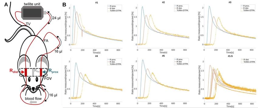
Quantitative measurement of the dynamic arterial blood concentration (arterial input function, AIF) is
a prerequisite for robust pharmacokinetic modeling in PET and MRI. However, for both modalities
AIF recordings are especially challenging in mice. In the current study, we simultaneously recorded
MRI AIFs and radioactive contrast agent analogs in an extracorporeal circulation approach.
12 intracranial tumor bearing nude mice were measured in a 9.4 T Bruker Biospec MRI. 35 mM Gd-
DO3A-butrol (7 mice) or Gd-DTPA (5 mice) were co-injected i.v. with their radioactive analog; 68Ga-
DO3A-butrol (7.2 Mbq ± 2.4) or 99mTc-DTPA (23.1 MBq ± 6.2). The extracorporeal circulation was
applied shunting the femoral artery to the tail vein. It featured 2 reservoirs in the MR field of view and
a MR compatible unit (Swisstrace, Twilite) for measurements of blood radioactivity (Figure 1A). A
novel Golden-angle Radial Sparse Parallel (GRASP) sequence was used for DCE-MRI. Compressed
sensing MP2RAGE was employed for T1 mapping.
Integrated simultaneous recordings of PET and DCE-MRI AIFs using the Twilite unit were technically
feasible. Dynamic acquisition demonstrated very little noise at 5s temporal resolution while allowing
3D isotropic whole brain coverage. Well quantitative correspondence on the whole range of dynamic
contrast agent and radiotracer concentrations was established when applying a fixed correction
factor of 1.8 (Figure 1B). Although our preliminary analysis does not yet feature dispersion correction,
the findings point towards a high precision across individual animals and establish a basis for
quantitative comparison of AIF recordings in hybrid small animal PET/MRI.
6PSI-BR02: „In vitro effects of the RNA-binding protein Musashi-1 on radiation and
chemotherapy in endometrial carcinoma“
Isabel Falke
Prof. Dr. Greve, Klinik für Strahlentherapie – Radioonkologie
Endometrial carcinoma is the most common gynecological cancer in Europe. Radiotherapy is
recommended for adjuvant treatment in patients with intermediate to high risk of recurrence based
on molecular classifications and patients with recurrent disease(1). Previous work of our group
showed increased Musashi-1 (Msi-1) expression in endometrial carcinoma compared to normal
endometrium(2) and identified a 20.8-fold higher Msi-1 expression in putative cancer stem cells(3).
High levels of Msi-1 are associated with decreased survival, indicating prognostic relevance(4). In
our study we characterized potential therapeutic effects and underlying molecular mechanisms of
Msi-1 in two endometrial carcinoma cell lines, Ishikawa and KLE. Specifically, we investigated effects
of siRNA-mediated Msi-1 knockdown regarding resistance to chemotherapy (via MTT assay) and
radiotherapy (via colony formation). qPCR and western blot studies were performed to characterize
the underlying mechanistic interplay. Colony formation and MTT cell viability assay revealed
significantly decreased cell viability after knockdown. While no additional chemosensitization was
seen, Msi-1 knockdown radiosensitized cancer cells. Telomerase, pathologically expressed in tumor
cells for immortality, and DNA-dependent protein kinase, responsible for DNA damage repair after
radiation, were downregulated after knockdown suggesting potential mechanistic explanations.
Notch pathway elements and cell cycle regulator p21 were also changed in expression, underlining
the antiproliferative effect. Given the convincing anti-proliferative and radiosensitizing effect after
knockdown, our results strongly indicate that Musashi-1 could be a potential therapeutic target for
treatment of endometrial carcinoma.
1. Concin N et al. ESGO/ESTRO/ESP guidelines for the management of patients with endometrial carcinoma. Int J
Gynecol Cancer. 2021; doi: 10.1136/ijgc-2020-002230
2. Götte M, Wolf M et al. Increased expression of the adult stem cell marker Musashi-1 in endometriosis and
endometrial carcinoma. J Pathol. 2008; doi: 10.1002/path.2364
3. Götte M, Greve B, Kelsch R et al. The adult stem cell marker Musashi-1 modulates endometrial carcinoma cell
cycle progression and apoptosis via Notch-1 and p21WAF1/CIP1. Int J Cancer. 2011; doi: 10.1002/ijc.25856
4. Ma L, Xu YL, Ding WJ, Shao HF, Teng YC. Prognostic value of Musashi-1 in endometrioid adenocarcinoma. Int J
Clin Exp Pathol. 2015
7PSI-BR03: „Effect of fluid shear stress on the membrane integrity of endothelial cells“
Ann Marleen Starke
Univ.-Prof. Dr. Gerke, Institut für Medizinische Biochemie
Eukaryotic cells depend on an intact plasma membrane to maintain cellular homeostasis, function,
and thus viability. Disruption of the plasma membrane is a common but serious threat to mammalian
cells and can be caused by bacterial toxins, chemicals, radiation, and mechanical stress. The latter
challenges various tissues in our body during physiological activities. Membrane damage in skeletal
muscle cells during stress has been well studied. Another tissue subjected to mechanical stress is
the endothelium of our blood vessels, which is subjected to hemodynamically generated mechanical
forces. To date, little is known about the membrane degrading effects of shear stress on endothelial
cells. I will present a new approach to study the membrane degrading effect of shear forces on
HUVECs using a flow-based live cell microscopy set up. Using this, we quantified the amount of
wounded cells in a cell monolayer exposed to fluid shear stress.
To survive membrane damage, mammalian cells have evolved various processes to restore
membrane integrity. As diverse as the causes of membrane injury are, so are the repair mechanisms,
but all require calcium as a trigger. Different processes leading to a repaired plasma membrane have
been proposed and studied for several decades. A key role in plasma membrane repair is exocytosis
of pre-existing intracellular vesicles. We were able to show that early and late endosomes undergo
exocytosis after shear stress induced membrane wounds, which possibly contribute to the process
of membrane repair.
PSI-BR04: „The role of PATJ in cilia maintenance“
Thomas Mönnig
Univ.-Prof. Dr. Dr. Krahn, Medizinische Klinik D
PATJ is a scaffold protein that is associated to tight junctions. Together with Pals1 and Crumbs it
forms the crumbs complex, which plays an important role in the development of apico basal polarity
in epithelial cells.
Prior research performed by our group showed that knockout of PATJ in MDCK cells leads to an
impaired maintenance of primary cilia and results in fewer ciliated cells. Primary cilia are non-motile
sensory organelles, which transmit extracellular signals into the cell. Defects in primary cilia have
been shown to lead to diseases called “ciliopathies”, an example being polycystic kidney disease.
One group of proteins, which have been shown to be involved in regulation of ciliogenesis, are
histone deacetylases (HDACs). A protein interaction screen performed by Luck et al. identified a
possible interaction between PATJ and HDAC71. In order to investigate whether this interaction might
8be the mechanism for the loss of cilia in MDCK dPATJ we treated MDCK cells with different HDAC
inhibitors, performed a knockdown of HDAC7 and measured the effect on ciliation. We also
performed coimmunoprecipitation experiments to try and validate the possible interaction between
PATJ and HDAC7.
1
Luck et al.(2020) A reference map of the human binary protein interactome. Nature 580, 402–408
PSI-BR05: „Non-genomic Action of Steroids on the Slo3 Potassium-Channel“
Johannes Lorenz
Univ.-Prof. Dr. Strünker, Centrum für Reproduktionsmedizin und Andrologie
Although the role of the sperm-specific potassium-channel Slo3 in fertilization is not yet well
understood, it is known that the channel is vital for a successful fertilization as demonstrated by
knock-out experiments. It has been assumed that the effects of the Slo3 channel in sperm-physiology
are mediated through its inhibition by steroid hormones like progesterone. Here we show that even
though steroid hormones do have an inhibitory effect on Slo3, the physiological response to local
influences is mediated by some other non-steroidal molecule.
Patch-clamp recordings of heterologously expressed Slo3 channels were performed to investigate
the effect of different substances on that very channel. We were able to show that six (Progesterone,
17-OH-Progesterone, Estradiol, Testosterone, DHEA and Corticosterone) out of the twelve steroid
hormones found in human follicular fluid (hFF) do have a distinct inhibitory effect on the Slo3
potassium-channel. However, the much stronger effect of native hFF on Slo3 could not be explained
by the compound effect of all steroids combined. To consolidate our hypothesis of another molecule
acting on the Slo3 channel, we performed measurements with hFF that had been stripped of lipid
mediators like steroids. This stripped hFF had an almost equal effect on the Slo3 channel, when
compared to native hFF strengthening our theory of a yet unknown molecule acting on the Slo3
potassium-channel.
9Postersession II:
PSII-BR01: „KCNQ1 and PI(3,5)P2 - a functional and structural analysis of potential binding
pockets“
Maurice Dellin
Univ.-Prof. Seebohm, Institut für Genetik von Herzerkrankungen, Abtl. Zelluläre Elektrophysiologie
The long QT syndrome (LQTS) is a condition in which the repolarization of the heart is prolonged,
resulting in an increased risk of an irregular heartbeat which can result in fainting, seizures, and/or
sudden cardiac death. The most common genetic basis for LQTS are mutations within the KCNQ1
channel (also known as Kv7.1), a classic slow delayed rectifying potassium channel in complex
with its ß-subunit KCNE1 mediates the IKS current in the human heart’s repolarization.
A vast majority of the known mutations is localized within the inner cell segments of the channel.
These regions are especially important for interactions with intracellular regulating pathways (e.g.
the interaction with PI(3,5)P2 and PI(4,5)P2 phospholipids). In our project we investigated the S0
alpha helix and the S2-3 loop, which both can contain LQT1 associated mutations. We created 11
mutants of a cysteine-removed variant of KCNQ1 by PCR mutagenesis (all single amino acid
substitution to cysteine) and expressed them in oocytes of Xenopus laevis. The cells were then
used in TEVC experiments, where we investigated the influence of the co-expressed kinase
PIKfyve (resulting in high intracellular levels of PI(3,5)P2) and the injection of cadmium on the
macroscopic current of the KCNQ1/KCNE1 complex. We found several mutants with varying
electrophysiological properties compared to the wild type indicating an important role of the
mutated amino acids in the regulation of KCNQ1/KCNE1.
To explore the molecular mechanisms of our measured results we are currently conducting
molecular dynamics simulations using the software YASARA.
PSII-BR02: „Characterization of TREK1 ion channel activators“
Lucas Spohler
Univ.-Prof. Dr. Dr. Dr. h. c. Meuth, Klinik für Neurologie mit Institut für Translationale Neurologie
In Multiple Sclerosis, a chronic autoimmune disease of the CNS, a reduced integrity of the blood
brain barrier plays an important role in the pathogenesis.[1] This then leads to the immigration of
leukocytes into the CNS, causing demyelination and axonal injury.
It was shown, that activating TREK1, a potassium channel belonging to the K2P family, reduces the
transmigration of immune cells across the blood brain barrier. [2] TREK1 activation consequently
might be a new therapeutical option in MS therapy.
This project aims to characterize a newly identified activator of TREK1 called A2 using the patch
clamp technique. Electrophysiological whole cell measurements were performed on murine thalamic
10neurons from freshly isolated brain slices. The effects of A2 were compared to the effects of already
known TREK1 modulators, called BL-1249 and Spadin.
BL-1249, an activator of TREK1, could increase the standing outward current (ISO) and, by mediating
an outward potassium flow, reduce the excitability of the cells.
Spadin is a specific blocker of TREK1. It showed a reduction of the ISO and could increase excitability.
As the newly discovered substance A2 is supposed to activate TREK1, an increase of the I SO was
expected. However, the opposite was observed in the experiments. Further electrophysiological
measurements, amongst others in TREK1 ko mice, point out, that the observed effects were not
TREK1 mediated. Thus, the substance possibly also interacts with a different, so far unknown target.
References:
[1] Weiss N, Miller F, Cazaubon S, Couraud PO. The blood-brain barrier in brain homeostasis and neurological diseases.
2009. Biochim Biophys Acta. 1788(4): 842-57
[2] Bittner S, Ruck T, Schuhmann MK, Herrmann AM, Moha ou Maati H, Bobak N, Göbel K, Langhauser F, Stegner D,
Ehling P, et al. 2013. Endothelial TWIK-related potassium channel-1 (TREK1) regulates immune-cell trafficking into the
CNS. Nat Med. 19(9): 1161-5
PSII-BR03: „Characterization of radial spoke defects in patients with Primary Ciliary
Dyskinesia“
Alina Biegemeier
Univ.-Prof. Dr. Omran, Klinik für Kinder- und Jugendmedizin - Allgemeine Pädiatrie
Primary Ciliary Dyskinesia (PCD) is a genetically heterogenous disease affecting the movement of
motile cilia and sperm, resulting in respiratory infections and subfertility. In nearly 50% of the patients,
situs abnormalities can occur.
The radial spoke (RS) is a protein complex and ubiquitous component of 9+2 axonema and flagella.
It consists of at least 23 proteins in Chlamydomonas reinhardtii, of which 13 human orthologues are
identified. The radial spoke serves as a mechanochemical transducer between the Central Pair
Complex (CPC) and axonemal dynein arms and probably modulates the beating pattern in cilia and
flagella. The diagnosis of PCD due to defects of the radial spoke is difficult to validate, as clinical
symptoms are often subtle and nonspecific. Patients with RS mutations have no situs inversus,
transmission electron microscopy is often normal, NO levels do not have to be abnormal. One of the
key diagnostic methods is Immunofluorescence staining (IF). Previously, the screening of RS-
mutants focused on RSPH9-abnormal stainings, as it was assumed, that RSPH9 was absent in
RSPH9, RSPH1 and RSPH4A-mutants. Contrary to this former belief, our research concluded, that
RSPH9 stainings are normal in RSPH1 mutants. Following this, a cohort including RSPH9-normal
patient samples was screened for RSPH1 abnormalities, but no new RSPH1-mutants could be
detected.
To improve the knowledge of RS defects and their interactions, other RS-proteins were examined,
resulting in some further insights. In conclusion, the analysis of a large number of patient samples
provides for a comprehensive understanding of the complex matter of RS defects.
11PSII-BR04: „Characterizing Inner Dynein Arm defects in motile cilia and flagella“
Greta Zweigart
Univ.-Prof. Dr. Omran, Klinik für Kinder- und Jugendmedizin - Allgemeine Pädiatrie
Primary Ciliary Dyskinesia (PCD) is a clinically and genetically heterogenous disease,
characterized by ultrastructural and/or functional defects of motile cilia. Typical symptoms are
recurrent upper and lower airway infections, caused by an impaired mucociliary clearance and an
altered left-right body asymmetry (Situs inversus) in 50% of the cases. Furthermore, PCD is
associated with male fertility problems, because motile cilia and sperm flagella share most
components of the axonemal ultrastructure.
The term MMAF (Multiple morphological abnormalities of the sperm flagellum) describes the most
severe form of morphological flagellar defects. It is characterized by a phenotype of short, coiled,
bent or absent sperm tails with near total immotility.
Today more than 20 genes are known to cause MMAF. Interestingly some of them are encoding for
Inner Dynein Arm (IDA) components (e.g. DNAH1), that are expressed in sperm flagella and in
respiratory cilia, but are not associated with PCD so far.
We here aimed to identify and characterize isolated IDA-defects by an Immunofluorescence-
screening on respiratory cilia of 100 individuals, with clinical PCD manifestations. For each subject
an anti-DNAH1-staining was performed, 34 of them received an extended IDA-screening (DNAH1,
DNAH6, DNAH7, DNAH10, DNALI1). Afterwards we proceed genetical analyses for 20 of them.
12 individuals of the extended group showed abnormal staining in at least one IDA-component, but
the applied genetic analyses did not detect mutations in IDA-associated genes. Bi-allelic mutations
in other PCD-associated genes were identified in 7 of the 20 individuals.
Genetic analyses such as Whole Genome Sequencing and linkage analyses will be initiated for
further clarification.
PSII-BR05: „Characterization of ciliogenesis defects and the impact of HAS3-Mutations on
congenital hydrocephalus“
Claus Seelmann-Eggebert
PD Dr. Schlingmann, Klinik für Kinder- und Jugendmedizin - Allgemeine Pädiatrie
Multiciliated Cells (MCC) can be found in various organ systems such as the upper and lower
airways, fallopian tubes and the ependyma. Lining on the apical cell membrane, motile cilia play an
important role for the accurate function of mucociliary clearance, the cerebrospinal fluid (CSF)
flow and multiple other organ systems. Therefore, the process of ciliogenesis is crucial for the
precise function and generation of motile cilia. Mutations in genes involved in ciliogenesis can
lead to dysfunctions in this complex process, resulting in severe motile ciliopathies. Depending
on the affected genes, the symptoms can range from upper and lower airway infections, across
situs inversus or female subfertitlity up to hydrocephalus. Numerous genes such as CCNO,
E2F4, TP73, FOXJ1 and RFX2 have been already described to play an important role in ciliogenesis.
12In order to better understand ciliogenesis defects immunofluorescence stainings
on air liquid interface (ALI)-cultures were performed. ALI-cultures from individuals suffering
from a confirmed or potential ciliogenesis defect were stained, targeting crucial transcription
factors and components of motile cilia. Attempting to replicate respiratory epithelium in-vitro,
ALI-cultures display a suitable cell culture model to study ciliogenesis defects. After identifying
various ALI-cultures with reduced number of motile cilia, one individual suffering from congenital
hydrocephalus was selected for further examinations. Finding a de-novo HAS3-Mutation, which
was confirmed by Sanger-Sequencing, led to additional experiments on ALI-cultures such as
immunofluorescence stainings and mucociliary clearance assays to investigate the effects on
ciliogenesis. The results implicate that the verified HAS3-Mutation has no significant effect on
generation and function of motile cilia.
13Postersession III:
PSIII-BR01: „Membrane potential dynamics during chemotaxis and respiratory burst“
Stina Becker
Univ.-Prof. Dr. Schwab, Institut für Physiologie II - Vegetative Physiologie
Neutrophil granulocytes are the major circulating white blood cells in humans. They play an essential
role in host defense against invading pathogens. It is known that the membrane potential of neutrophil
granulocytes may vary between -60mV and +60mV.
The project follows up on the observations that the respiratory burst of neutrophils is accompanied
by an enormous depolarization of the membrane potential. It has also been observed that the
neutrophils migrate less when they are stimulated with the synthetic activator PMA, a strong stimulus
for reactive oxygen species (ROS) production.
First, I quantified ROS production with a fluorescent ROS indicator. The ROS production increases
more strongly when the cells are primed with TNFα and stimulated with C5a as compared to cells
that are only treated with C5a.
Afterwards, 3D chemotaxis assays were performed with the most potent inducers of ROS production.
The unprimed neutrophils migrate significantly faster and in a more directed way towards the
chemoattractant (C5a) gradient.
Summing up, I could show, using physiological stimuli, that the neutrophil migration is strongly
impeded when neutrophils are primed. Instead, they produce more ROS.
Finally, we performed membrane potential measurements to investigate the role of the membrane
potential for this phenomenon. Different stimuli cause completely different patterns of depolarization.
The depolarization caused by PMA is slower and reaches a plateau whereas the depolarization after
stimulation with C5a is faster and directly followed by a hyperpolarization. However, the exact
mechanisms and channels that cause these patterns remain to be determined.
PSIII-BR02: „Function of Dead End Protein in controlling cell fate of Primordial Germ Cells“
Solveig Reinecke
Univ.-Prof. Dr. Raz, Institut für Zellbiologie
In early embryogenesis primordial germ cells (PGCs) – precursors of sperm and egg – migrate
through developing somatic tissues to reach the gonad region while preserving pluripotency to
transmit genetic information to the next generation. A key regulator for this process is the RNA-
binding protein Dead End (Dnd).
Recent studies show that Dnd ensures fertility among others by repressing somatic gene
expression and maintaining a dormant state of pluripotency. Depletion of Dnd leads to a trans-
differentiation of PGCs into somatic fates. However, the exact function and the functional time
window of Dnd is still unknown.
14Using zebrafish as a vertebrate in vivo model, we established an inducible late knock down system
for Dnd protein to reveal its functional time window. Induction of Dnd knock down after PGC fate
specification led to a clear reduction of PGCs arriving at the gonad, which confirms the role of Dnd
in PGC fate maintenance. Intriguingly, PGCs, in which Dnd is depleted shortly after specification,
did not primarily undergo trans-differentiation but instead were strongly decreased in cell numbers.
The later PGCs were deprived of Dnd the more they recovered in cell numbers and in arrival at the
gonad. These findings suggest a narrow time window in early development, in which Dnd is
required for fate maintenance.
Dnd protein is highly conserved among vertebrates. Consequently, deeper understanding of its
function regarding PGC fate will shed light onto the pathological mechanisms underlying male
infertility and formation of germ cell tumors.
PSIII-BR03: „Role of Neuregulin1-dependend circRNA´s in the functional network of the
brain of the mouse“
Helen Haupt
Univ.-Prof. Dr. Zhang, Klinik für Psychische Gesundheit
Circular RNA (circRNA) are a heterogeneous group of none coding RNA transcripts and functioning
amongst others as miRNA sponges regulating target mRNA, but the entirety of their function and
regulation of their biogenesis remains poorly understood. Since highly abundant in brain- especially
in synaptic areas - and often derived from host genes coding for synaptic function circRNA seem to
play a role in brain function. Their expression patterns in brain change during neuronal development
and after induced synaptic plasticity independently of their linear isoforms (1, 2,3). Moreover,
circRNA are related to several diseases (4), including neuropsychiatric disorders like Schizophrenia
(SZ) – which is characterized by positive and negative symptoms as well as cognitive deficits and
associated with impairments of the dopamine system as well as the NRG1-ErbB4 pathway.
Our aim is to elucidate the differentiated regulation as well as the role in brain function of circRNA
derived from genes of the NRG1 family and synaptic relevant host genes in different mouse models.
Therefore, we chose 12 circRNA candidates as well as their linear transcripts to quantify via RT-q-
PCR in PFC and Hippocampus tissue using a SYBR Green Assay. In our ongoing project brain slices
from WT (BL6) mice are incubated with dopamine (DA) and DA plus high concentrated potassium
solution to induce depolarization. Activity changes of PFC pyramidal cells are measured using Patch-
Clamp.
Later we plan to perform these experiments on HANI mice (NRG1-III overexpression) to further
investigate the regulation of circRNA expression in the context of NRG1-ErbB4 pathway.
References:
1) “Neural circular RNAs are derived from synaptic genes and regulated by development and plasticity” - You et al.,
2015
2) “The role of circRNA in the functional regulation of the neuronal network in the mouse brain” – Janis Hötzel, 2019,
(Master Thesis)
3) “Circular RNAs in the Mammalian Brain Are Highly Abundant, Conserved, and Dynamically Expressed” – Rybak-Wolf
et al., 2015
154) “Circular RNA biogenesis is decreased in postmortem cortical gray matter in schizophrenia and may alter the
bioavailability of associated miRNA” -Mahmoudi et al., 2019
PSIII-BR04: „Longitudinal rs-fMRI and graph theoretical analysis reveal brain network
changes in the GAERS rat model of absence epilepsy”
Leo Hebbelmann
Univ.-Prof. Dr. Faber, Klinik für Radiologie
Absence epilepsy is a non-convulsive type of childhood epilepsy. Patients suffer from short periods
of impaired consciousness and behavioral arrest during seizures which occur up to several
hundred times per day. The Genetic Absence Epilepsy Rats from Strasbourg (GAERS) reproduce
many features of the human disease. Whether learning and attention deficits in humans [1] and
rodents [2] represent consequences of frequently occurring absences remains unclear. In this
longitudinal study we performed resting-state (rs-) fMRI in 12 GAERS and 12 non-epileptic controls
(NEC) from 3 to 8 months followed by graph-theoretical analysis to investigate brain network
differences between epileptic and non-epileptic animals and potential changes with age. Another
aim was to identify potential targets for modulation of seizures.
The data was preprocessed, registered to an anatomical rat atlas template with MagnAN [3] and
graph theoretical analysis was performed. Gephi [4] was used to calculate brain network
community structure. Network-based statistics [5] were applied to compare GAERS and NEC and
different timepoints.
The overall brain network structure was similar in NEC and GAERS, indicating preserved
functionality. Statistically stronger connections within sensory input regions stood out in NEC.
Stronger connections within association and sensorimotor cortex indicated cortex segregation in
GAERS. Increasing numbers of significantly stronger connections with age in both strains likely
represent the rising level of network complexity during brain maturation. The number of
connections formed by sensory input regions increased with age in NEC, but remained lower in
GAERS, suggestive of impaired development of sensory perception and cognition in epileptic
animals.
References:
[1] Killory BD, Bai X, Negishi M, Vega C, Spann MN, Vestal M, Guo J, Berman R, … Blumenfeld H (2011) Impaired
attention and network connectivity in childhood absence epilepsy. Neuroimage 56, 2209–2217.
[2] Marques-Carneiro JE, Faure JB, Barbelivien A, Nehlig A, Cassel JC (2016) Subtle alterations in memory systems and
normal visual attention in the gaers model of absence epilepsy. Neuroscience 316, 389–401.
[3] Kreitz, S., Alonso, B. de C., Uder, M., & Hess, A. (2018). A new analysis of resting state connectivity and graph theory
reveals distinctive short-term modulations due to whisker stimulation in rats. Frontiers in Neuroscience, 12(MAY), 1–19.
[4] Bastian Mathieu, Heymann S., J. M. (2009). Gephi: an open source software for exploring and manipulating networks.
International AAAI Conference on Weblogs and Social Media.
[5] Zalesky, A., Fornito, A., & Bullmore, E. T. (2010). Network-based statistic: Identifying differences in brain networks.
NeuroImage, 53(4), 1197
16PSIII-BR05: „Studies on colibactin polyketide expression in E. coli”
Ann-Kathrin Alraun
Univ.-Prof. Dr. Dobrindt, Institut für Hygiene
The bacterial polyketide colibactin is a cyclomodulin, i.e. a bacterial toxin that interferes with the
eukaryotic cell cycle. It is produced by several members of the Enterobacteriaceae family harbouring
the polyketide synthesis island (pks), mainly by E. coli but also C. koseri and K. pneumoniae.
Colibactin expression can be correlated with DNA damage by inducing double strand breaks and
DNA cross-linking activity. In addition, it activates DNA damage pathways, leading to inhibition of cell
cycle progression. Moreover, pks+ E. coli isolates are frequently detected in biopsies of patients with
colorectal carcinoma. Thus, colibactin could have a major impact on human health. Little is known
about the regulation of colibactin expression.
One part of this work was to establish a DNA cross-linking assay to investigate differences in
phenotypic colibactin production between different pks+ isolates. First results showed that the amount
of cross-linking activity differs, so we hypothesized that this may correlate with differences in gene
expression. Using qRT-PCR we quantified and compared gene expression levels. To further extend
this analysis, we compare DNA damage levels in a HeLa cell culture model using phosphorylated
gamma-H2AX as a molecular marker.
Furthermore, this project aims to identify conditions which lead to an increase or decrease of
colibactin expression. We analyzed the effect of various conditions by a broad screening using a
yellow fluorescent protein (yfp)- based reporter gene fusion in E. coli. So far, we could not find any
remarkable conditions under which colibactin expression was strongly induced or repressed.
17Sie können auch lesen