"Angiodysplasie: high flow, low flow" - Robert Clemens Spital Männedorf - SSUM Cours final Echo-Doppler des artères et veines périphériques
←
→
Transkription von Seiteninhalten
Wenn Ihr Browser die Seite nicht korrekt rendert, bitte, lesen Sie den Inhalt der Seite unten
SSUM Cours final Echo-Doppler des artères et veines périphériques
                                Bâle 2022

         „Angiodysplasie: high flow,
                 low flow“
                         Robert Clemens
                         Spital Männedorf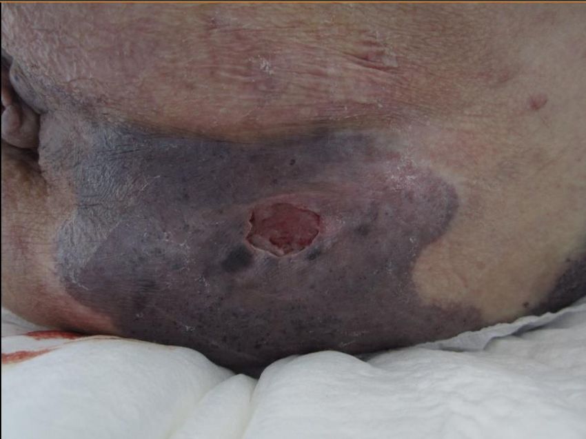
Duplexsonographie
Primäre (oder ergänzende) Bildgebung
Vaskuläre Malformation oder vaskulärer Tumor
Kompressibel oder nicht
Schneller oder langsamer Fluss
hohe PRF, PW-Doppler
Beurteilung umgebender Strukturen
Zu-/abführende Gefässe
Zugang für Behandlung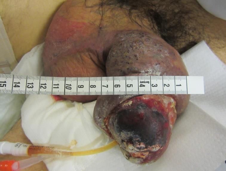
Vascular Anomalies
                Tumors                              Malformations
•  Benign:                                   •   Low flow:
   Infantile Hemangioma (IH)                 •   Capillary malformation (CM)
• Rapidly involuting congenital hemangioma
                                             •   Venous malformation (VM)
   (RICH)
                                             •   Lymphatic malformation (LM)
• Non-involuting congenital hemangioma
  (NICH)
                                             •   High flow:
•    Borderline:                             •    Arteriovenous malformation
•    Kaposiform Hemangioendothelioma             (AVM)
    (KHE)
                                             •   Arteriovenous fistula (AVF)

•   Malignant:
•   Angiosarcoma                             •   Combined and syndromic
•   Epithelioid Hemangioendothelioma (EHE)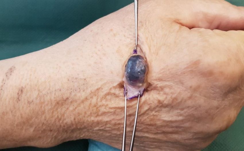
Angiology     Radiology       Pathology     Dermatology

Lymphatic Malformation        Cavernous hemangioma

               Lymphangioma                 Cystic hygroma

Orthopedics Tumor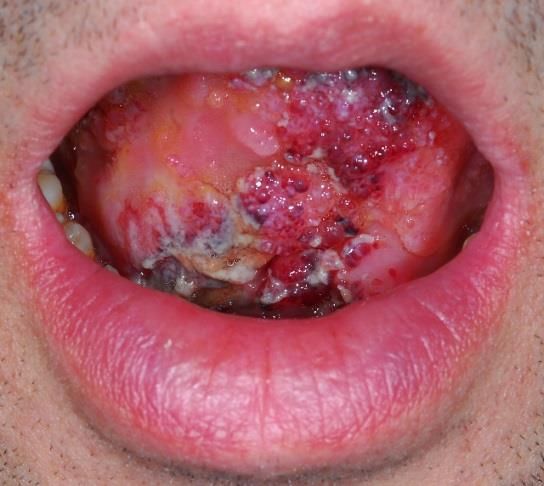
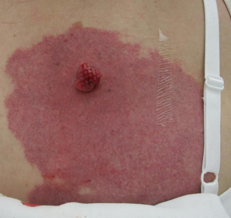
Malformation vs. Tumor?
•   “Pyogenes Granuloma”
•   Gestielter Tumor
•   Zentrale Gefässe
•   Schneller Fluss
•   Elektrischer Schlinge
•   Histologie
MRI:15 x 8 x 16 mm large arterio-venous malformation

                                             Dg: Myoperizytom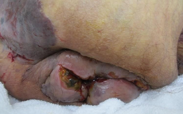
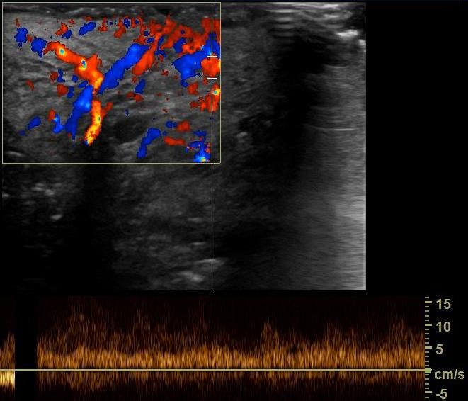
Arteriovenöse Fisteln (AVF)
- PRF hoch
- Volumenmessung
- arterialisierter Fluss
- Zu/abführende Gefässe
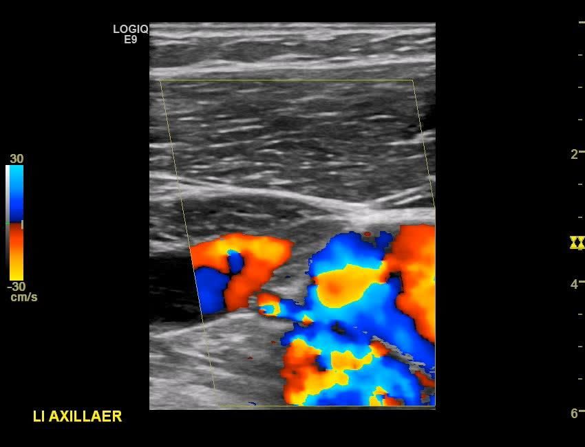
High flow in low flow
High flow and low flow?
High- vs. low-flow

   Capillary-arteriovenous Malformation
   «Parkes-Weber-Syndrome»
   RASA1-Mutation
Capillary-arteriovenous Malformation
«Parkes-Weber-Syndrome»
RASA1-Mutation
Überwuchs
Schnell fliessend/Mikroshunts
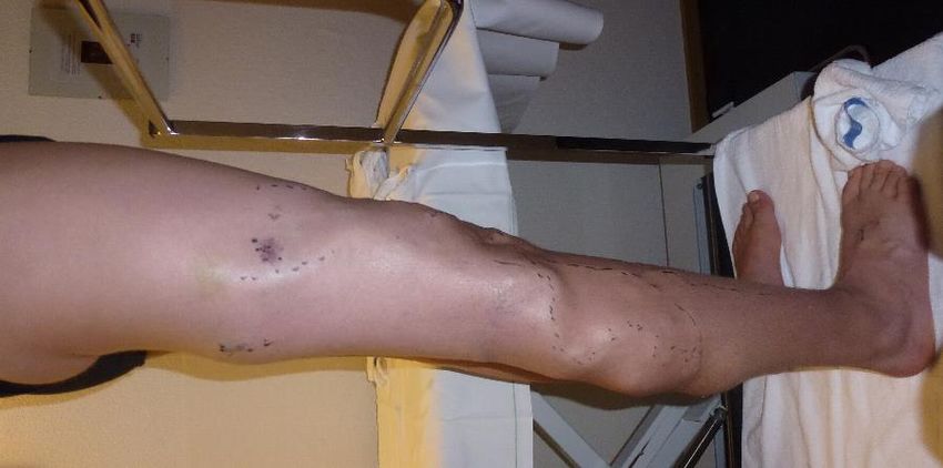
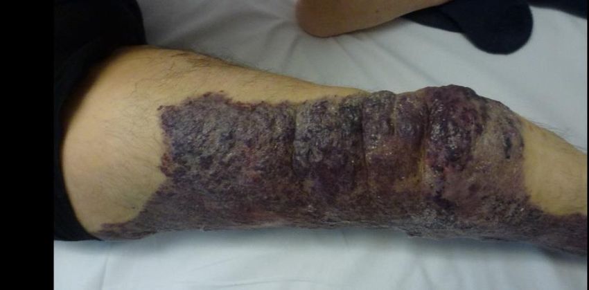
Seitenvergleich

Distal gelegene arteriovenöse Malformation
(AVM) Fingerendglied
High flow
Low flow
Arteriovenöse Malformation (AVM)
Arteriovenous Malformation (AVM)
Vor Embolisation

Post Embolisation

3 Monate später

AVM Unterarm
Malformation veineuse
• Malformation à flux lent la plus fréquente
• Congénitale
• 90% avec participation cutanée
• Spongiforme, phlébectasique, anévrismale, réticulaire
• Mutation somatique
Exemples cliniques
Progression VM

HASSANEIN AH, MULLIKEN JB, FISHMAN SJ ET AL. VENOUS   MALFORMATION: RISK OF PROGRESSION DURING CHILDHOOD AND ADOLESCENCE.

                                        ANN PLAST SURG. 2012 FEB;68(2):198-201.
Type anévrismal
• Risque de TVP et EP
Traitement actif
AnticoagulationType phlébectasique
Capillary-venous-lymphatic Malformation
Klippel-Trénaunay-SyndromeKapillär-lymphatisch-venöse Malformation (KTS)
Vulnerable Haut, SepsisType phlébectasique
Antikoagulation?Rezidivierende Thrombosen - Aktive Behandlung: Intervention, Kompression, Antikoagulation
Réseau veineux profond
•   présent?
•   perméable?
•   continent?
•   écho-Doppler, IRM,
    scanner
Type spongiforme
Malformation glomuveineuse (MGV)
Blue rubber bleb nevus syndrome (BRBNS)
•   Venöse Malformation

•   Haut, Weichteile, Darm, u.a.

•   Korrelation zwischen Hautläsionen + Intestinal
•   Chronische Blutung (selten akut und massiv)
     •    Spontane Sistierung, multiple Blutungen
     •    Kapselendoskopie

•    Auftreten neuer Läsionen

•    Zunahme/Auftreten Blutungen

                   Fishman S et al. Blue rubber bleb nevus syndrome: surgical eradication of gastrointestinal bleeding. Ann Surg. 2005 Mar;241(3):523-8.
Kompressibilität: VM vs. LM
Malformation lymphatique macrozystique
Malformation lymphatique microzystique
Central Conducting Lymphatic Anomalies
Stärken der Duplexsonographie
•   Diagnosestellung: «besser» als MRI/CT oder andere «statische» Verfahren

•   Differenzierung der venösen Malformation nach Typ

•   Funktionelle Beurteilung tiefes Venensystem (incl. Durchgängigkeit)

•   Beurteilung des Zugangswegs

• Festlegung des Zugangswegs während Intervention

• Gezielte Beurteilung
Synthèse
• Echo-Doppler en première intention ou comme
  complément
• Image mode B: tumeur ou malformation vasculaire
• Echo-Doppler: flux rapide ou lent
• CEUS: lésions profondes, DD
• Diagnostic descriptif
• Comparaison controlatérale
   • Diagnostic complémentaire (imagerie + labo)
   • Question des possibilités de traitement
   • Pathologie chronique, traitements multiples
Sie können auch lesen


