Evaluation von Risikofaktoren für die Entwicklung des akut-auf-chronischen Leberversagens bei dekompensierter Leberzirrhose
←
→
Transkription von Seiteninhalten
Wenn Ihr Browser die Seite nicht korrekt rendert, bitte, lesen Sie den Inhalt der Seite unten
Aus der
Medizinischen Klinik und Poliklinik I – Allgemeine Innere Medizin mit den
  Schwerpunkten Gastroenterologie und Hepatologie, Nephrologie,
            Infektiologie, Endokrinologie und Diabetologie
                    des Universitätsklinikums Bonn
       Direktor: Herr Univ.-Prof. Dr. med. Christian P. Strassburg

 Evaluation von Risikofaktoren für die Entwicklung
    des akut-auf-chronischen Leberversagens bei
              dekompensierter Leberzirrhose

                           Habilitationsschrift
                   zur Erlangung der Venia Legendi
                   der Hohen Medizinischen Fakultät
         der Rheinischen-Friedrich-Wilhelms-Universität Bonn
                          Für das Lehrgebiet
                            „Innere Medizin“

                             Vorgelegt von

                 Dr. med. Michael Praktiknjo
                      Wissenschaftlicher Assistent
                        an der Universität Bonn
                               Bonn 2021
Datum des Habilitationskolloquiums: 28.Oktober 2021
Übersicht

Der vorliegenden Habilitationsschrift mit dem Titel „Evaluation von Risikofaktoren für
die Entwicklung des akut-auf-chronischen Leberversagens bei dekompensierter
Leberzirrhose“ liegen folgende publizierte Arbeiten zu Grunde:

1. Praktiknjo M, Simón-Talero M, Römer J, Roccarina D, Martínez J, Lampichler K,
Baiges A, Low G, Llop E, Maurer MH, Zipprich A, Triolo M, Maleux G, Fialla AD, Dam
C, Vidal-González J, Majumdar A, Picón C, Toth D, Darnell A, Abraldes JG, López M,
Jansen C, Chang J, Schierwagen R, Uschner F, Kukuk G, Meyer C, Thomas D,
Wolter K, Strassburg CP, Laleman W, La Mura V, Ripoll C, Berzigotti A, Calleja JL,
Tandon P, Hernandez-Gea V, Reiberger T, Albillos A, Tsochatzis EA, Krag A,
Genescà J, Trebicka J; Baveno VI-SPSS group of the Baveno Cooperation. Total
area of spontaneous portosystemic shunts independently predicts hepatic
encephalopathy and mortality in liver cirrhosis.
Erschienen in Journal of Hepatology 2020 Jun;72(6):1140-1150.

2. Praktiknjo M*, Torner J*, Simón-Talero M, Gu W, Perez-Poch A, Torre C, Val ID,
Alpiste F, Genescà J, Trebicka J. Reply to: “Definition of SPSS: we need to speak the
same language”: Computer-assisted image processing for better quantification.
Erschienen in Journal of Hepatology 2020 Aug;73(2):464-465.

3. Praktiknjo M, Clees C, Pigliacelli A, Fischer S, Jansen C, Lehmann J, Pohlmann
A, Lattanzi B, Krabbe VK, Strassburg CP, Arroyo V, Merli M, Meyer C, Trebicka J.
Sarcopenia Is Associated With Development of Acute-on-Chronic Liver Failure in
Decompensated Liver Cirrhosis Receiving Transjugular Intrahepatic Portosystemic
Shunt.
Erschienen in Clinical and Translational Gastroenterology. 2019
Apr;10(4):e00025.

4. Praktiknjo M*, Book M*, Luetkens J, Pohlmann A, Meyer C, Thomas D, Jansen C,
Feist A, Chang J, Grimm J, Lehmann J, Strassburg CP, Abraldes JG, Kukuk G,
Trebicka J. Fat-free muscle mass in magnetic resonance imaging predicts acute-on-
chronic liver failure and survival in decompensated cirrhosis.
Erschienen in Hepatology. 2018 Mar;67(3):1014-1026.

5. Praktiknjo M*, Monteiro S*, Grandt J, Kimer N, Madsen JL, Werge MP, William P,
Brol MJ, Turco L, Schierwagen R, Chang J, Klein S, Uschner FE, Welsch C, Moreau
R, Schepis F, Bendtsen F, Gluud LL, Møller S, Trebicka J. Cardiodynamic state is
associated with systemic inflammation and fatal acute-on-chronic liver failure.
Erschienen in Liver International 2020 Jun;40(6):1457-1466.

*contributed equally as first author
Inhaltsverzeichnis

                                                                                                                        Seite

1.    Einleitung .............................................................................................................. 5

1.1   Die Leberzirrhose und ihre Komplikationen ............................................................ 5

1.2   Das akut-auf-chronische Leberversagen ................................................................ 8

1.3   Die systemische Inflammation in der Leberzirrhose ............................................... 9

1.4   Die Sarkopenie in der Leberzirrhose ...................................................................... 9

1.5   Spontane portosystemische Shunts ..................................................................... 11

1.6   Fragestellung ........................................................................................................ 13

2.    Ergebnisse .......................................................................................................... 14

2.1   Spontane portosystemische Shunts als Risikofaktor der Leberzirrhose
      Praktiknjo et al. J Hepatol 2020 Jun;72(6):1140-1150 .......................................... 14

2.2   Die Rolle der CT-definierten Sarkopenie als Risikofaktor bei Leberzirrhose
      Praktiknjo et al. Clin Transl Gastroenterol 2019 Apr;10(4):e00025 ...................... 31

2.3   Die Rolle der MRT-definierten Sarkopenie als Risikofaktor bei Leberzirrhose
      Praktiknjo et al. Hepatology. 2018 Mar;67(3):1014-1026 ..................................... 43

2.4   Systemische Inflammation als Risikofaktor bei Leberzirrhose
      Praktiknjo et al. Liver Int. 2020 Jun;40(6):1457-1466 ........................................... 59

3.    Diskussion .......................................................................................................... 72

4.    Zusammenfassung ............................................................................................. 76

5.    Überlappung mit anderen Habilitationsschriften ............................................. 78

6.    Bibliographie....................................................................................................... 79

7.    Danksagung ........................................................................................................ 86
5

1. Einleitung

1.1 Die Leberzirrhose und ihre Komplikationen

In den weltweiten Gesundheitssystemen spielen chronische Lebererkrankungen eine
wachsende Rolle. Durch kontinuierliche Schädigung der Leber kommt es zu einer
fortschreitenden Vernarbung (Fibrogenese) des Lebergewebes. Unter Fibrogenese
versteht man den Prozess der reaktiven Bindegewebsvermehrung bzw. der Ansammlung
von extrazellulärer Matrix, was zu Fibrose und Zirrhose führt (Friedman, 2008). Die
Leberzirrhose   ist   das   gemeinsame      Endstadium    der    meisten   chronischen
Lebererkrankungen und wird definiert als Zerstörung der physiologischen Läppchen- und
Gefäßstruktur der Leber, begleitet von einer Bindegewebsvermehrung.

Die Leberzirrhose stellt ein wachsendes klinisches und volkswirtschaftliches Problem dar
(Pimpin et al., 2018). Im Allgemeinen hat Europa die weltweit größte Anzahl an
chronischen Lebererkrankungen (Global Health Data Exchange., 2020). In Deutschland
im Speziellen leiden etwa 2 % der Bevölkerung an einer Leberzirrhose und deren
Komplikationen. Bei den Betroffenen führt die Leberzirrhose zu einer entsprechend hohen
Morbidität und Mortalität (Chirapongsathorn et al., 2016). Die Zahl an vollstationären
Behandlungen der Leberzirrhose in Deutschland ist in den Jahren 2000 bis 2017 von
79.647 auf 89.613 angestiegen (Gesundheitsberichterstattung des Bundes, 2019). Die
mittlere Überlebenszeit von Patienten mit einer klinisch kompensierten Leberzirrhose
beträgt nur etwa sieben Jahre. Die Wahrscheinlichkeit einer Dekompensation beträgt bis
zu 10% pro Jahr bei diesen Patienten. Ist sie eingetreten, sinkt das Zwei-Jahres-
Überleben auf unter 50% (Talwalkar and Kamath, 2005).

Komplikationen der Leberzirrhose

Durch den Anstieg des vaskulären Widerstandes in der Leber kommt es zu einem
Druckanstieg in den Gefäßen des portalvenösen Systems und zur Ausbildung der
sogenannten Portalen Hypertension. Während des Progresses der Zirrhose kann es zu
akuten Dekompensationen wie Varizenblutungen, Aszitesentwicklung oder hepatischer
Enzephalopathie kommen (Angeli et al., 2018).        Eine fortschreitende Vernarbung,
Entzündungen und permanente Leberschädigungen sind als treibende Kräfte dieser
6

akuten Dekompensationen, Morbidität und Mortalität beschrieben (Bataller and Brenner,
2005; Friedman, 2008).

Interessanterweise neigen Patienten mit dekompensierter Leberzirrhose dazu weitere
Dekompensationen zu entwickeln, ohne dass hierfür ein genauer Pathomechanismus
bekannt ist (Gustot et al., 2015). Neben der Behandlung der Grunderkrankung werden die
Komplikationen der Leberzirrhose und portalen Hypertension zunächst konservativ-
medikamentös bzw. endoskopisch und in fortgeschrittenen Stadien auch interventionell
behandelt (Angeli et al., 2018; Gerbes et al., 2019).

Ösophagusvarizen / Varizenblutung

Mit Ausbildung von Ösophagusvarizen besteht auch das Risiko der Varizenblutung, die
eine hohe Mortalität hat (de Franchis and Baveno VI Faculty, 2015; Götz et al., 2017).
Primär ist zur Prophylaxe einer Varizenblutung eine medikamentöse Behandlung mit
nicht-selektiven Betablockern empfohlen, die eine Reduktion des Blutungsrisikos von
11% und der Mortalität von 9% erreicht (Cheng et al., 2003). Alternativ zur
medikamentösen Therapie kann eine endoskopische Varizenligatur erfolgen, die in einer
Cochrane-Analyse vergleichbare Ergebnisse zu nicht-selektiven Betablockern zeigt
(Gluud and Krag, 2012). Bei Patienten, die bereits eine Varizenblutung überlebten,
besteht ein hohes Risiko (60%) für eine Rezidivblutung mit hoher (33%) Mortalität (Bari
and Garcia-Tsao, 2012), sodass eine Sekundärprophylaxe unerlässlich für das Outcome
dieser   Patienten   ist.     In   dieser   Situation    ist   nach    aktueller   Datenlage   eine
Kombinationstherapie         aus    nicht-selektiver     Betablockade       und    endoskopischer
Ligaturtherapie angezeigt (Puente et al., 2014; Thiele et al., 2012). In ausgewählten Fällen
(Child-Pugh Stadium C < 14 Punkte oder Child-Pugh Stadium B mit aktiver Blutung) kann
ein sogenannter transjugulärer intrahepatischer portosystemischer Shunt (TIPS)
innerhalb von 72 Stunden implantiert werden. Der TIPS ist ein interventionell implantierter
Shunt, der endovaskulär den hepatischen Ausflusstrakt mit dem Pfortadersystem
verbindet (Rössle, 2013). Der Hochdruck des portalvenösen Systems wird durch die
teilweise Umgehung des physiologischen hepatischen Pfortaderflusses gesenkt und
somit    die   Varizen      entlastet.   Hierdurch      werden   bei    ausgewählten     Patienten
Rezidivblutungen nahezu eliminiert und das Überleben verbessert (García-Pagán et al.,
2010; Hernández-Gea et al., 2019; Lv et al., 2019).
7

Aszites

Der klinische Nachweis von Aszites zeigt eine schwere fortgeschrittene Erkrankung bzw.
portalen Hypertension an und definiert ein Voranschreiten der Leberzirrhose mit einer
Verschlechterung der Prognose (D’Amico et al., 2006; Guardiola et al., 2002; Planas et
al., 2006), vor allem durch die sekundäre Entwicklung von spontan bakteriellen
Peritonitiden oder eines hepatorenalen Syndroms (Ginès et al., 2004).
In   der   ersten   Linie   ist   eine   medikamentöse      (diuretische)   Therapie      mit
Aldosteronantagonisten und bei unzureichender Aszitesmobilisation die Kombination mit
einem Schleifendiuretikum angezeigt (Gerbes et al., 2019).
Im Falle einer weiter fortschreitenden Leberzirrhose kann therapierefraktärer oder
intraktabler Aszites entstehen. Hier können wiederholte großvolumige Parazentesen und
der transjuguläre intrahepatische portosystemische Shunt (TIPS)               therapeutisch
eingesetzt werden (Bureau et al., 2017a; Solà et al., 2017; Thomas et al., 2015).
Großvolumige Parazentesen können zu hämodynamischen Veränderungen führen; die
sogenannte zirkulatorische Dysfunktion nach Parazentese (Ginès et al., 1996, 1988;
Pozzi et al., 1994). Interessanterweise besteht bei Patienten mit Leberzirrhose oft bereits
eine gewisse zirkulatorische Dysfunktion, die möglicherweise bereits einen Einfluss auf
das Outcome hat (Turco et al., 2018a).
Die TIPS-Anlage reduziert den portalen Druck als zugrundeliegende Pathologie und
verbessert durch Verbesserung der renalen Perfusion die Nierenfunktion (Allegretti et al.,
2016; Brensing et al., 2000; Lebrec et al., 1996; Wong et al., 1995). Im Vergleich zur
wiederholten großvolumigen Parazentese ist die TIPS-Anlage effektiver, verbessert die
Lebensqualität und ist auch bezüglich der Mortalität überlegen (Allegretti et al., 2016;
Bureau et al., 2017b; Gülberg et al., 2002; Rössle et al., 2000; Salerno et al., 2007).

Hepatische Enzephalopathie

Die hepatische Enzephalopathie (HE) ist ein Syndrom, welches die Summe aller
Störungen des Zentralnervensystems, die als Komplikation akuter oder chronischer
Lebererkrankungen und/oder portosystemischer Kollateralkreisläufe auftreten können,
umfasst (Ferenci et al., 2002; Vilstrup et al., 2014). Pathophysiologisch liegt der HE eine
reduzierte Kapazität der erkrankten Leber zur Ammoniakentgiftung zugrunde. Die
8

Skelettmuskulatur des Körpers ist in der Lage durch eine Glutamin-Synthetase-Aktivität
relevante Mengen von Ammoniak in Glutamin zu konvertieren und so zu eliminieren
(Jindal and Jagdish, 2019; Wright et al., 2011).
Klinisch zeigt sich eine charakteristische zunehmende psychomotorische Verlangsamung
bis hin zum Koma sowie ein breites Spektrum variabel auftretender intellektueller,
emotionaler, kognitiver, psychischer und motorischer Störungen (Ferenci et al., 2002).
Selbst bei klinisch unauffälligen Patienten lassen sich bei 20 – 85 % kognitive
Einschränkungen feststellen (Ferenci et al., 2002; Labenz et al., 2017; Poordad, 2007;
Romero-Gómez et al., 2001; Saunders et al., 1981; Vilstrup et al., 2014).
Das Auftreten einer HE bei Patienten mit Leberzirrhose ist ein Prädiktor für die Mortalität
mit einer Ein-Jahres-Sterblichkeit von 64 % (Bustamante et al., 1999; Hartmann et al.,
2000; Jepsen et al., 2010).

1.2 Das akut-auf-chronische Leberversagen (ACLF)

Die akut dekompensierte Leberzirrhose und das akut-auf-chronische Leberversagen
(ACLF) sind zwei wichtige Zustände, die bei Patienten mit dekompensierter Leberzirrhose
beobachtet werden können. Die akut dekompensierte Leberzirrhose umfasst die
Entwicklung von Aszites, HE, gastrointestinaler Blutung und jedwede Kombination dieser
Komplikationen bei Patienten mit Zirrhose (Moreau et al., 2013a; Sarin et al., 2019). Das
Konzept des ACLF entstand aus Studien, die die Entwicklung eines Syndroms mit hoher
Kurzzeit-Sterblichkeit (Tod innerhalb von 28 Tagen nach Hospitalisation) in Patienten mit
akut dekompensierter Zirrhose zeigten. Drei Hauptmerkmale charakterisieren dieses
Syndrom: Es tritt im Kontext einer ausgeprägten systemischen Inflammation auf,
entwickelt sich in engem zeitlichen Zusammenhang mit proinflammatorischen
auslösenden Ereignissen (z.B. Infektionen, Alkoholhepatitis, gastrointestinale Blutung)
und ist mit dem Versagen mindestens eines Organsystems verbunden (Arroyo et al.,
2020; Trebicka et al., 2020).
Aufgrund der äußerst hohen Mortalität des ACLF von bis über 90% in 90 Tagen und den,
mit Ausnahme der Lebertransplantation, noch fehlenden therapeutischen Optionen, ist
ein frühes Erkennen von Risikopatienten entscheidend (Moreau et al., 2013b).
9

Die Evaluation von Biomarkern zur Risikostratifizierung von Patienten mit Leberzirrhose
für die Entwicklung eines ACLF ist eine zentrale Fragestellung dieser Habilitation. Die
einzelnen untersuchten Aspekte werden im Folgenden eingeführt.

1.3 Die systemische Inflammation in der Leberzirrhose

In der Erarbeitung der pathophysiologischen Mechanismen des ACLF kommt der
systemischen Inflammation eine besondere Rolle zu (Clària et al., 2016). Diese
systemische Inflammation kann chronisch, durch Translokation von proinflammatorischen
Signalen aus dem Intestinum in die systemische Zirkulation, auftreten (Albillos et al., 2014;
Bernardi et al., 2015; Medzhitov, 2008; Úbeda et al., 2010). Es konnte bereits gezeigt
werden, dass Infektionen besonders häufige Auslöser von ACLF sind (Piano et al., 2019;
Trebicka et al., 2020). Bei Patienten mit ACLF findet sich im Besonderen jedoch ein Sturm
proinflammatorischer Mediatoren (Laleman et al., 2018; Monteiro et al., 2020; Trebicka et
al., 2019a). Bei Patienten mit portaler Hypertension wurde vor Kurzem gezeigt, dass
Serum C-reaktives Protein (CRP, als Surrogat für systemische Inflammation) und eine
hypo- oder hyperdyname Kreislaufsituation (jeweils definiert als cardiac index (CI)
4.2 L/min/m2) mit der Entwicklung von Aszites und erhöhter
Mortalität assoziiert sind (Turco et al., 2018b). Zudem wurde in einer Kohorte von
ambulanten Zirrhosepatienten gezeigt, dass ein niedriger Blutdruck, als Ausdruck der
kardiozirkulatorischen Dysfunktion, unabhängig mit der Entwicklung von ACLF assoziiert
ist (Piano et al., 2017). Das Zusammenspiel von Markern der systemischen Inflammation
(außer CRP) und systemischer Hämodynamik mit der Entwicklung von ACLF bei
Patienten mit Leberzirrhose ist nicht untersucht und daher eine Fragestellung dieser
Habilitationsarbeit.

1.4 Die Sarkopenie in der Leberzirrhose

Patienten mit Leberzirrhose sind häufig mangelernährt und zeigen dabei einerseits
Eiweißmangel und Muskelschwund (Sarkopenie) sowie andererseits einen Überschuss
an extrazellulärem Wasser (Peng et al., 2007). Eine Sarkopenie kann bei 40 – 60 % der
Patienten mit Leberzirrhose nachgewiesen werden.
10

Ein normaler oder erhöhter Body Mass Index (BMI) schließt eine Mangelernährung nicht
aus und ist bei Zirrhosepatienten durch die Wassereinlagerung oft irreführend (Peng et
al., 2007). Die Sarkopeniediagnostik aus einem Schnittbild (Computertomographie oder
Magentresonanztomographie) zur Quantifizierung der Muskelmasse ist in Leitlinien
aufgrund mangelnder Datenlage noch nicht konsentiert (Durand et al., 2014; European
Association for the Study of the Liver., 2019; Montano-Loza et al., 2012, 2016).
Schnittbildgebungen werden jedoch bei diesen Patienten häufig aufgrund anderer
Indikationen durchgeführt, z.B. im Rahmen einer Evaluation zur Lebertransplantation, zur
TIPS-Implantation oder beim Screening auf ein hepatozelluläres Karzinom (HCC), sodass
aus diesen Daten gleichzeitig auch Daten zur Sarkopenie erhoben werden können.
Einige   Methoden     und    Grenzwerte    zur    Quantifizierung    der     Sarkopenie    aus
Schnittbildgebungen wurden vorgeschlagen (Carey et al., 2017; Cruz-Jentoft et al., 2010;
Durand et al., 2014; Giusto et al., 2015; Golse et al., 2017; Praktiknjo et al., 2018a). Die
meisten Methoden beschreiben spezifisch Muskelparameter aus CT-Untersuchungen,
wobei zumeist spezielle Software benötigt wird, welche die Messungen im klinischen
Alltag zu zeitaufwendig machen könnten (Golse et al., 2017; Montano-Loza, 2014).

Eine einfache und schnell erfassbare Methode ist die Messung der transversalen
Psoasmuskeldicke (TPMT) normalisiert auf die Körpergröße, welche bereits als Prädiktor
der Sterblichkeit von Patienten auf der Warteliste für eine Lebertransplantation
beschrieben wurde (Durand et al., 2014; Huguet et al., 2018). Die Muskulatur ist abhängig
von einer Vielzahl an Faktoren. Insbesondere das Geschlecht beeinflusst die
Muskelstruktur und -masse (Moctezuma-Velázquez et al., 2018). Die verfügbare Literatur
deutet zunehmend darauf hin, dass unterschiedliche Grenzwerte für die Definition der
Sarkopenie    für   Männer    und    Frauen      herangezogen       werden     sollten.   Klare
geschlechtsspezifische Grenzwerte existieren jedoch noch nicht für Patienten mit
dekompensierter Zirrhose (Giusto et al., 2015; Praktiknjo et al., 2018a).

Ein Zusammenhang der altersbedingten Sarkopenie mit einer erhöhten systemischen
Inflammation ist bereits beschrieben, ebenso wie ein Zusammenhang von systemischer
Inflammation mit der Entwicklung von ACLF (Dalle et al., 2017; Laleman et al., 2018).
Während bereits eine Reihe von Risikofaktoren für die Entwicklung von ACLF diskutiert
11

wurden, ist die Beziehung von Sarkopenie mit der Entwicklung von ACLF noch unbekannt
(Gustot et al., 2015; Moreau et al., 2013b; Praktiknjo et al., 2018b).

Die Evaluation der Rolle der CT-definierten Sarkopenie, via geschlechts-spezifischer
TPMT, auf die Entwicklung von ACLF von Patienten mit dekompensierter Leberzirrhose
ist daher ein weiterer Fokus dieser Habilitationsarbeit.

Die CT hat einige Nachteile wie die methodenbedingte Strahlenexposition der Patienten
oder das Risiko der Auslösung oder Aggravation einer Niereninsuffizienz durch das
entsprechende     iodhaltige   Kontrastmittel       (Kontrastmittel-induzierte   Nephropathie)
(Rudnick et al., 1995). Bei Patienten mit Kontraindikationen gegen CT-Kontrastmittel und
auch für spezifische medizinische Fragestellungen (z.B. genauere Detektion von HCC)
findet die MRT heutzutage immer häufiger Anwendung (Lee et al., 2015). Neben der
Gesamtmuskelmasse scheint auch die Qualität und Funktion der Muskulatur von
Bedeutung für das Outcome der Patienten zu sein, was in bestimmten Myokinen, wie dem
Follistatin, als Surrogatparameter reflektiert wird (Dasarathy et al., 2011; Tsuchida, 2008;
Wagner, 2005). Mittels MRT kann nicht nur die Gesamtmuskelfläche, sondern auch der
Fettanteil der Muskulatur identifiziert und quantifiziert werden.

MRT-definierte    Muskelparameter      zur   Prognoseeinschätzung         bei    Patienten    mit
dekompensierter      Leberzirrhose     wurden        im    Gegensatz      zu     CT-definierten
Muskelparametern bislang noch überhaupt nicht untersucht.

Die Evaluation neuartiger Muskelparameter aus MRT-Untersuchungen und ihre
prognostische    Bedeutung     für   die   Entwicklung      von   ACLF    bei    Patienten    mit
dekompensierter Leberzirrhose ist daher eine Fragestellung dieser Habilitationsschrift.

1.5 Spontane portosystemische Shunts (SPSS)

Im Verlauf der Leberzirrhose kann die portale Hypertension zur Ausbildung von spontanen
portosystemischen Shunts (SPSS) als Umgehungskreisläufe zur Reduktion des
portalvenösen      Druckes      führen.      Eine       Assoziation      von     SPSS        oder
chirurgisch/interventionell angelegter Shunts mit der Entwicklung von HE ist lange
12

bekannt und die ersten Embolisationen von SPSS als Therapieoptionen wurden bereits
in den 1980er Jahren beschrieben (Henderson, 1989; Ohnishi et al., 1986; Uflacker et al.,
1987). Seitdem wurden wenige Studien zu SPSS als Therapieziel bei Patienten mit
Leberzirrhose veröffentlicht (Aseni et al., 1986; Miyamoto et al., 2003; Shioyama et al.,
1996; Tarantino et al., 2009; Zidi et al., 2007). Eine große multizentrische Studie bestätigte
eine Assoziation von großen SPSS (> 8mm Durchmesser) mit dem Auftreten von HE
(Simón-Talero et al., 2018).

Eine große multizentrische Studie bestätigte eine Assoziation von großen SPSS (> 8mm
Durchmesser) mit dem Auftreten von HE (Simón-Talero et al., 2018). In dieser Studie
wurde der Nachweis von SPSS, ähnlich wie die oben beschriebenen Muskelparameter,
aus CT-Untersuchungen erhoben, die aus medizinischer Indikation erfolgten.

Aufgrund der mangelnden Datenlage fehlt es in aktuellen Leitlinien an Empfehlungen zum
Management von SPSS (American Association for the Study of Liver Diseases and
European Association for the Study of the Liver, 2014; Angeli et al., 2018; de Franchis
and Baveno VI Faculty, 2015; Sarin et al., 2014). Der Einfluss von SPSS im Allgemeinen
und ihrer kumulativen Durchmesser im Speziellen auf das Überleben von Patienten wurde
noch nicht untersucht. Aus pathophysiologischer Sicht nahmen wir an, dass die Summe
der Querschnittsflächen aller SPSS (Total SPSS Area, TSA) das portosystemisch
„geshuntete“ Blutvolumen besser abbildet als die einfachen Durchmesser (Gao and Drew,
2014).

Der Einfluss der TSA auf die Mortalität und insbesondere ACLF-Entwicklung von
Patienten mit Leberzirrhose war daher ein Aspekt der Fragestellung dieser
Habilitationsarbeit.
13

1.6 Fragestellung

In den vorangegangenen Abschnitten wurde die besondere klinische Schwere des ACLF
für Patienten mit Leberzirrhose dargelegt. Außer der Lebertransplantation als ultima ratio
stehen aktuell noch kaum therapeutische Optionen zur Verfügung.
Verlässliche Biomarker zur Identifizierung von Zirrhosepatienten mit hohem Risiko für die
Entwicklung von ACLF sind hierfür notwendig, aber noch wenig untersucht. In dieser
Habilitationsschrift werden vor allem nicht-invasive Biomarker evaluiert.
Zum einen wird TSA, ein neuartiger Parameter des portosystemischen Shuntings, der
zusätzlich aus bereits vorhandenen CT-Untersuchungen ermittelt werden kann, bezüglich
der prognostischen Wertigkeit auf das Outcome von Patienten mit Leberzirrhose
untersucht. Zum anderen wird die Möglichkeit der Quantifizierung der Muskelmasse aus
CT- und MRT-Untersuchungen bei Patienten mit Leberzirrhose und deren Bedeutung als
Prädiktor für die Entwicklung von ACLF evaluiert.
Abschließend werden zirkulierende Marker der systemischen Inflammation im Kontext der
Kreislaufsituation für die Prädiktion von ACLF evaluiert.
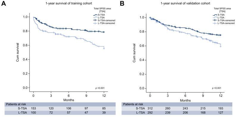
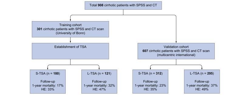
14

2. Ergebnisse

2.1 Spontane portosystemische Shunts als Risikofaktor der Leberzirrhose
“Total area of spontaneous portosystemic shunts independently predicts hepatic
encephalopathy and mortality in liver cirrhosis.”
Praktiknjo M, Simón-Talero M, Römer J, Roccarina D, Martínez J, Lampichler K, Baiges
A, Low G, Llop E, Maurer MH, Zipprich A, Triolo M, Maleux G, Fialla AD, Dam C, Vidal-
González J, Majumdar A, Picón C, Toth D, Darnell A, Abraldes JG, López M, Jansen C,
Chang J, Schierwagen R, Uschner F, Kukuk G, Meyer C, Thomas D, Wolter K, Strassburg
CP, Laleman W, La Mura V, Ripoll C, Berzigotti A, Calleja JL, Tandon P, Hernandez-Gea
V, Reiberger T, Albillos A, Tsochatzis EA, Krag A, Genescà J, Trebicka J; on behalf of the
Baveno VI-SPSS group of the Baveno Cooperation.
Erschienen in Journal of Hepatology 2020 Jun;72(6):1140-1150.

“Reply to: “Definition of SPSS: we need to speak the same language”: Computer-assisted
image processing for better quantification.“
Praktiknjo M, Torner J, Simón-Talero M, Gu W, Perez-Poch A, Torre C, Val ID, Alpiste
F, Genescà J, Trebicka J.
Erschienen in Journal of Hepatology 2020 Aug;73(2):464-465.

Zur Messung der Gesamtfläche aller SPSS (total SPSS area, TSA) wurden retrospektiv
in CT-Untersuchungen sämtliche SPSS manuell identifiziert und vermessen. Hieraus
wurde dann die TSA berechnet und anschließend mit Daten des klinischen Verlaufes der
Patienten korreliert. Wir untersuchten zunächst 301 Patienten mit Leberzirrhose aus der
eigenen Kohorte am Universitätsklinikum Bonn und bestätigten die Ergebnisse in einer
internationalen, multizentrischen (12 Zentren aus 9 Ländern) Kohorte von 607 Patienten
der internationalen Baveno-Studiengruppe (Abb. 2.1.1). Primärer Endpunkt war das Ein-
Jahres-Überleben.

Überleben stratifiziert nach TSA

Zunächst wurde in der Trainingskohorte mittels Receiver Operating Characteristics (ROC)
Analyse ein optimaler Grenzwert der TSA von 83 mm 2 berechnet und die Patienten
entsprechend in L-TSA (große TSA, > 83 mm2) und S-TSA (kleine TSA, ≤ 83 mm2)
eingeteilt. Die Kaplan-Meier Kurve für das Ein-Jahresüberleben zeigt eine signifikant
höhere Sterblichkeit in der L-TSA im Vergleich zu der S-TSA Gruppe (Abb. 2.1.2 A).
Dieses Ergebnis konnte in der Validierungskohorte bestätigt werden (Abb. 2.1.2 B). Die
häufigste Ursache in beiden Kohorten war ACLF in jeweils 63% und 55% der Todesfälle.
15

Abbildung. 2.1.1: Flowchart der eingeschlossenen Patienten. Es wurde 301 Patienten aus der eigenen Kohorte in Bonn
als Trainingskohorte untersucht und die gewonnenen Ergebnisse in einer internationalen, multizentrischen
Validierungskohorte bestätigt. SPSS (spontane portosystemische Shunts), CT (Computertomographie), TSA
(Gesamtfläche aller SPSS), S-TSA (Kleines TSA), L-TSA (große TSA), HE (Hepatische Enzephalopathie).

Abbildung. 2.1.2: Kaplan-Meier Kurve für das Ein-Jahresüberleben. (A) In der Trainingskohorte zeigte die L-TSA Gruppe
(hellblau) eine signifikant höhere Sterblichkeit im Vergleich zu der S-TSA Gruppe (dunkelbau). (B) Dieses Ergebnis
zeigt sich auch in der Validierungskohorte. P durch log-rank Test. TSA (Gesamtfläche aller SPSS), S-TSA (Kleines
TSA), L-TSA (große TSA).
16

               Die Patienten wurden ebenfalls mittels einfachem Durchmesser der SPSS eingeteilt, da
               diese klinisch einfacher zu bestimmen ist. Allerdings konnten die Patienten hierdurch nicht
               signifikant stratifiziert werden.

               Uni- und multivariate Cox Regressionsanalysen bestätigten das Vorhandensein von L-
               TSA als unabhängigen Prädiktor für das Ein-Jahresüberleben in beiden Kohorten mit
               ACLF als häufigster Todesursache (Tabelle 2.1.1)

               Tabelle. 2.1.1: Univariate und multivariate Cox Regressionsanalyse der Trainingskohorte für 1-Jahressterblichkeit.

1-year mortality                                         univariate Cox regression                          multivariate Cox regression
Parameter                                          p            HR                CI                  p            HR                CI
age1                                             0.025        1.027       1.003        1.051
17
Research Article
Cirrhosis and Liver Failure
Total area of spontaneous portosystemic shunts
independently predicts hepatic encephalopathy and
mortality in liver cirrhosis
Graphical abstract Authors
Spontaneous Porto-Systemic Shunt (SPSS) Michael Praktiknjo, Macarena Simón-Talero,
cross-section measured in CT scan
at largest diameter Julia Römer, ., Aleksander Krag,
Joan Genescà, Jonel Trebicka
Improved survival stratification
by total SPSS area
Correspondence
Survival stratified by diameter
jonel.trebicka@kgu.de (J. Trebicka), jge-
1.0 nesca@vhebron.net (J. Genescà).
Multiple SPSS
0.8
5 mm
0.6 Lay summary
Cum survival
5mm Diameter: 110 mm2
Total area: 220 mm2
0.4 Total SPSS Area
[TSA]
S-TSA
The prevalence of spontaneous portosys-
L-TSA
S-TSA-censored
0.2 L-TSA-censored
temic shunts (SPSS) is higher in patients
0.0
Single SPSS 0 2 4 6
Months
8 10 12 with more advanced chronic liver disease.
Survival stratified by area The presence of more than 1 SPSS is
common in advanced chronic liver disease
1.0
0.8
Diameter: 110 mm2
10 mm
Total area: 280 mm2 and is associated with the development of
Cum survival
0.6
0.4 hepatic encephalopathy. This study shows
0.2
that total cross-sectional SPSS area (rather
0.0
0 2 4 6
Months
8 10 12
than diameter of the single largest SPSS)
predicts survival in patients with
advanced chronic liver disease. Our results
support the clinical use of total cross-
Highlights
sectional SPSS area for risk stratification
Total cross-sectional SPSS area (TSA) predicts survival in
and decision-making in the management
patients with advanced chronic liver disease.
of SPSS.
The cut-off for TSA that is associated with worse survival
corresponds to a single shunt of >10 mm diameter.
This study may impact on the clinical use of TSA/SPSS for risk
stratification and decision-making in the management of
patients with cirrhosis.
https://doi.org/10.1016/j.jhep.2019.12.021
© 2020 European Association for the Study of the Liver. Published by Elsevier B.V. This is an open access article under the CC BY-NC-ND license
(http://creativecommons.org/licenses/by-nc-nd/4.0/). J. Hepatol. 2020, 72, 1140–115018
Research Article
Cirrhosis and Liver Failure
Total area of spontaneous portosystemic shunts independently
predicts hepatic encephalopathy and mortality in liver cirrhosisq
Michael Praktiknjo1,†, Macarena Simón-Talero2,†, Julia Römer1, Davide Roccarina3,
Javier Martínez4, Katharina Lampichler5, Anna Baiges6, Gavin Low7, Elba Llop8,
Martin H. Maurer9, Alexander Zipprich10, Michela Triolo11, Geert Maleux12,
Annette Dam Fialla13, Claus Dam13, Judit Vidal-González2, Avik Majumdar3, Carmen Picón14,
Daniel Toth5, Anna Darnell15, Juan G. Abraldes16, Marta López8, Christian Jansen1,
Johannes Chang1, Robert Schierwagen23, Frank Uschner23, Guido Kukuk17, Carsten Meyer17,
Daniel Thomas17, Karsten Wolter17, Christian P. Strassburg1, Wim Laleman18,
Vincenzo La Mura19,20, Cristina Ripoll10, Annalisa Berzigotti21, José Luis Calleja8,
Puneeta Tandon16, Virginia Hernandez-Gea6, Thomas Reiberger22, Agustín Albillos4,
Emmanuel A. Tsochatzis3, Aleksander Krag13, Joan Genescà2,*, Jonel Trebicka13,23,24,25,*, for the
Baveno VI-SPSS group of the Baveno Cooperation
1
Department of Internal Medicine I, University of Bonn, Bonn, Germany; 2Liver Unit, Department of Internal Medicine, Hospital Universitari
Vall d’Hebron, VHIR, Universitat Autònoma de Barcelona, CIBERehd, Barcelona, Spain; 3Sheila Sherlock Liver Unit and UCL Institute for Liver
and Digestive Health, Royal Free Hospital and UCL, London, United Kingdom; 4Department of Gastroenterology and Hepatology, Hospital
Universitario Ramón y Cajal, IRICYS, Universidad de Alcalá, CIBERehd, Spain; 5Department of Biomedical Imaging and Image-guided Therapy,
Medical University of Vienna, Austria; 6Hepatic Hemodynamic Laboratory, Liver Unit, Hospital Clinic, IDIBAPS, Universitat de Barcelona,
CIBERehd, Spain; 7Department of Radiology, University of Alberta, Edmonton, Canada; 8Liver Unit, Hospital U. Puerta de Hierro, Universidad
Autónoma de Madrid, Madrid, Spain; 9Department of Radiology, Inselspital, University of Berne, Berne, Switzerland; 10First Department of
Internal Medicine, Martin Luther University Halle-Wittenberg, Halle (Saale), Germany; 11Medicina Interna, Istituto di Ricovero e Cura a
Carattere Scientifico (IRCCS) San Donato, Università Degli Studi di Milano, San Donato Milanese (MI), Italy; 12Department of Interventional
Radiology, University Hospitals Leuven, KU Leuven, Belgium; 13Department of Gastroenterology and Hepatology, Odense University Hospital,
Odense, Denmark; 14Department of Radiology, Hospital Universitario Ramón y Cajal, IRICYS, Universidad de Alcalá, CIBERehd, Spain;
15
Department of Radiology, Hospital Clinic, IDIBAPS, Universitat de Barcelona, Barcelona, Spain; 16Cirrhosis Care Clinic, University of Alberta,
Edmonton, Canada; 17Department of Radiology, University of Bonn, Bonn, Germany; 18Department of Gastroenterology and Hepatology,
University Hospitals Leuven, Leuven, Belgium; 19Fondazione IRCCS Ca’ Granda, Ospedale Maggiore Policlinico, U.O.C. Medicina Generale-
Emostasi e Trombosi, Milano, Italy; 20Dipartimento di Scienze biomediche per la Salute and Centro di Ricerca Coordinata “A. M. e A.
Migliavacca” per lo Studio e la Cura delle Malattie del Fegato, Università degli Studi di Milano, Milano, Italy; 21Hepatology, Inselspital,
University of Berne, Berne, Switzerland; 22Vienna Hepatic Hemodynamic Lab, Division of Gastroenterology and Hepatology, Medical
University of Vienna, Vienna, Austria; 23Department of Internal Medicine I, University of Frankfurt, Frankfurt, Germany; 24European
Foundation for the Study of Chronic Liver Failure - EF CLIF, Barcelona, Spain; 25Institute for Bioengineering of Catalonia, Barcelona, Spain
Background & Aims: Spontaneous portosystemic shunts (SPSS) Methods: In this retrospective international multicentric study,
frequently develop in liver cirrhosis. Recent data suggested that CT scans of 908 cirrhotic patients with SPSS were evaluated for
the presence of a single large SPSS is associated with complica- TSA. Clinical and laboratory data were recorded. Each detected
tions, especially overt hepatic encephalopathy (oHE). However, SPSS radius was measured and TSA calculated. One-year survival
the presence of >1 SPSS is common. This study evaluates the was the primary endpoint and acute decompensation (oHE,
impact of total cross-sectional SPSS area (TSA) on outcomes in variceal bleeding, ascites) was the secondary endpoint.
patients with liver cirrhosis. Results: A total of 301 patients (169 male) were included in the
training cohort. Thirty percent of all patients presented with >1
Keywords: Spontaneous portosystemic shunt; Ascites; TIPS; SPSS; Computed to-
mography; Cirrhosis; Liver; Acute decompensation; Portal hypertension; Hepatic SPSS. A TSA cut-off of 83 mm2 was used to classify patients with
encephalopathy; Acute-on-chronic liver failure; ACLF. small or large TSA (S-/L-TSA). Patients with L-TSA presented with
Received 19 July 2019; received in revised form 12 December 2019; accepted 21 December higher model for end-stage liver disease score (11 vs. 14) and
2019; available online 15 January 2020
more commonly had a history of oHE (12% vs. 21%, p19
p 5 mm was considered by our radiologist to provide
support the clinical use of TSA/SPSS for risk stratification and accurate SPSS size. The date of CT scan was defined as baseline.
decision-making in the management of patients with cirrhosis. Exclusion criteria were presence of hepatocellular carcinoma
Lay summary: The prevalence of spontaneous portosystemic beyond Milan criteria, previous transjugular intrahepatic porto-
shunts (SPSS) is higher in patients with more advanced chronic systemic shunt (TIPS) or surgical shunt, any medical condition with
liver disease. The presence of more than 1 SPSS is common in expected survival of less than 6 months, presence of neurologic, or
advanced chronic liver disease and is associated with the psychiatric disorder preventing a proper hepatic encephalopathy
development of hepatic encephalopathy. This study shows that evaluation and absence of critical information in the medical his-
total cross-sectional SPSS area (rather than diameter of the single tory.15 The validation cohort was formed of 607 consecutive pa-
largest SPSS) predicts survival in patients with advanced chronic tients, identified between 2010 and 2015, with the same selection
liver disease. Our results support the clinical use of total cross- criteria as the training cohort from the rest of the participating
sectional SPSS area for risk stratification and decision-making centers in the previously published multicenter study.15 Although
in the management of SPSS. excluding small SPSS of less than 5 mm was not an original criterion
© 2020 European Association for the Study of the Liver. Published by in the prior multicenter study, it was applied to the validation
Elsevier B.V. This is an open access article under the CC BY-NC-ND cohort for consistency. In all patients, cross-sectional area of all
license (http://creativecommons.org/licenses/by-nc-nd/4.0/). detectable SPSS was assessed and calculated in CT scans. Clinical
and laboratory blood analysis data was followed up until end of
Introduction follow-up, death or liver transplantation (LT).
In the course of liver cirrhosis, the development of portal hyper- The primary endpoint was 1-year survival and secondary
tension is a major driver of complications and therefore a frequent endpoints were acute decompensations (hepatic encephalopa-
cause of acute decompensation (AD).1,2 AD may lead to a systemic thy, variceal bleeding and ascites) during follow-up.
inflammatory response and progress to acute-on-chronic liver The local ethics committee of the participating centers
failure (ACLF), a syndrome with high short-term mortality.3–6 approved the study. The study was performed in accordance
Portal hypertension also drives the development of spontaneous with the Helsinki Declaration.
portosystemic shunts (SPSS) in patients with cirrhosis.
The association of SPSS or surgical/interventional shunting Assessment of SPSS parameters
with hepatic encephalopathy is well-known and the first em- All CT scans were reviewed by radiologists with expertise in liver
bolizations of SPSS, aimed at limiting the complications of portal diseases. SPSS were defined as previously described.15 The
hypertension, were reported more than 30 years ago.7–9 How- radiological study protocol is shown in the supplementary
ever, since then, few reports on the role of SPSS in cirrhosis and materials and methods. All CT scans were screened for any
their possible treatment have been published.10–14 A large mul- SPSS by scrolling through the abdominal CT scan in the axial
ticentric study confirmed the association of a single large plane. If available, portal venous phase was preferred. The radi-
(diameter >8 mm) SPSS with the occurrence of hepatic en- ologists looked for any additional veins leaving the inferior vena
cephalopathy.15 Other reports have also demonstrated that cava, portal vein, splenic vein, right/left renal vein and superior/
interventional embolization of SPSS can improve refractory he- inferior mesenteric vein. The presence of SPSS was verified in the
patic encephalopathy and liver failure in selected patients.16,17 coronal and sagittal plane.
Since the procedure of SPSS-embolization is invasive and in The position of the SPSS with the largest diameter was then
many cases requires direct portal venous access, there is an open identified. At this position the short-axis diameter was recon-
discussion as to whether or when the procedure is indi- structed and measured between both walls of the vessel.
cated.12,18–20 As a result, recommendations for the management The 607 CTscans from the validation cohort were reviewed again
of SPSS are still missing from current guidelines.2,21–23 to measure the total cross-sectional SPSS area (TSA) for the present
The presence of SPSS and especially their cumulative size has study by the same radiologists who evaluated them in the prior
not been associated with hard endpoints such as survival. From a study.15 We have chosen to measure the cross-sectional area
pathophysiological point of view the total cross-sectional shunt instead of the diameter because more than 1 SPSS can occur in pa-
area of an SPSS (or cumulative area of several SPSS) may reflect tients with liver cirrhosis and portal hypertension.15 Though the sum
the portosystemically shunted blood volume24 more accurately of diameters of all SPSS can be the same, the sum of cross-sectional
than SPSS diameter. With the improved quality of imaging, areas can be vastly different as shown in Fig. S1. We hypothesized
especially with CT, the detection of SPSS in clinical routine is that TSA reflects the shunted blood volume better than diameters.
feasible and reliable. This present study aimed to evaluate the For each SPSS we calculated the area by the formula pr2. All SPSS
role of the combined cross-sectional area of all SPSS, as a sur- areas were then summed up to calculate the TSA for each patient.
rogate marker of portosystemically shunted blood volume, in the The diameters of the SPSS were measured twice (initial data
natural course of patients with liver cirrhosis. were collected from the previous study by Simón-Talero et al.;15
for the current work, all the CTs were reviewed again by the
Patients and methods same expert radiologists). Therefore, the intra-rater variability of
Study population the measurement has been calculated, with an intraclass corre-
For this retrospective study, a total of 301 patients from the Uni- lation coefficient of 0.95 (95% CI 0.94–0.96).
versity Hospital of Bonn were identified for inclusion as a training Esophageal and gastric varices were documented, but not
cohort. Inclusion criteria were age 18 years or older, diagnosis of measured. Rectal varices were neither measured nor
Journal of Hepatology 2020 vol. 72 j 1140–1150 1141Research Article 20 Cirrhosis and Liver Failure
documented. This is because, in both cases, the shunts are more most common etiology of cirrhosis (57% of patients), while 20%
of a network than a single vessel that can be determined. of patients had chronic viral hepatitis B and/or C infection. Other
etiologies were present in 23% of patients. Most of the patients
Statistical analysis were decompensated (Child-Pugh B or C in 59%) with 64% of the
We performed descriptive statistics for all variables. Non- patients exhibiting ascites at time of CT scan; 16% had experi-
parametric testing was used to compare different groups when enced at least 1 episode of hepatic encephalopathy and 26% had
suitable. Paired non-parametric testing was used to compare hepatic encephalopathy at baseline. A history of variceal
data of baseline and follow-up of the same patients. Correlation bleeding was present in 28% of the patients. Median MELD score
of metric variables was performed using Spearman’s correlation. was 13 (6-40). Detailed general characteristics are displayed in
For the selection of cut-off values of TSA, receiver-operating Table 1. Of note, high platelet counts >250×109/L were found in
characteristics analysis with 1-year survival as the endpoint 26 patients, of whom 9 had infection, 3 recent bleeding and 2
was calculated. To examine the impact of TSA on survival we iron deficiency, as likely causes for high platelet counts. Median
used a Kaplan-Meier curve with log-rank test. Univariate and follow-up time was 15 (0–117) months. Median time from
multivariate risk factor analyses were performed with Cox diagnosis of liver cirrhosis to CT scan was 17 months (0–1,322).
regression for 1-year mortality and episodes of hepatic enceph- Indications for CT scans are displayed in Table S1.
alopathy as endpoints. Univariate analysis included general Follow-up data on survival status was available in 254 pa-
characteristics (age, sex) and clinical conditions (hepatic tients (Table 1). During follow-up MELD decreased slightly, while
encephalopathy, hepatorenal syndrome, ascites, spontaneous other prognostic scores (MELD-Na, Child-Pugh) did not change
bacterial peritonitis) as well as prognostic score (model for end- significantly. Compared to baseline, the rate of patients devel-
stage liver disease [MELD]) and laboratory parameters (Na, oping hepatorenal syndrome (23%) and episodes of hepatic en-
creatinine, bilirubin, international normalized ratio [INR]) at cephalopathy (38%) increased significantly. The rate of patients
baseline. Multivariate analysis included all values with p21 Table 1. General characteristics of the training cohort (n = 301). Parameter History Baseline Follow-up Median (range) or absolute (percentage) General Age [years] 58 (28-85) Gender [male/female] 169/132 (56/44%) Etiology of cirrhosis [alcohol, viral, other] 173/60/68 (57/20/23%) Number of shunts [1/2/3] 213/86/2 (71/29/1%) Total SPSS area [mm2] 59 (6-881) Clinical events Ascites 143 (48%) 194 (64%) 116 (53%) Variceal Bleeding 85 (28%) 48 (16%) 29 (13%) Spontaneous Bacterial Peritonitis 20 (7%) 32 (11%) 20 (9%) Hepatorenal Syndrome 30 (10%) 49 (16%) 50 (23%)*** Hepatic Encephalopathy 47 (16%) 78 (26%) 84 (38%)*** Scores MELD 13 (6-40) 12.5 (6-40)* MELD-Na 15 (6-40) 14 (6-40) Child-Pugh 7 (5-13) 7 (5-12) Child-Pugh class A/B/C 103/143/34 (34/48/11%) 90/68/32 (41/31/15%) CLIF-C AD 20.65 (10-29) 20.58 (9-32) Laboratory Sodium [mmol/L] 138 (119-154) 140 (119-163)*** Creatinine [mg/dl] 0.97 (0.3-6.04) 1 (0.1-9.39)*** Bilirubin [mg/dl] 1.86 (0.21-48.44) 1.75 (0.19-42.49) AST [U/L] 52 (12-653) 44.5 (9-5,644) ALT [U/L] 31 (8-349) 33 (6-1,952) Albumin [g/L] 29.2 (3.2-59.9) 32.8 (3.2-55)*** INR 1.2 (0.9-4.6) 1.2 (0.9-5.3) WBC [103/ll] 5.86 (1.02-37.17) 5.795 (0.04-36.22) Platelets [×109/L] 105.5 (11-653) 107.5 (14-479) ALT, alanine aminotransferase; AST, aspartate aminotransferase; CLIF-C AD, Chronic Liver Failure Consortium acute decompensation; INR, international normalized ratio; MELD, model of end-stage liver disease; SPSS, spontaneous portosystemic shunts; WBC, white blood cell count. *p
Research Article                                                                22                                           Cirrhosis and Liver Failure

Table 2. General characteristics of external validation cohort (n = 607).
Parameter                                                             History                                          Baseline                          Follow-up
Median (range) or absolute (percentage)
General
  Age [years]                                                                                                       58 (18-87)
  Sex male/female                                                                                            397/210 (65/35%)
  Etiology of cirrhosis alcohol/viral/others                                                          259/164/184 (43/27/30%)
  Number of shunts 1/2/3/4                                                                           480/110/14/3 (79/18/2/1%)
  Total SPSS area [mm2]                                                                                          79 (13-2205)
Clinical events
  Ascites                                                          345 (58%)                                          321 (53%)                           341 (57%)
  Variceal bleeding                                                151 (25%)                                           65 (11%)                            96 (16%)
  Spontaneous bacterial peritonitis                                 65 (11%)                                            39 (7%)                            72 (12%)
  Hepatorenal syndrome                                               18 (3%)                                            23 (4%)                          63 (11%)***
  Hepatic encephalopathy                                           183 (30%)                                          152 (25%)                         247 (42%)***
Scores
  MELD                                                                                                              13 (6-37)
  MELD-Na                                                                                                           15 (6-40)
  Child-Pugh                                                                                                          8 (5-15)
  Child-Pugh class A/B/C                                                                              195/238/147 (34/41/25%)
Laboratory
  Sodium [mmol/L]                                                                                                  138 (95-164)
  Creatinine [mg/dl]                                                                                                0.8 (0.3-9.2)
  Bilirubin [mg/dl]                                                                                                1.8 (0.1-45.2)
  Albumin [g/L]                                                                                                       32 (10-50)
  INR                                                                                                               1.4 (0.9-5.2)
  Platelets [×109/L]                                                                                                87 (13-436)
ALT, alanine aminotransferase; AST, aspartate aminotransferase; CLIF-C AD, Chronic Liver Failure Consortium acute decompensation; INR, international normalized ratio;
MELD, model of end-stage liver disease; SPSS, spontaneous portosystemic shunts; WBC, white blood cell count.
*p
23 Table 3. Clinical and laboratory characteristics of training cohort stratified for total shunt area. Parameter S-TSA L-TSA Median (range) or absolute (percentage) n = 180 n = 121 Baseline general Age [years] 57 (28-85) 58 (31-78) Sex male/female 99/81 (55/45%) 70/51 (58/42%) Etiology of cirrhosis alcohol/viral/others 103/41/36 (57/23/20%) 70/19/32 (58/16/26%) Number of shunts 1/2/3 162/18/0 (90/10/0%) 51/68/2 (42/56/2%)*** Total SPSS area [mm2] 34.72 (5.72-82.34) 141.46 (83.29-880.65)*** History of clinical events Ascites 89 (49%) 54 (45%) Variceal bleeding 48 (27%) 37 (31%) Spontaneous bacterial peritonitis 12 (7%) 8 (7%) Hepatorenal syndrome 19 (11%) 11 (9%) Hepatic encephalopathy 22 (12%) 25 (21%)* Baseline clinical events Ascites 126 (70%) 68 (56%)* Variceal bleeding 34 (19%) 14 (12%) Spontaneous bacterial peritonitis 18 (10%) 14 (12%) Hepatorenal syndrome 26 (14%) 23 (19%) Hepatic encephalopathy 42 (23%) 36 (30%) Baseline scores MELD 11 (6-35) 14 (6-40)*** MELD-Na 14 (6-36) 16 (6-40)** Child-Pugh 7 (5-11) 7 (5-13) Child-Pugh class A/B/C 63/91/13 (35/51/7%) 40/52/21 (33/43/17%) Baseline laboratory Sodium [mmol/L] 138 (119-148) 139 (122-154) Creatinine [mg/dl] 0.96 (0.3-6.04) 0.99 (0.42-5.09) Bilirubin [mg/dl] 1.56 (0.21-19.9) 2.45 (0.26-48.44)*** Albumin [g/L] 29.4 (3.2-51.6) 28.9 (4.8-59.9) INR 1.2 (0.9-2.8) 1.3 (1-4.6)*** Parameter S-TSA L-TSA Median (range) or absolute (percentage) n = 180 n = 121 FU Survival FU 1 year [months] 12 (0-12) 8.5 (0-12)* FU state 1-year dead/LT 22 / 9 (17%) 29 /10 (32%)** Lost to FU 36 (20%) 23 (19%) FU clinical events Ascites 76 (55%) 40 (49%) Variceal bleeding 22 (16%) 7 (9%) Spontaneous bacterial peritonitis 14 (10%) 6 (7%) Hepatorenal syndrome 33 (24%) 17 (21%) Hepatic encephalopathy 46 (33%) 38 (47%)* FU scores MELD 12 (6-40) 15 (6-40) ** MELD-Na 13 (6-40) 16 (6-40)* Child-Pugh 6 (5-12) 7 (5-12)* Child-Pugh class A/B/C 63/41/14 (46/30/10%) 27/27/18 (33/33/22%)* ALT, alanine aminotransferase; AST, aspartate aminotransferase; CLIF-C AD, Chronic Liver Failure Consortium acute decompensation; FU, follow-up; INR, international normalized ratio; LT, liver transplantation; L-TSA, large total SPSS area; MELD, model of end-stage liver disease; SPSS, spontaneous portosystemic shunts; S-TSA, small total SPSS area; WBC, white blood cell count. *p
Research Article                                                                                 24                                                Cirrhosis and Liver Failure

A                                    1-year HE occurrence training cohort
                                                                                                  B                                  1-year HE occurrence validation cohort
                           1.0                                              Total SPSS area                                1.0                                            Total SPSS area
                                                                                  [TSA]                                                                                         [TSA]
                                                                                S-TSA                                                                                         S-TSA
                                                                                L-TSA                                                                                         L-TSA
                                                                                S-TSA-censored                                                                                S-TSA-censored
                           0.8                                                  L-TSA-censored                             0.8                                                L-TSA-censored
       Cum HE occurrence

                                                                                                       Cum HE occurrence
                           0.6                                                                                             0.6

                           0.4                                                                                             0.4

                           0.2                                                                                             0.2

                           0.0                                                p
25 Table 4. Clinical and laboratory characteristics of validation cohort stratified for total shunt area. Parameter S-TSA L-TSA Median (range) or absolute (percentage) n = 312 n = 295 Baseline general Age [years] 59 (18-87) 57 (20-84) Sex male/female 209/103 (67/33%) 188/107 (64/36%) Etiology of cirrhosis alcohol/viral/others 129/86/97 (41/28/31%) 130/78/87 (44/26/30%) Number of shunts 1/2/3 283/27/2/0 (91/8/1/0%) 67/28/12/3 (67/28/4/1%)*** Total SPSS area [mm2] 38 (13-79) 201 (89-2205)*** History of clinical events Ascites 180 (58%) 165 (57%) Variceal bleeding 75 (25%) 76 (26%) Spontaneous bacterial peritonitis 37 (12%) 28 (10%) Hepatorenal syndrome 9 (3%) 9 (3%) Hepatic encephalopathy 71 (23%) 112 (38%)*** Baseline clinical events Ascites 176 (56%) 145 (49%) Variceal bleeding 42 (14%) 23 (8%)* Spontaneous bacterial peritonitis 22 (7%) 17 (6%) Hepatorenal syndrome 15 (5%) 8 (3%) Hepatic encephalopathy 64 (21%) 88 (30%)** Baseline scores MELD 12 (6-37) 14 (6-33)** MELD-Na 15 (6-37) 15 (6-40) Child-Pugh 8 (5-15) 8 (5-15)* Child-Pugh class A / B / C 109/120/73 (36/40/24%) 86/118/74 (31/42/27%) Baseline laboratory Sodium [mmol/L] 137 (117-164) 138 (95-148) Creatinine [mg/dl] 0.8 (0.3-3.8) 0.8 (0.4-9.2) Bilirubin [mg/dl] 1.5 (0.1-42.9) 2.1 (0.3-45.2)* Albumin [g/L] 32 (10-50) 32 (15-50) INR 1.4 (0.9-5.2) 1.4 (1.0-4.1) Parameter S-TSA L-TSA Median (range) or absolute (percentage) n = 312 n = 295 FU Survival FU 1 year [months] 12 (0-12) 11 (0-12)* FU state 1-year dead/LT 45/28 (23%) 78/31 (37%)*** Lost to FU 42 (13%) 56 (19%) FU clinical events Ascites 182 (59%) 159 (56%) Variceal bleeding 55 (18%) 41 (14%) Spontaneous bacterial peritonitis 37 (12%) 35 (12%) Hepatorenal syndrome 34 (11%) 29 (10%) Hepatic encephalopathy 107 (35%) 140 (49%)*** ALT, alanine aminotransferase; AST, aspartate aminotransferase; CLIF-C AD, Chronic Liver Failure Consortium acute decompensation; FU, follow-up; INR, international normalized ratio; LT, liver transplantation; L-TSA, large total SPSS area; MELD, model of end-stage liver disease; SPSS, spontaneous portosystemic shunts; S-TSA, small total SPSS area; WBC, white blood cell count. *p
Research Article                                                                                    26                                               Cirrhosis and Liver Failure

    A                                     1-year survival of training cohort                         B                                  1-year survival of validation cohort
                                                                               Total SPSS area                                                                                     Total SPSS area
                                                                                     [TSA]                                                                                               [TSA]
                                                                                   S-TSA                                                                                               S-TSA
                              1.0                                                  L-TSA                                     1.0                                                       L-TSA
                                                                                   S-TSA-censored                                                                                      S-TSA-censored
                                                                                   L-TSA-censored                                                                                      L-TSA-censored

                              0.8                                                                                            0.8
               Cum survival

                              0.6                                                                                            0.6

                                                                                                              Cum survival
                              0.4                                                                                            0.4

                              0.2                                                                                            0.2

                              0.0                                               p
27
Table 6. Univariate and multivariate Cox regression analysis of validation cohort with 1-year mortality as endpoint.
Univariate Cox regression Multivariate Cox regression
Parameter HR 95% CI p value HR 95% CI p value
Age1 0.148 0.148 1.020 1.006 1.034 0.004
Sex 1.407 1.016 0.040 0.040
L-TSA 1.724 1.276Research Article 28 Cirrhosis and Liver Failure
retrograde embolization of a coexisting spontaneous splenorenal shunt. [31] Spina G, Santambrogio R. The role of portosystemic shunting in the
Cardiovasc Intervent Radiol 1996;19(1):53–55. management of portal hypertension. Baillieres Clin Gastroenterol
[12] Zidi SH, Zanditenas D, Gelu-Siméon M, Rangheard A-S, Valla DC, 1992;6(3):497–515.
Vilgrain V, et al. Treatment of chronic portosystemic encephalopathy in [32] Riggio O, Nardelli S, Moscucci F, Pasquale C, Ridola L, Merli M. Hepatic
cirrhotic patients by embolization of portosystemic shunts. Liver Int Off J encephalopathy after transjugular intrahepatic portosystemic shunt. Clin
Int Assoc Study Liver 2007;27(10):1389–1393. Liver Dis 2012;16(1):133–146.
[13] Tarantino G, Citro V, Conca P, Riccio A, Tarantino M, Capone D, et al. What [33] Fonio P, Discalzi A, Calandri M, Doriguzzi Breatta A, Bergamasco L,
are the implications of the spontaneous spleno-renal shunts in liver Martini S, et al. Incidence of hepatic encephalopathy after transjugular
cirrhosis? BMC Gastroenterol 2009;9:89. intrahepatic portosystemic shunt (TIPS) according to its severity and
[14] Miyamoto Y, Oho K, Kumamoto M, Toyonaga A, Sata M. Balloon-occluded temporal grading classification. Radiol Med 2017;122(9):713–721.
retrograde transvenous obliteration improves liver function in patients [34] Borentain P, Soussan J, Resseguier N, Botta-Fridlund D, Dufour J-C,
with cirrhosis and portal hypertension. J Gastroenterol Hepatol Gérolami R, et al. The presence of spontaneous portosystemic shunts
2003;18(8):934–942. increases the risk of complications after transjugular intrahepatic por-
[15] Simón-Talero M, Roccarina D, Martínez J, Lampichler K, Baiges A, Low G, tosystemic shunt (TIPS) placement. Diagn Interv Imaging
et al. Association between portosystemic shunts and increased compli- 2016;97(6):643–650.
cations and mortality in patients with cirrhosis. Gastroenterology [35] He C, Lv Y, Wang Z, Guo W, Tie J, Li K, et al. Association between non-
2018;154(6):1694–1705.e4. variceal spontaneous portosystemic shunt and outcomes after TIPS in
[16] Laleman W, Simon-Talero M, Maleux G, Perez M, Ameloot K, Soriano G, cirrhosis. Dig Liver Dis 2018;50(12):1315–1323.
et al. Embolization of large spontaneous portosystemic shunts for re- [36] Sauerbruch T, Mengel M, Dollinger M, Zipprich A, Rössle M, Panther E,
fractory hepatic encephalopathy: a multicenter survey on safety and ef- et al. Prevention of rebleeding from esophageal varices in patients with
ficacy. Hepatol Baltim Md 2013;57(6):2448–2457. cirrhosis receiving small-diameter stents versus hemodynamically
[17] Mukund A, Rajesh S, Arora A, Patidar Y, Jain D, Sarin SK. Efficacy of controlled medical therapy. Gastroenterology 2015;149(3):660–668.e1.
balloon-occluded retrograde transvenous obliteration of large sponta- [37] Wang Q, Lv Y, Bai M, Wang Z, Liu H, He C, et al. Eight millimetre covered
neous lienorenal shunt in patients with severe recurrent hepatic en- TIPS does not compromise shunt function but reduces hepatic encepha-
cephalopathy with foam sclerotherapy: initial experience. J Vasc Interv lopathy in preventing variceal rebleeding. J Hepatol 2017;67(3):508–516.
Radiol JVIR 2012;23(9):1200–1206. [38] Praktiknjo M, Fischer S, Pieper C, Jansen C, Pohlmann A, Lehmann J, et al.
[18] Trebicka J. Emergency TIPS in a Child-Pugh B patient: when does the Sub maximally dilated Viatorr CX improves one-year survival compared
window of opportunity open and close? J Hepatol 2017;66(2):442–450. to conventional covered TIPS: a case-control study. J Hepatol
[19] Perricone G, Vangeli M, De Nicola S, Airoldi A, Belli LS. Adding emboli- 2018;68:S696–S697.
zation to TIPS implantation: a better therapy to control bleeding from [39] Schepis F, Vizzutti F, Garcia-Tsao G, Marzocchi G, Rega L, De Maria N, et al.
ectopic varices? J Hepatol 2017;67(1):200–201. Under-dilated TIPS associate with efficacy and reduced encephalopathy in
[20] Trebicka J, Gluud LL. Reply to: “Adding embolization to TIPS implantation: a prospective, non-randomized study of patients with cirrhosis. Clin
a better therapy to control bleeding from ectopic varices?” J Hepatol Gastroenterol Hepatol 2018;16(7):1153–1162.e7.
2017;67(1):202–203. [40] Trebicka J, Bastgen D, Byrtus J, Praktiknjo M, Terstiegen S, Meyer C, et al.
[21] American Association for the Study of Liver Diseases, European Associa- Smaller-diameter covered transjugular intrahepatic portosystemic shunt
tion for the Study of the Liver. Hepatic encephalopathy in chronic liver stents are associated with increased survival. Clin Gastroenterol Hepatol
disease: 2014 practice guideline by the European Association for the 2019;17(13):2793–2799.e1.
Study of the Liver and the American Association for the Study of Liver [41] Nery F, Chevret S, Condat B, de Raucourt E, Boudaoud L, Rautou P-E, et al.
Diseases. J Hepatol 2014;61(3):642–659. Causes and consequences of portal vein thrombosis in 1,243 patients with
[22] Sarin SK, Kedarisetty CK, Abbas Z, Amarapurkar D, Bihari C, Chan AC, et al. cirrhosis: results of a longitudinal study. Hepatol Baltim Md
Acute-on-chronic liver failure: consensus recommendations of the Asian 2015;61(2):660–667.
Pacific Association for the Study of the Liver (APASL) 2014. Hepatol Int [42] Nery F, Correia S, Macedo C, Gandara J, Lopes V, Valadares D, et al.
2014;8(4):453–471. Nonselective beta-blockers and the risk of portal vein thrombosis in pa-
[23] de Franchis R, Baveno VI Faculty. Expanding consensus in portal hyper- tients with cirrhosis: results of a prospective longitudinal study. Aliment
tension: report of the Baveno VI Consensus Workshop: stratifying risk Pharmacol Ther 2019;49(5):582–588.
and individualizing care for portal hypertension. J Hepatol [43] La Mura V, Braham S, Tosetti G, Branchi F, Bitto N, Moia M, et al. Harmful
2015;63(3):743–752. and beneficial effects of anticoagulants in patients with cirrhosis and portal
[24] Gao Y-R, Drew PJ. Determination of vessel cross-sectional area by vein thrombosis. Clin Gastroenterol Hepatol 2018;16(7):1146–1152.e4.
thresholding in Radon space. J Cereb Blood Flow Metab [44] Intagliata NM, Caldwell SH, Tripodi A. Diagnosis, development, and
2014;34(7):1180–1187. treatment of portal vein thrombosis in patients with and without
[25] Berzigotti A, Rossi V, Tiani C, Pierpaoli L, Zappoli P, Riili A, et al. Prognostic cirrhosis. Gastroenterology 2019;156(6):1582–1599.e1.
value of a single HVPG measurement and Doppler-ultrasound evaluation [45] Pettinari I, Vukotic R, Stefanescu H, Pecorelli A, Morelli M, Grigoras C,
in patients with cirrhosis and portal hypertension. J Gastroenterol et al. Clinical impact and safety of anticoagulants for portal vein throm-
2011;46(5):687–695. bosis in cirrhosis. Am J Gastroenterol 2019;114(2):258–266.
[26] Zardi EM, Uwechie V, Caccavo D, Pellegrino NM, Cacciapaglia F, Di [46] Praktiknjo M, Book M, Luetkens J, Pohlmann A, Meyer C, Thomas D,
Matteo F, et al. Portosystemic shunts in a large cohort of patients with et al. Fat-free muscle mass in magnetic resonance imaging predicts acute-
liver cirrhosis: detection rate and clinical relevance. J Gastroenterol on-chronic liver failure and survival in decompensated cirrhosis. Hepatol
2009;44(1):76–83. Baltim Md 2018;67(3):1014–1026.
[27] Berzigotti A, Merkel C, Magalotti D, Tiani C, Gaiani S, Sacerdoti D, et al. [47] Praktiknjo M, Clees C, Pigliacelli A, Fischer S, Jansen C, Lehmann J, et al.
New abdominal collaterals at ultrasound: a clue of progression of portal Sarcopenia is associated with development of acute-on-chronic liver fail-
hypertension. Dig Liver Dis 2008;40(1):62–67. ure in decompensated liver cirrhosis receiving transjugular intrahepatic
[28] Riggio O, Efrati C, Catalano C, Pediconi F, Mecarelli O, Accornero N, et al. portosystemic shunt. Clin Transl Gastroenterol 2019;10(4):e00025.
High prevalence of spontaneous portal-systemic shunts in persistent [48] Tsien C, Shah SN, McCullough AJ, Dasarathy S. Reversal of sarcopenia
hepatic encephalopathy: a case-control study. Hepatol Baltim Md predicts survival after a transjugular intrahepatic portosystemic stent. Eur
2005;42(5):1158–1165. J Gastroenterol Hepatol 2013;25(1):85–93.
[29] Lam KC, Juttner HU, Reynolds TB. Spontaneous portosystemic shunt: [49] Nardelli S, Lattanzi B, Torrisi S, Greco F, Farcomeni A, Gioia S, et al. Sar-
relationship to spontaneous encephalopathy and gastrointestinal hem- copenia is risk factor for development of hepatic encephalopathy after
orrhage. Dig Dis Sci 1981;26(4):346–352. transjugular intrahepatic portosystemic shunt placement. Clin Gastro-
[30] Sakurabayashi S, Sezai S, Yamamoto Y, Hirano M, Oka H. Embolization enterol Hepatol 2016;15(6):934–936.
of portal-systemic shunts in cirrhotic patients with chronic recurrent [50] Gioia S, Merli M, Nardelli S, Lattanzi B, Pitocchi F, Ridola L, et al. The
hepatic encephalopathy. Cardiovasc Intervent Radiol 1997;20(2): modification of quantity and quality of muscle mass improves the
120–124. cognitive impairment after TIPS. Liver Int 2019;39(5):871–877.
1150 Journal of Hepatology 2020 vol. 72 j 1140–1150Sie können auch lesen