PTCL Peripheres T-Zell-Lymphom - Tumorzentrum Oberösterreich
←
→
Transkription von Seiteninhalten
Wenn Ihr Browser die Seite nicht korrekt rendert, bitte, lesen Sie den Inhalt der Seite unten
Gültigkeitsbereich:                                                       Leitlinie
                                             Tumorzentrum Oberösterreich                                                     PTCL

PTCL
Peripheres T-Zell-Lymphom

Diese Leitlinie ist gültig für das periphere T-Zell-Lymphom, nicht anders spezifiziert (PTCL, NOS), das
Angioimmunoblastische T-Zell-Lymphom (AITL), die ALK-positiven und ALK-negativen anaplastisch
großzelligen Lymphome (ALCL) und die extranodalen NK/T-Zell-Lymphome (ENKTL).

Medizinische Leitlinie

Leitlinie erstellt von:              OÄ Dr. Emine Kaynak (OKL), Dr. Petra Hasengruber (OKL)

                                     OA PD Dr. Johannes Clausen (OKL), OA Dr. Michael Girschikofsky (OKL),
Leitlinie geprüft von:
                                     Univ. Doz. Dr. Ansgar Weltermann (TZ)
                                     OÄ Dr. Emine Kaynak (OKL), Dr. Petra Hasengruber (OKL)
Fachliche Freigabe:
                                     Revision v. 01.02.2021

Diese Leitlinie ist eine Grundlage für die Diagnostik und Therapie innerhalb des Tumorzentrums Oberösterreich und erhebt
nicht den Anspruch auf Vollständigkeit.
Darüberhinaus von den jeweiligen Fachgesellschaften festgelegte Qualitätsstandards sind dem Stand der Wissenschaft
entsprechend einzubeziehen.

                                                                                                             Seite 1 von 23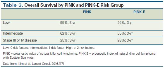
Gültigkeitsbereich:                                                                         Leitlinie
                                                         Tumorzentrum Oberösterreich                                                                       PTCL

INHALTSVERZEICHNIS

1   Allgemeines ........................................................................................................................................... 3
2   Diagnostik und Scoring .......................................................................................................................... 3
  2.1    Diagnostik ...................................................................................................................................... 3
  2.2    Fertilität ......................................................................................................................................... 4
  2.3    WHO-Klassifikation ........................................................................................................................ 5
  2.4    Typische Befundkonstellationen verschiedener Subtypen ........................................................... 5
  2.5 Stadieneinteilung nach den modifizierten Ann-Arbor-Kriterien ......................................................... 6
  2.6    Risikostratifizierung ....................................................................................................................... 7
    2.6.1      IPI ........................................................................................................................................... 7
    2.6.2      PINK-E Risikoscore ................................................................................................................. 8
3 Behandlungsplan ................................................................................................................................... 9
  3.1    T-Zell-Lymphome - fit for transplant ............................................................................................. 9
  3.2    T-Zell-Lymphome – not fit for transplant .................................................................................... 10
  3.3    T-Zell-Lymphome – Rezidiv ......................................................................................................... 11
  3.4    Extranodale NK/T-Zell-Lymphome – Erstdiagnose...................................................................... 12
  3.5    Extranodale NK/T-Zell-Lymphome - Rezidiv................................................................................ 13
  3.6    Strahlentherapie.......................................................................................................................... 14
  3.7    Supportive Therapie mit G-CSF ................................................................................................... 14
  3.8    Infektionsprophylaxe während der Immunochemotherapie...................................................... 15
         Corona-Infektion (COVID-19) bei PTCL-Patienten ....................................................................... 15
4 Besondere klinische Situationen ......................................................................................................... 15
  4.1    Hohe Tumorlast ........................................................................................................................... 15
  4.2    ZNS-Befall .................................................................................................................................... 15
  4.3    Eingeschränkte Linksventrikelfunktion / erhöhtes kardiovaskuläres Risiko ............................... 16
5 Verlaufskontrolle und Nachsorge........................................................................................................ 17
  5.1    Responsebeurteilung .................................................................................................................. 17
  5.2    Verlaufskontrollen unter Therapie und nach Therapieabschluss ............................................... 19
6 Literatur/Quellenangaben................................................................................................................... 20
Anhang: Therapieprotokolle ....................................................................................................................... 22
Anhang: Studienblatt .................................................................................................................................. 23
Anhang: Wirtschaftliche Analyse (optional)................................................................................................ 23

                                                                                                                                            Seite 2 von 23
Gültigkeitsbereich: Leitlinie
Tumorzentrum Oberösterreich PTCL
1 Allgemeines
Unter dem Begriff periphere T-Zell Lymphome (PTCL) werden die klinisch meist aggressiv verlaufenden
nodalen und extranodalen Lymphome mit dem Phänotyp reifer post-thymischer NK- und T-Zellen
zusammengefasst. Nach der überarbeiteten WHO-Klassifikation von 2017 sind sie von unreifen T-Zell
Neoplasien (lymphoblastisches T-Zell-Lymphom, T-ALL), sowie den primär kutanen (Cutaneous T-Cell
lymphoma = CTCL, Mycosis fungoides u.a.) und den primär leukämisch verlaufenden reifen T/NK-Zell
Neoplasien (T-PLL, T-LGL) abzugrenzen. PTCL machen etwa 10 % aller neu diagnostizierten NHL aus.
Die Inzidenz der einzelnen Entitäten ist regional abhängig. In Europa ist das PTCL ohne weitere
Spezifizierung (PTCL, NOS), das angioimmunoblastische T-Zell Lymphom (AITL), sowie die anaplastisch
großzelligen Lymphome (ALCL) mit und ohne ALK-Expression (Anaplastic lymphoma kinase) die häufigsten
Formen. Die extranodalen NK/T-Zell Lymphome hingegen kommen im asiatischen Raum gehäuft vor.
Zur sicheren Diagnosestellung ist die Beurteilung eines histologischen Präparates (Lymphknoten-
Exstirpation) notwendig. Eine Feinnadelaspiration oder Stanzbiopsie ist nicht ausreichend um eine sichere
Subklassifizierung vorzunehmen. Nicht selten lässt sich die Diagnose nur aus Zusammenschau von
Zytomorphologie, Immunhistochemie und Klonalitätsnachweis (T-Zellrezeptor-Gen-Rearrangement, TCR)
stellen.
2 Diagnostik und Scoring
2.1 Diagnostik
Anamnese, u.a. B-Symptomatik, Risiko bzgl. NHL wie Immunsuppression, Familienanamnese
insbesondere bzgl Krebserkrankungen
Klinischer Status, ECOG
Histologie: Lymphknotenexstirpation (Stanzbiopsie nur, wenn Gewebegewinnung nicht anders
möglich; Feinnadelaspiration nur zur Sicherung eines Rezidivs) oder Hautbiopsie je nach Klinik bzw.
Befall
Labor (BB + Diff., LDH, Harnsäure, ß2-Mikrogobulin, Eiweißelektropherese, Immunglobuline
quantitativ), Schwangerschaftstest (ß-HCG im Blut), TSH, proBNP (bei Anthrazyklintherapie)
HIV- und Hepatitis-Serologie (inkl. HBs Ag: Impfung falls Hepatitis B naiv)
Anamnese/Information bzgl. Influenza- und Covid-19 Impfung und Nutzen einer Umgebungsimpfung
(Influenza, Covid-19)
EBV-PCR
EKG, Echokardiographie
PET-CT (mit diagnostischem CT): Bei dringlicher Notwendigkeit einer Therapieeinleitung und
fehlender rascher Verfügbarkeit eines PET-CTs ist eine CT Thorax/Abdomen +/- Hals ausreichend.
Über 90% der PTCL sind FDG-avid (Gurion R, 2018): Das PET-CT besitzt bei extranodalen nicht-kutanen
Manifestationen, insbesondere im GI-Trakt, eine der CT überlegene Sensitivität. Die Beurteilung
erfolgt nach Deauville-Score (5-Punkte-Skala: 1: no uptake above background; 2: uptake ≤
mediastinum; 3: uptake > mediastinum but ≤ liver; 4: uptake moderately > liver; 5, uptake markedly
higher than liver and/or new lesions; X, new areas of uptake unlikely to be related to lymphoma.)
Knochenmarksaspiration + -biopsie (Morphologie, IHC, FACS, Zytogenetik und Molekularbiologie)
Seite 3 von 23Gültigkeitsbereich: Leitlinie
Tumorzentrum Oberösterreich PTCL
Eine CCT bzw. MRT-Untersuchung des Schädels oder eine Lumbalpunktion werden nur bei klinischem
Verdacht empfohlen. Bei höherer Zellzahl (> 5/µl) ist eine FACS-Diagnostik aus dem Liquor
durchzuführen.
Fertilität siehe 2.2
2.2 Fertilität
Männer: Bei einem Teil mit dem CHOP-Protokoll behandelter Männer kommt es innerhalb von 5 bis
7 Jahren zu einer Erholung der Spermatogenese. Da dies im Einzelfall nicht vorhersehbar ist, sollte
bei Kinderwunsch vor der Chemotherapie eine Samenkonservierung erfolgen.
Bei Frauen kommt es im Anschluss an eine Behandlung mit CHOP nur selten zu bleibender
Amenorrhoe. Häufig ist dagegen eine Verminderung der Ovarialreserve mit vorzeitiger Menopause
(letzter Zyklus vor dem 40. Lebensjahr). Das zeitliche Fenster für die Erfüllung eines Kinderwunsches
ist insbesondere für Patientinnen, die zum Zeitpunkt der Chemotherapie das 30. Lebensjahr
überschritten haben, kurz.
Patientinnen mit prospektivem Kinderwunsch sollten soweit vertretbar vor Einleitung der Therapie
einem reproduktionsmedizinischen Zentrum vorgestellt werden.
GnRH Analoga: Der Einsatz von GnRH Analoga zur Ovarialprotektion unter Chemotherapie ist keine
Standardtherapie, kann jedoch die ovarielle Funktion und das Risiko einer langfristigen
Chemotherapie-induzierten Amenorrhoe senken (Goserelin (Zoladex®) 3.6 mg subkutan alle 4
Wochen: Start 1 Woche vor dem 1. Zyklus Chemotherapie bis 2 Wochen vor oder nach Ende des
letzten Zyklus Chemotherapie)
Kryokonservierung von Ovarialgewebe: Die Kryokonservierung von laparoskopisch entnommenem
Ovarialgewebe ist ein neuer, experimenteller Ansatz. Aufgrund der mit dem Alter abnehmenden
Follikeldichte im Ovar wird diese fertilitätsprotektive Technik nur bei Frauen bis zu einem Alter von
35 Jahren empfohlen. Die Maßnahme ist partnerunabhängig und würde bei einem späteren
erfolgreichen Angehen des Transplantates auch die endokrinologische Situation der Patientin
verbessern. Der Zeitbedarf beträgt ca. 2 Tage.
Kontakt: Kepler-Universitätsklinikum, Kinderwunschzentrum, Tel.: +43 (0)5 7680 84 – 24630
Seite 4 von 23Gültigkeitsbereich: Leitlinie
Tumorzentrum Oberösterreich PTCL
2.3 WHO-Klassifikation
Mature T- and NK-cell neoplasms:
T-cell prolymphocytic leukaemia Primary cutaneous CD30-positive T-cell
T-cell large granular lymphocytic leukaemia lymphoproliferative disorders
Chronic lymphoproliferative disorder of NK - Lymphomatoid papulosis
cells - Primary cutaneous anaplastic large cell
Aggressive NK-cell leukaemia lymphoma
Systemic EBV-positive T-cell lymphoma of Primary cutaneous gamma delta T-cell
childhood lymphoma
Chronic active EBV infection of T- and NK-cell Primary cutaneous CD8-positive aggressive
type, systemic form epidermotropic cytotoxic T-cell lymphoma
Hydroa vacciniforme-like Primary cutaneous acral CD8-positive T-cell
lymphoproliferative disorder lymphoma
Adult T-cell leukaemia/lymphoma Primary cutaneous CD4-positive
Extranodal NK/T-cell lymphoma, nasal type small/medium T-cell lymphoproliferative
Enteropathy-associated T-cell lymphoma disorder
Monomorphic epitheliotropic intestinal T- Peripheral T-cell lymphoma, NOS
cell lymphoma Angioimmunoblastic T-cell lymphoma
Intestinal T-cell lymphoma, NOS Follicular T-cell lymphoma
Indolent T-cell lymphoproliferative disorder Nodal peripheral T-cell lymphoma with T
of the gastrointestinal tract follicular helper phenotype
Hepatosplenic T-cell lymphoma Anaplastic large cell lymphoma, ALK-positive
Subcutaneous panniculitis-like T-cell Anaplastic large cell lymphoma, ALK-
lymphoma negative
Mycosis fungoides Breast implant-associated anaplastic large
Sézary syndrome cell lymphoma
2.4 Typische Befundkonstellationen verschiedener Subtypen
Der Klonalitätsnachweis eines T-Zellrezeptor-Gen-Rearrangements gelingt mittels PCR bei allen Entitäten
außer bei den extranodalen NK/T-Zell-Lymphomen. (Miyata-Takata T, 2018; Takayama T 2018)
Das ALK-positive ALCL ist mit der Translokation t(2;5) (p23;q35) assoziiert, dies führt zur aberranten
Expression eines NPM-ALK-Fusionsproteins und geht mit einer deutlich günstigeren Prognose einher
(Amin HM). Der Nachweis von CD30, ALK-Expression bei allen ALCL sowie DUSP22/IRF4-Rearrangement
bei ALK-negativen ALCL ist ein wichtiger prognostischer Indikator hinsichtlich einer günstigeren Prognose.
Wohingegen ein TP53-Rearrengement mit einem aggressiveren Verlauf vergesellschaftet ist (Parrilla
Castellar ER, 2014).
Die Tumorzellen des AITL leiten sich von den sogenannten follikulären T-Helferzellen ab und exprimieren
typischerweise CD10, BCL6, PD1 und CXCL13. Die häufigsten zytogenetischen Abnormalitäten sind
Trisomie 3, Trisomie 5 und/oder ein zusätzliches X-Chromosom (Schlegelberger B 1990). Zudem können
Mutationen in TET2, IDH2, DNMT3A und RHOA aufweisen. Die TET2-Mutation Die klinische Signifikanz
dieser Mutationen ist jedoch aktuell noch unklar.
Typsich für die extranodalen NK/T-Zell-Lymphome ist der Nachweis von EBV-DNA, welcher auch als
Verlaufsparameter prognostische Relevanz hat, und die CD56-Positivität (Au WY 2004; Kwong YL 2014).
Da ENKTL meist P-Glykoprotein in hoher Konzentration expremieren, welches eine Multidrug-Resistenz
vermitteln, sind anthrazylin basierte Chemotherapien nicht wirksam (Yamaguchi M, Miyazaki K 2017).
Seite 5 von 23Gültigkeitsbereich: Leitlinie
Tumorzentrum Oberösterreich PTCL
2.5 Stadieneinteilung nach den modifizierten Ann-Arbor-Kriterien
Die Stadieneinteilung der T-Zell Lymphome erfolgt analog zu den aggressiven B-Zell-Lymphomen nach den
Ann-Arbor-Kritierin, d.h. anhand des PET-CT bei FDG-aviden Tumoren und anhand des CT bei nicht FDG-
aviden Tumoren. Tonsillen, Waldeyer’s Ring und Milz werden als „nodales“ Gewebe gewertet. Die
Stadieneinteilung unterscheidet in „Limited Disease“ (Stadium I/II ohne „bulky“) und „Advanced Disease“
(Stadium III/IV). Ein Patient im Stadium II mit bulky ist in Abhängigkeit von Histologie und anderen
Risikofaktoren (z.B. IPI) entweder als limited oder advanced disease zu behandeln.
Modifizierte Ann Arbor Klassifikation (Lugano Classification)
Befall eines Lymphknotens oder einer Lymphknotenregion (I)
oder
Stadium I (IE)
lokalisierter Befall eines einzigen extralymphatischen Organs (IE) ohne
Lymphknotenbefall
Befall von zwei oder mehr Lymphknotenregionen auf der gleichen Seite des Zwerchfells
(II)
Stadium II (IIE) oder
Kriterien von LK-Befall von Stadium I/II mit unifokalem („zusammenhängend“) Befall
eines extralymphatischen Gewebes (IIE)
Befall von Lymphknotenregionen auf beiden Seiten des Zwerchfells (III)
Stadium III (III) oder
Lymphknotenbefall oberhalb des Zwerchfells und Milzbefall (III)
Multifokaler Befall eines oder Befall mehrerer extralymphatischer Organe oder Gewebe
mit oder ohne Befall von lymphatischen Gewebe
Stadium IV
Als diffuser Befall gelten auch mehrere lokale Manifestationen in einer extranodalen
Lokalisation, Leber- und/oder KM-Befall
bei Stadium II-IV definiert als RF ≥ 10 cm (Die Größe von Raumforderungen sollte in
„bulky“ „cm“ angegeben werden, da ab einer Größe von 7,5 cm eine Strahlentherapie in
Erwägung zu ziehen ist)
Lymphknotenregionen
Seite 6 von 23Gültigkeitsbereich: Leitlinie
Tumorzentrum Oberösterreich PTCL
2.6 Risikostratifizierung
Im Allgemeinen besteht bei Patienten mit peripheren T-Zell-Lymphomen bei der Diagnose ein höherer IPI-
Score, die Prognose ist zudem bei sämtlichen peripheren T-Zell-Lymphomen mit Ausnahme des AKL-
positiven ALCL’s eine signifikant schlechtere als bei B-Zell-Lymphomen. Sie weisen ein schlechteres
Ansprechen auf die gängigen Therapieprotokolle sowie eine sehr hohe Rezidivrate auf.
Aus diesem Grund sollte eine primäre Hochdosischemotherapie mit autologer bzw. je nach Ansprechen
evtl. auch mit allogener Stammzelltransplantation bei fitten Patienten im Alter von unter 60 Jahren mit
entsprechender Risikokonstellation (erhöhte LDH, Stadium >III und mehr als ein extranodaler Befall)
und zu erwartendem schlechtem Outcome in jedem Fall in Erwägung gezogen werden (d’Amor F 2012).
Für eine Risikostratifizierung und Abschätzung der Prognose wird der „International Prognostic Index“ (IPI)
verwendet. Zudem erfolgt für die EKNTL eine Risikostratifizierung bzgl. Stammzellharvest.
2.6.1 IPI
International Prognostic Index (IPI) IPI-Score (Summe)
Age >60 Low 0 or 1
LDH > normal Low intermediate 2
Performance status 2-4
High intermediate 3
Stage III or IV
High 4 or 5
Extranodal involvement > 1 site
Tabelle: Prognose nach IPI bei Patienten mit verschiedenen T-Zell-Lymphomen (ALCL/ALK+, ALCL/ALK-,
AITL und PTCL, NOS) mit Anthrazyklin-basierter Chemotherapie (Schmitz N, Blood 2010)
IPI EFS 3-Year rate OS 3-Year rate
Low 65% 78%
low-intermediate 43% 61%
high-intermediate 41% 50%
High 10% 27%
Seite 7 von 23Gültigkeitsbereich: Leitlinie
Tumorzentrum Oberösterreich PTCL
Tabelle: Prognose je nach Entität bei Patienten mit Anthrazyklin-basierter Chemotherapie (Schmitz N,
Blood 2010)
Entität EFS (3-Year rate) OS (3-Year rate)
ALCL, ALK+ 76% 90%
ALCL, ALK- 46% 62%
AITL 50% 67%
PTCL, NOS 41% 54%
ENKTL 36% 46%
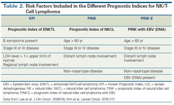
2.6.2 PINK-E Risikoscore
Seite 8 von 23Gültigkeitsbereich: Leitlinie
Tumorzentrum Oberösterreich PTCL
3 Behandlungsplan
3.1 T-Zell-Lymphome - fit for transplant
* Definierte T-Zell-Lymphome
Definierte T-Zell-Lymphome* Peripheral T-cell lymphoma (PTCL), NOS
Biolog. Alter 40 Jahre
1 Risikofaktor 5 CR mit 1 Risikofaktor 5
und 3 Risikofaktoren 5
beta2MG > 3mg/l beta2MG > 3mg/l
beta2MG > 3mg/l
> 1 extranodal site > 1 extranodal site
> 1 extranodal site
Erhöhte LDH Erhöhte LDH
Erhöhte LDH
St III/IV Ja Ja St III/IV
St III/IV CR/PR
Ja Nein
Ja
Nein Nein Nein
Bridging to Tx
ABSCT ABSCT
(1-2 Zyklen GDP q3w 4)
RESTAGING Allogene 8 RESTAGING
Palliative Therapie 7
PET-CT Stammzelltransplantation PET-CT
Ja
Fit für
CR Nein Nein CR
Allogene SZT
Nein
Ja
Ja
RTX
überlegen
Nachsorge
1 Horwitz S, Lancet 2019; 19;393:229-240 (ECHELON 2)
2 Primär vorbeugende oder sekundäre Zyklusintervallverlängerung auf q3w oder Abkürzungen (alphabetisch)
Dosiseskalation auf CHOP q3w, wenn Allgemeinzustand die Durchführung der ABSCT Autologe Stammzelltransplantation
Standardtherapie (CHEOP q2w) nicht erlaubt; Pfreundschuh M, Blood 2004 104:634-641 sALCL systemic Anaplastic Large Cell Lymphoma
3 Stammzellharvest aus dem 3. (falls kein KM-Befall) oder 4. Zyklus ALK anaplastic lymphoma kinase
4 Skamene T, Leuk Lymphoma. 2017;58: 2319 (Alternativen: DEXA-BEAM, DHAP, ICE, GemOx) BV Brentuximab vedotin
5 Parrilla Castellar ER, Blood 2014; 124(9):1473 CHP CHOP ohne Vinristin
6 Schmitz N, Blood 2010;116: 3418 GDP Gemcitabine, dexamethasone, cisplatin
7 Je nach AZ: Palliative Care +/- Lymphomtherapie (Lenolidomid, Gem, Strahlentherapie ) RTX Lokale Strahlentherapie
8 Allogene SZT bevorzugt vor Autologer SZT
Seite 9 von 23Gültigkeitsbereich: Leitlinie
Tumorzentrum Oberösterreich PTCL
3.2 T-Zell-Lymphome – not fit for transplant
* Definierte T-Zell-Lymphome
Definierte T-Zell Lymphome* Peripheral T-cell lymphoma (PTCL), NOS
Biolog. Alter >70 Jahre oder ECOG = 2 1 Angioimmunoblastic T-cell lymphoma (AITL)
not fit for transplant Anaplastic large cell lymphoma (ALCL), ALK positive
Anaplastic large cell lymphoma (ALCL), ALK negative
Nein Fit for CHOP Ja
CD30-Expression CD30-Expression
Ja Nein Ja Nein
10% 10%
3 Zyklen 3 Zyklen 3 Zyklen 3 Zyklen
BV mono 2 Mini-CHOP q3w BV-CHP 3 CHOP q3w
CR/PR Nein Nein CR/PR CR/PR Nein Nein CR/PR
Ja Ja Ja Ja
bis 16 Zyklen 1-3 Zyklen 3 Zyklen 3 Zyklen
Brentuximab 2 Mini-CHOP q3w BV-CHP 3 CHOP q3w 4
CR Nein Nein CR CR Nein Nein CR
Ja Ja Ja Ja
Nachsorge Nachsorge Nachsorge Nachsorge
Lokale Radiato Lokale Radiato
Ja RTX 5 Ja
sinnvoll? sinnvoll?
Nein Nein
Ja
Reinduktionstherapie
Nein
sinnvoll?
Zweitlinientherapie Palliative Care ohne
(palliativ) Lymphomtherapie
1 Bei vorbestehendem ECOG > 2 rein symptomatische Cortisontherapie
2 Prince HM, Lancet. 2017;390:555-566 (ALCANZA) Abkürzungen (alphabetisch)
3 Horwitz S, Lancet 2019; 19;393:229-240 (ECHELON-2) BV Brentuximab-Vedotin
4 Bei ALCL für Stadium I-II: optional nur 4 Zyklen CHOP und anschließend Radiatio (Ellin CHP CHOP ohne Vincristin
F., Blood. 2014 Sep;124(10):1570-7. RTX Lokale Strahlentherapie
5 Keine etablierte Standarddosis für die lokale Strahlentherapie. In der Literatur sind
Dosierungen von 40-50 Gy beschrieben.
Seite 10 von 23Gültigkeitsbereich: Leitlinie
Tumorzentrum Oberösterreich PTCL
3.3 T-Zell-Lymphome – Rezidiv
Seite 11 von 23Gültigkeitsbereich: Leitlinie
Tumorzentrum Oberösterreich PTCL
3.4 Extranodale NK/T-Zell-Lymphome – Erstdiagnose
Seite 12 von 23Gültigkeitsbereich: Leitlinie
Tumorzentrum Oberösterreich PTCL
3.5 Extranodale NK/T-Zell-Lymphome - Rezidiv
Seite 13 von 23Gültigkeitsbereich: Leitlinie
Tumorzentrum Oberösterreich PTCL
3.6 Strahlentherapie
Die peripheren T-Zell-Lymhome mit Ausnahme der NK-/T-Zell-Lymphome sind etwas weniger
strahlenempfindlich als die aggressiven B-Zell-Lymphome und benötigen daher höhere Strahlendosen.
Obwohl es in NCCN / UpToDate und Onkopedia die isRT als Empfehlung in lokalisierten Stadien optional
enthalten ist bzw. empfohlen wird, ist die Evidenz eines Nutzens sehr gering. Daher Vorgehen wie bei
anderen Lymphomen in Abhängigkeit vom PET-CT und Restbulk.
Eine lokale Bestrahlung ist zu empfehlen:
bei lokalem Befall – Stadium I-II, die mit Chemotherapie eine CR erreicht haben
bei Patienten mit PR und singulärem oder symptomatischem Restlymphom, welche nicht für eine
ASZT geeignet sind
als Symptomkontrolle bei Patienten, die nicht für Chemotherapie geeignet sind
Empfohlene Strahlendosen abhängig vom Therapieansprechen, IPI-Score und Patienten-Fitness:
Konsolidierung: 36-40 Gy
Komplementär bei PR oder therapeutisch bei primär Refraktären oder für nicht HD-geeignete
Patienten: 40-55 Gy
In Kombination mit HD-Therapie, je nach Lokalisation und Vorbehandlung mit Strahlentherapie:
20-36 Gy
3.7 Supportive Therapie mit G-CSF
Grundsätzlich wird eine Primärprophylaxe mit G-CSF bei Chemotherapieschemata empfohlen, die
ein > 20 %iges Risiko einer febrilen Neutropenie haben.
Eine Primärprophylaxe sollte außerdem erfolgen bei Patienten ≥ 65 Jahre sowie Patienten mit
zusätzlichen Risikofaktoren für infektiöse Komplikationen, wie frühere intensive Chemotherapien,
Vorbestrahlung im Beckenbereich oder zusätzliche Infektionsprobleme.
Eine Sekundärprophylaxe ist indiziert, wenn im zuvor durchgeführten Chemotherapiezyklus in der
neutropenischen Phase Fieber auftrat oder die Erhaltung der Dosisintensität sowie die zeitgerechte
Gabe der Chemotherapie für den Behandlungserfolg entscheidend ist.
Folgende Empfehlungen gelten für die Primärprophylaxe:
BV-CHP q3w G-CSF fix ab d4
CHOEP q2w/q3w G-CSF fix ab d4
CHOP q3w G-CSF nur bei zusätzlichem Risikofaktor* ab d4
Mini-CHOP G-CSF nur bei zusätzlichem Risikofaktor (außer Alter)* ab d4
BV mono G-CSF nur bei zusätzlichem Risikofaktor* ab d4
DDGP G-CSF fix ab d6
2/3 DeVIC G-CSF fix ab d4
GDP G-CSF fix ab d4
SMILE G-CSF fix ab d6
* Alter > 65 Jahre, Niedriger Performancestatus (niedriger Karnofsky Index, hoher ECOG), Mukositis,
Komorbiditäten (COPD, Herzinsuffizienz NYHA III-IV, HIV-Infektion, Autoimmunerkrankung, deutlich
eingeschränkte Nierenfunktion), weit fortgeschrittene, symptomatische Tumorerkrankung, V.a
eingeschränkte hämatopoetische Reserve, z.B. bei kürzlich zurückliegender Chemotherapie oder
Chemotherapie ab 3. Therapielinie, Laborparameter (Anämie, LymphozytopenieGültigkeitsbereich: Leitlinie
Tumorzentrum Oberösterreich PTCL
3.8 Infektionsprophylaxe während der Immunochemotherapie
Es gelten die Prophylaxen gemäß der Leitlinie antimikrobielle Prophylaxe und Therapie.
Corona-Infektion (COVID-19) bei PTCL-Patienten
Aufgrund der Leukozytopenie besteht grundsätzlich ein erhöhtes Risiko für einen schweren
Infektionsverlauf COVID-19 unter Immuno-/Chemotherapie mit (CHOEP), Brentuximab, Gemcitabin,
Bendamustin, Altemtuzumab, Carboplatin, Etoposid, Ifosfamid.
Ausführliche und aktuelle Empfehlungen sind auf der Homepage von Onkopedia aus der Covid-19-Leitlinie
zu entnehmen.
Impfempfehlung
Weder das hämatologische Grundleiden, in diesem Fall PTCL, noch eine spezifische Therapie (Immuno-/
Chemotherapie) stellen eine Kontraindikation gegen eine Schutzimpfung. Allerdings ist bei
immunsupprimierten Patienten von einer geringen Schutzwirkung auszugehen. Eine Auffrischung der
Impfung sollte nach Beendigung der Therapie erneut in Erwägung gezogen werden.
Sowohl nach B-Zell-Depletion als auch nach einer Stammzelltransplantation ist von einer fehlenden bzw.
deutlich reduzierten Impfantwort auszugehen. Auch hier stellt eine Schutzimpfung frühestens nach 3 bis
>6 Monaten nach der Therapie keine Kontraindikation dar.
Um das Infektionsrisiko des immunsupprimierten Patienten zu minimieren, sollten auch die
Familienmitglieder auf die Covid-19-Impfung hingewiesen werden.
Eine ausführliche Impfempfehlung ist auf der Homepage von Onkopedia aus der Covid-19-Schutzimpfung-
Leitlinie zu entnehmen.
4 Besondere klinische Situationen
4.1 Hohe Tumorlast
Bei Patienten mit hoher Tumorlast (hohe LDH, Bulk > 7,5cm, Organkompression,…) ist an die
Möglichkeit einer Vorphase (Prednisolon 100 mg (d1-5) zu denken.
Allopurinol/Rasbirucase
Hydrierung!
4.2 ZNS-Befall
Die Häufigkeit eines ZNS-Befalls bei den peripheren T-Zell-Lymphomen und den extranodalen NK-/T-Zell-
Lymphomen wird in der Literatur mit 2-6% beziffert (Chihara D 2018; Pro B 2010).
In Gegensatz zum diffus großzelligen B-Zell-Lymphom liegen keine Daten zu Risikofaktoren für einen
ZNS-Befall bzw. für ein erhöhtes Risiko eines ZNS-Rezidivs vor. Es kann somit aktuell auch keine
Empfehlung bezüglich einer ZNS-Prophylaxe gegeben werden.
Seite 15 von 23Gültigkeitsbereich: Leitlinie
Tumorzentrum Oberösterreich PTCL
4.3 Eingeschränkte Linksventrikelfunktion / erhöhtes kardiovaskuläres Risiko
Bei erhöhtem kardiovaskulärem Risiko, vor allem eingeschränkter Linksventrikelfunktion sollte folgende
Therapieadaption durchgeführt werden:
Myocet anstelle von Doxorubicin
Etoposid anstelle von Doxorubicin bei fortgeschrittener Herzinsuffizienz (CEOP)
Seite 16 von 23Gültigkeitsbereich: Leitlinie
Tumorzentrum Oberösterreich PTCL
5 Verlaufskontrolle und Nachsorge
5.1 Responsebeurteilung
Es stehen derzeit 2 international gültige Klassifikationen zur Responsebeurteilung zur Auswahl (The
Lugano Classification 2014; RECIL 2017). In der vorliegenden Leitlinie wird die Lugano-Klassifikation
(Cheson BD, 2014) verwendet (siehe Folgeseite).
Zum Restaging unter laufender Therapie (nach 3 Zyklen) werden alle initial positiven klinischen und
bildgebenden Verfahren neuerlich angewendet, jedoch kein PET-CT und keine KM-Untersuchung.
Wenn keine fixe Indikation für eine Strahlentherapie besteht, erfolgt das Restaging bei
Therapieabschluss frühestens 6 Wochen nach Ende der Chemotherapie:
o PET CT (inkl. diagnostisches CTs)
o KM (falls initial positiv)
o Echokardiographie
Response and Site PET-CT–Based Response CT-Based Response
Complete Remission Complete metabolic response Complete radiologic response (all of the following)
Lymph nodes and Score 1, 2 or 3 (1) with or without a residual mass on Target nodes/nodal masses must regress to ≤ 1.5 cm in
extralymphatic 5PSjavascript:popRef('TF3-2') (2) LDi
sites It is recognized that in Waldeyer's ring or extranodal No extralymphatic sites of disease
sites with high physiologic uptake or with activation
within spleen or marrow (eg, with chemotherapy or
myeloid colony-stimulating factors), uptake may be
greater than normal mediastinum and/or liver. In this
circumstance, complete metabolic response may be
inferred if uptake at sites of initial involvement is no
greater than surrounding normal tissue even if the
tissue has high physiologic uptake
Nonmeasured Not applicable Absent
lesion
Organ enlargement Not applicable Regress to normal
New lesions None None
Bone marrow No evidence of FDG-avid disease in marrow Normal by morphology; if indeterminate, IHC negative
Partial Remission Partial metabolic response Partial remission (all of the following)
Lymph nodes and Score 4 or 5javascript:popRef('TF3-2') (2) with ≥ 50% decrease in SPD of up to 6 target measurable
extralymphatic sites reduced uptake compared with baseline and residual nodes and extranodal sites
mass(es) of any size
At interim, these findings suggest responding disease When a lesion is too small to measure on CT, assign 5
mm × 5 mm as the default value
At end of treatment, these findings indicate residual When no longer visible, 0 × 0 mm
disease
For a node > 5 mm × 5 mm, but smaller than normal,
use actual measurement for calculation
Nonmeasured lesions Not applicable Absent/normal, regressed, but no increase
Organ enlargement Not applicable Spleen must have regressed by > 50% in length beyond
normal
New lesions None None
Bone marrow Residual uptake higher than uptake in normal marrow Not applicable
but reduced compared with baseline (diffuse uptake
compatible with reactive changes from chemotherapy
allowed). If there are persistent focal changes in the
marrow in the context of a nodal response,
consideration should be given to further evaluation
with MRI or biopsy or an interval scan
Fortsetzung der Tabelle und Legende zur Tabelle auf der nächsten Seite
Seite 17 von 23Gültigkeitsbereich: Leitlinie
Tumorzentrum Oberösterreich PTCL
Response and Site PET-CT–Based Response CT-Based Response
Stable disease No metabolic response Stable disease
Target nodes/nodal Score 4 or 5 (2) with no significant change in FDG < 50% decrease from baseline in SPD of up to 6
masses, extranodal uptake from baseline at interim or end of treatment dominant, measurable nodes and extranodal sites; no
lesions criteria for progressive disease are met
Nonmeasured lesions Not applicable No increase consistent with progression
Organ enlargement Not applicable No increase consistent with progression
New lesions None None
Bone marrow No change from baseline Not applicable
Progressive disease Progressive metabolic disease Progressive disease requires at least 1 of the following
Individual target Score 4 or 5 (2) with an increase in intensity of uptake PPD progression:
nodes/nodal masses from baseline and/or
Extranodal lesions New FDG-avid foci consistent with lymphoma at An individual node/lesion must be abnormal with:
interim or end-of-treatment assessment LDi > 1.5 cm and
Increase by ≥ 50% from PPD nadir and
An increase in LDi or SDi from nadir
0.5 cm for lesions ≤ 2 cm
1.0 cm for lesions > 2 cm
In the setting of splenomegaly, the splenic length must
increase by > 50% of the extent of its prior increase
beyond baseline (eg, a 15-cm spleen must increase to >
16 cm). If no prior splenomegaly, must increase by at
least 2 cm from baseline
New or recurrent splenomegaly
Nonmeasured lesions None New or clear progression of preexisting nonmeasured
lesions
New lesions New FDG-avid foci consistent with lymphoma rather Regrowth of previously resolved lesions
than another etiology (eg, infection, inflammation). If A new node > 1.5 cm in any axis
uncertain regarding etiology of new lesions, biopsy or A new extranodal site > 1.0 cm in any axis; if < 1.0 cm in
interval scan may be consideredl any axis, its presence must be unequivocal and must be
attributable to lymphoma
Assessable disease of any size unequivocally
attributable to lymphoma
Bone marrow New or recurrent FDG-avid foci New or recurrent involvement
Abbreviations:
5PS 5-point scale CT computed tomography
FDG fluorodeoxyglucose IHC immunohistochemistry
LDi longest transverse diameter of a lesion MRI magnetic resonance imaging
PET positron emission tomography PPD cross product of the LDi and perpendicular diameter
SDi shortest axis perpendicular to the LDi SPD sum of the product of the perpendicular diameters for multiple lesions
(1) A score of 3 in many patients indicates a good prognosis with standard treatment, especially if at the time of an
interim scan. However, in trials involving PET where de-escalation is investigated, it may be preferable to consider a score
of 3 as inadequate response (to avoid undertreatment). Measured dominant lesions: Up to six of the largest dominant
nodes, nodal masses, and extranodal lesions selected to be clearly measurable in two diameters. Nodes should preferably
be from disparate regions of the body and should include, where applicable, mediastinal and retroperitoneal areas. Non-
nodal lesions include those in solid organs (eg, liver, spleen, kidneys, lungs), GI involvement, cutaneous lesions, or those
noted on palpation. Nonmeasured lesions: Any disease not selected as measured, dominant disease and truly assessable
disease should be considered not measured. These sites include any nodes, nodal masses, and extranodal sites not
selected as dominant or measurable or that do not meet the requirements for measurability but are still considered
abnormal, as well as truly assessable disease, which is any site of suspected disease that would be difficult to follow
quantitatively with measurement, including pleural effusions, ascites, bone lesions, leptomeningeal disease, abdominal
masses, and other lesions that cannot be confirmed and followed by imaging. In Waldeyer's ring or in extranodal sites
(eg, GI tract, liver, bone marrow), FDG uptake may be greater than in the mediastinum with complete metabolic
response, but should be no higher than surrounding normal physiologic uptake (eg, with marrow activation as a result of
chemotherapy or myeloid growth factors).
Fortsetzung der Legende zur Tabelle auf der nächsten Seite
Seite 18 von 23Gültigkeitsbereich: Leitlinie
Tumorzentrum Oberösterreich PTCL
(2) PET 5 – Point Scale (Deauville Criteria)
1 no uptake above background
2 uptake ≤ mediastinum
3 uptake > mediastinum but ≤ liver
4 uptake moderately > liver
5 uptake markedly higher than liver and/or new lesions
X new areas of uptake unlikely to be related to lymphoma
5.2 Verlaufskontrollen unter Therapie und nach Therapieabschluss
Restaging nach der Hälfte der Therapiezyklen und frühestens 6 Wochen nach Abschluss einer
zytostatischen Therapie bzw. 12 Wochen nach Beendigung der Radiatio (bei klinischem Ansprechen nur
die initial pathologischen Untersuchungen).
Die ersten 2 Jahre vierteljährlich
Anamnese und klinische Untersuchung
Labor (BB, Chemie inkl. LDH, EBV-PCR)
Ab dem Jahr 2 sind klinische Kontrolle (Anamnese und klinische Untersuchung anhand des
Nachsorgebogens) alle 6 Monate für weitere 2 Jahre, ab dem 5. Jahr in jährlichen Abständen.
Seite 19 von 23Gültigkeitsbereich: Leitlinie
Tumorzentrum Oberösterreich PTCL
6 Literatur/Quellenangaben
Grundlage der Empfehlungen der vorliegenden Leitlinie sind die zum Zeitpunkt der Freigabe aktuell
gültigen internationalen Empfehlungen von Onkopedia, ESMO und NCCN sowie Übersichtsarbeiten, u.a.
aus UpToDate. Die nachfolgenden Quellenangaben zur Leitlinie stellen nur eine Auswahl der
Literaturquellen dar, die für die Erkrankung bedeutsam sind. Weitere Literaturquellen sind den
internationalen Leitlinien zu entnehmen.
1. Gurion R et al, Utility of PET-CT for Evaluation of Patients With Peripheral T-cell Lymphoma. Clin
Lymphoma Myeloma Leuk. 2018;18(10):687-691
2. Miyata-Takata T et al, Expression of T-cell receptor signalling pathway components in extranodal NK/T-
cell lymphoma. Histopathology. 2018;73(6):1030-1038
3. Takayama T et al, Identification of T-cell receptor expression in EBV-positive neoplastic cells in
extranodal NK/T-cell lymphoma, nasal-type, and comparison with T-cell receptor gene rearrangement by
BIOMED-2 assay. Hum Pathol. 2018;73:51-58
4. Amin HM, Lai R. Pathobiology of ALK+ anaplastic large-cell lymphoma. Blood 2007; 110:2259
5. Parrilla Castellar ER et al, ALK-negative anaplastic large cell lymphoma is a genetically heterogeneous
disease with widely disparate clinical outcomes. Blood 2014; 124(9): 1473–1480.
6. Schlegelberger B et al, Stepwise development of chromosomal abnormalities in angioimmunoblastic
lymphadenopathy. Cancer Genet Cytogenet 1990; 50(1):15-29
7. Au WY, Pang A, Choy C, Chim CS, Kwong YL. Quantification of circulating Epstein-Barr virus (EBV) DNA in
the diagnosis and monitoring of natural killer cell and EBV-positive lymphomas in immunocompetent
patients. Blood 2004;104(1):243-249
8. Kwong YL, Pang AW, Leung AY, Chim CS, Tse E. Quantification of circulating Epstein-Barr virus DNA in
NK/T-cell lymphoma treated with the SMILE protocol: diagnostic and prognostic significance. Leukemia
2014;28:865–70
9. d’Amor F et al, Up-front autologous stem-cell transplantation in peripheral T-cell lymphoma: NLG-T-01. J
Clin Oncol 2012; 30(25):3093-9
10. Schmitz N et al. Treatment and prognosis of mature T-cell and NK-cell lymphoma: an analysis of patients
with T-cell lymphoma treated in studies of the German High-Grade Non-Hodgkin Lymphoma Study
Group. Blood 2010; 116:3418.
11. Chihara D et al, The risk of central nervous system relapses in patients with peripheral T-cell lymphoma.
PLoS ONE 2018; 13(3): e0191461
12. Pro B, Central nervous system prophylaxis in peripheral T-cell lymphoma. Blood 2010; 115 (26): 5427.
13. Horwitz S et al, Brentuximab vedotin with chemotherapy for CD30-positive peripheral T-cell lymphoma
(ECHELON-2): a global, double-blind, randomised, phase 3 trial. Lancet. 2019; 393(10168):229-240
14. Yamaguchi et al, Concurrent Chemoradiotherapy for Localized Nasal Natural Killer/T-Cell Lymphoma: An
Updated Analysis of the Japan Clinical Oncology Group Study JCOG0211, J Clin Oncol 2012
15. Kim et al, Treatment of localized extranodal NK/T cell lymphoma, nasal type: a systematic review Journal
of Hematology & Oncology (2018) 11:140
Seite 20 von 23Gültigkeitsbereich: Leitlinie
Tumorzentrum Oberösterreich PTCL
16. Yamaguchi et al, Phase II study of SMILE chemotherapy for newly diagnosed stage IV, relapsed, or
refractory extranodal natural killer (NK)/T-cell lymphoma, nasal type: the NK-Cell Tumor Study Group
study, J Clin Oncol. 2011
17. Kwong YL et al., SMILE for natural killer/T-cell lymphoma: analysis of safety and efficacy from the Asia
Lymphoma Study Group. Blood. 2012;120(15):2973
18. Wang JJ et al., GDP (Gemcitabine, Dexamethasone, and Cisplatin) Is Highly Effective and Well-Tolerated
for Newly Diagnosed Stage IV and Relapsed/Refractory Extranodal Natural Killer/T-Cell Lymphoma, Nasal
Type. Medicine (Baltimore). 2016;95(6):e2787.
19. Wang X, Zhang L, Liu X, et al. Efficacy and survival in newly diagnosed advanced extranodal natural
killer/T-cell lymphoma: a randomized, controlled, multicenter and open-labeled study with DDGP
regimen versus SMILE regimen. Abstract #463. Presented at the 2019 American Society of Hematology
Annual Meeting, December 8, 2019; Orlando,
20. FLLee J et al, Autologous hematopoietic stem cell transplantation in extranodal natural killer/T cell
lymphoma: a multinational, multicenter, matched controlled study; Biol Blood Marrow Transplant.
2008;14(12):1356
21. Kim SJ, A prognostic index for natural killer cell lymphoma after non-anthracycline-based treatment: a
multicentre, retrospective analysis, Lancet Oncol. 2016;17:389-400
22. Jaccard A, Gachard N, Marin B, et al. Efficacy of L-asparaginase with methotrexate and dexamethasone
(AspaMetDex regimen) in patients with refractory or relapsing extranodal NK/T-cell lymphoma, a phase
2 study. Blood 2011; 117:1834-1839
23. Ennishi D et al, Allogeneic hematopoietic stem cell transplantation for advanced extranodal natural
killer/T-cell lymphoma, nasal type. Leuk Lymphoma 2011;52(7):1255.
24. Tse E et al, Allogeneic haematopoietic SCT for natural killer/T-cell lymphoma: a multicentre analysis from
the Asia Lymphoma Study Group, Bone Marrow Transplant. 2014; 49(7):902-6
25. Wang JH et al, Analysis of the efficacy and safety of a combined gemcitabine, oxaliplatin and
pegaspargase regimen for NK/T-cell lymphoma, Oncotarget, Vol. 7, No. 23, 35412-35422.
26. Yamaguchi M, Miyazaki K 2017, Current treatment approaches for NK/T-cell lymphoma, Journal of
clinical and experimental hematopathology Vol. 57 No.3, 98-108, 2017.
Seite 21 von 23Gültigkeitsbereich: Leitlinie
Tumorzentrum Oberösterreich PTCL
Anhang: Therapieprotokolle
BV-CHP GDP
Tag 1: Brentuximab-Vedotin 1,8 mg/kg Tag 1: Cisplatin 75 mg/m2 (+Hydratation)
Tag 1: Cyclophosphamid 750 mg/m² Tag 1+8: Gemcitabine 1000 mg/m²
Tag 1: Doxorubicin 50 mg/m² Tag 1–4: Fortecortin 40mg, p.o.
Tag 1-5: Prednisolon 100 mg, p.o. Wiederholung alle 3 Wochen
Wiederholung alle 3 Wochen Dexamethason: bei p.o. Gabe von Dexamethason und
gleichzeitiger Gabe von Akynzeo Dosisreduktion von
BV mono Dexamethason um 50%
Tag 1: Brentuximab-Vedotin 1,8 mg/kg
Wiederholung alle 3 Wochen Mini-CHOP
Tag 1: Cyclophosphamid 400 mg/m²
CHOP Tag 1: Doxorubicin 25 mg/m²
Tag 1: Cyclophosphamid 750 mg/m² Tag 1: Vincristin 1 mg absolut
Tag 1: Doxorubicin 50 mg/m² Tag 1-5: Prednisolon 40 mg/m², p.o.
Tag 1: Vincristin 1,4 mg/m² (max 2 mg) Wiederholung alle 3 Wochen
Tag 1-5: Prednisolon 100 mg, p.o.
Wiederholung alle 3 Wochen 2/3 DeVIC
Tag 1: Carboplatin 200 mg/m²
CHOEP Tag 1 bis 3: Etoposid 67 mg/m²
Tag 1: Cyclophosphamid 750 mg/m² Tag 1 bis 3: Ifosfamid 1000 mg/m²
Tag 1: Doxorubicin 50 mg/m² Tag 1 bis 3: Dexamethason 40 mg i.v.
Tag 1: Vincristin 1,4 mg/ m² (max. 2 mg) Wiederholung alle 3 Wochen
Tag 1-3: Etoposid 100 mg/m² Evtl. gleichzeitig Radiatio
Tag 1-5: Prednisolon 100 mg, p.o.
Wiederholung alle 2 Wochen (falls erforderlich mSMILE
Intervallverlängerung auf alle 3 Wochen möglich) Tag 1: Methotrexat 2 g/m²
Tag 2 bis 4: Etoposid 100 mg/m²
DDGP Tag 2 bis 4: Ifosfamid 1500 mg/m² (+Mesna)
Tag 1-4: Cisplatin 20 mg/m2 (+Hydratation) Tag 2 bis 4: Dexamethason 40 mg i.v.
Tag 1–5: Dexamethason 15 mg/m2, p.o. Tag 8: peg-Asparaginase 2.500 IU/m²
Tag 1+8: Gemcitabine 800 mg/m² Wiederholung alle 4 Wochen
Tag 1: peg-Aparaginase 2.500 IU/m2
Wiederholung alle 3 Wochen
Dosisreduktion von Brentuximab-Vedotin je nach Polyneuropathie (Horwitz S; Lancet 2019)
Neuropathie Monotherapie Kombinationstherapie
(sensorisch up
motorisch)
Grad 1 Fortsetzen der Therapie Fortsetzen der Therapie
Grad 2 Evtl. Therapieunterbrechung bis Toxizität ≤1, Evtl. Therapieunterbrechung bis Toxizität ≤1,
dann Dosisreduktion auf 1,2 mg/kg max. alle dann Dosisreduktion auf 0,9 mg/kg max. alle
3 Wochen 2 Wochen (hier auch Vincristin reduzieren!)
Grad 3 Therapieunterbrechung bis Toxizität ≤1 oder Therapieunterbrechung bis Toxizität ≤2,
Ausgangsbefund, dann Dosisreduktion auf dann Dosisreduktion auf 0,9 mg/kg max. alle
1,2 mg/kg max. alle 3 Wochen 2 Wochen (hier auch Vincristin reduzieren!)
Grad 4 Therapieabbruch Therapieabbruch
Grading anhand der Common toxicity criteria (CTCAE) v5.0
Grad 1 Grad 2 Grad 3 Grad 4 Grad 5
Periphere Asymptomatisch; Moderate Schwere Symptome; Lebensbedrohlich Tod
sensotische Verlust der tiefen Symptome; Einschränkung der e Folgen;
Neuropathie Sehnenreflexe Einschränkung selbstversorgenden Intervention
oder Parästhesie der ATL dringend
instrumentellen angezeigt
ATL
Periphere Asympatomatisch; Moderate Schwere Symptome; Lebensbedrohlich Tod
motorische lediglich klinische Symptome; Einschränkung der e Folgen;
Neuropathie oder Einschränkung selbstversorgenden Intervention
diagnostische der ATL; mechanische dringend
Beobachtung; instrumentellen Assistenz angezeigt angezeigt
Intervention nicht ATL
angezeigt
Seite 22 von 23Gültigkeitsbereich: Leitlinie
Tumorzentrum Oberösterreich PTCL
Anhang: Studienblatt
Studie DSHNHL 2015-1_NIVEAU
Ordensklinikum Elisabethinen
PI: Prim. Univ. Prof. Dr. Andreas Petzer, 0732 7676 4000
Rekrutierungszeit erwartet bis Q4 2022
http://agmt.at/wp-content/uploads/2019/04/AGMT_NIVEAU_Poster_OeGHO_2019_web-1.pdf
Studie LYSARC - ORACLE
Kepler-Universitätsklinikum-Linz
PI: Prim. Univ. Prof. Dr. Clemens Schmitt, Tel. +43 (0)5 7680 82 - 0
Rekrutierungszeit erwartet bis Q1 2022
Anhang: Wirtschaftliche Analyse (optional)
---
Seite 23 von 23Sie können auch lesen