Regulation of septum formation by RHO4 GTPase signalling in Neurospora crassa
←
→
Transkription von Seiteninhalten
Wenn Ihr Browser die Seite nicht korrekt rendert, bitte, lesen Sie den Inhalt der Seite unten

Regulation of septum formation by RHO4 GTPase
         signalling in Neurospora crassa

                          Dissertation
                 zur Erlangung des Doktorgrades
       der Mathematisch-Naturwissenschaftlichen Fakultäten
            der Georg-August-Universität zu Göttingen

                          vorgelegt von
                     Daniela Justa-Schuch
                        aus Quakenbrück

                         Göttingen 2010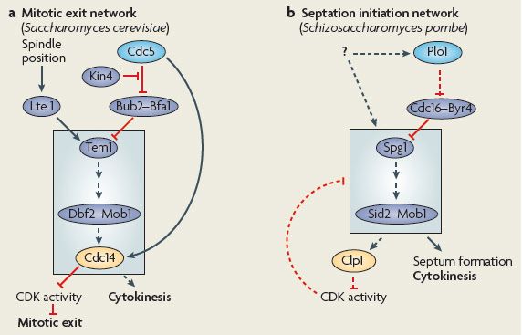
Die vorliegende Arbeit wurde von Januar 2006 bis März 2010 in der Abteilung Molekulare
Mikrobiologie und Genetik unter Anleitung von Prof. Dr. Gerhard H. Braus am Institut für
Mikrobiologie und Genetik der Georg-August-Universität zu Göttingen angefertigt.

D7
Referent:                          Prof. G. H. Braus
Korreferent:                       Prof. S. Pöggeler
Tag der mündlichen Prüfung:        30. April 2010
Teile dieser Arbeit wurden veröffentlicht in:

Justa-Schuch, D., Heilig, Y., Richthammer, C., Seiler, S. (2010) Septum formation is
       regulated by the RHO4-specific exchange factors BUD3 and RGF3 and by the
       landmark protein BUD4 in Neurospora crassa. Mol. Microbiol. 76: 220-235.

Si, H., Justa-Schuch, D., Seiler, S., Harris, SD. Regulation of septum formation by the Bud3-
        Rho4-GTPase module in Aspergillus nidulans. Genetics. 185: 165-176.
I

Table of Contents

Summary                                                                                       1

Zusammenfassung                                                                               2

1.     Introduction                                                                           3

     1.1       Cytokinesis                                                                    3

     1.2       Selection of the division site                                                 5

       1.2.1        Division site selection in Saccharomyces cerevisiae                      5
       1.2.2        Division site selection in Schizosaccharomyces pombe                     6
       1.2.3        Division site selection in filamentous fungi                             7

     1.3       Coordination of cytokinesis with the cell cycle                                8

       1.3.1        Coordination of cytokinesis with the cell cycle in S. cerevisiae          9
       1.3.2        Coordination of cytokinesis with the cell cycle in S. pombe              11
       1.3.3        Coordination of cytokinesis with the cell cycle in filamentous fungi     12

     1.4       Formation of the actomyosin ring, ring contraction and septum formation       14

       1.4.1       Formation of the actomyosin ring, ring contraction and septum formation
                   in S. cerevisiae                                                          14
       1.4.2       Formation of the actomyosin ring, ring contraction and septum formation
                   in S. pombe                                                               16
       1.4.3       Formation of the actomyosin ring, ring contraction and septum formation
                   in filamentous fungi                                                      18

     1.5    Rho GTPases                                                                      21

       1.5.1        Rho GTPases and cytokinesis/septation                                    23
       1.5.2        Rho4 GTPase family members and septation                                 24

     1.6.      Aims of this work                                                             24

2.     Materials and methods                                                                 26

     2.1       Media and growth conditions                                                   26

     2.2       Plasmid construction                                                          27

       2.2.1        MYC-tagged overexpression constructs of rgf-3 and bud-3                  27
       2.2.2        GFP constructs for localization studies                                  27
       2.2.3        Maltose binding protein (MBP) constructs for in vitro GEF assays         28
       2.2.4        Deletion cassettes for the double knockout Δrgf-3;Δbud-3                 29
       2.2.5        DBF2T671E plasmid                                                        30
II

     2.3       Strains                                                               32

       2.3.1       E. coli                                                           32
       2.3.2       S. cerevisiae                                                     32
       2.3.3       N. crassa                                                         33

     2.4       General molecular biological methods                                  36

       2.4.1       Genomic DNA preparation and hybridisation                         36
       2.4.2       PCR                                                               36
       2.4.3       DNA isolation                                                     37
       2.4.4       Ligation and restriction digests                                  37
       2.4.5       DNA transformation in E. coli                                     37
       2.4.6       Sequence analysis                                                 37

     2.5       Biochemical methods                                                   38

       2.5.1       Protein expression and purification in E. coli                    38
       2.5.2       In vitro GEF assays                                               38
       2.5.3       Protein isolation and analysis                                    39
       2.5.4       Immunoprecipitation                                               40

     2.6       Microscopy                                                            40

       2.6.1       Fluorescence microscopy                                           41

3.     Results                                                                       42

     3.1       Septation markers in Neurospora crassa                                42

     3.2       The two distinct exchange factors BUD3 and RGF3 control
               RHO4-dependent septum formation                                       44

     3.3       Aspergillus nidulans AnBud3 acts as an AnRho4 GEF and can also
               stimulate N. crassa RHO4 activity                                     50

     3.4       BUD3 and RGF3 define two independent functions of RHO4
               during septation                                                      52

     3.5       The two RHO4 modules BUD3-RHO4 and RGF3-RHO4 are acting
               prior to actomyosin ring formation                                    57

     3.6       The localization of BUD4 at the cortex is independent of BUD3
               and RGF3                                                              59

     3.7       CDC12-GFP and CLA4-GFP localize as constricting rings at forming septa 64

4.     Discussion                                                                    66

     4.1       The cell division machinery is conserved in N. crassa                 66
III
4.2 The two RHO4 modules BUD3-RHO4 and RGF3-RHO4 are involved in
septation 66
4.3 BUD3 functions in site selection and the actual septation process 67
4.4 The anillin-related protein BUD4 marks future septation sites 68
4.5 BUD3 and BUD4 do not seem to have a prominent role in polar growth 69
4.6 Expression of dominant-active RHO4 suppressed the septation defect of
Δbud-3 but not of Δrgf-3 70
4.7 BUD3-RHO4 and RGF3-RHO4 are required for the formation of the
contractile actomyosin ring and act downstream of BUD4 71
4.8 Outlook 73
5. References 76
Acknowledgements 91
CURRICULUM VITAE 921 Summary Cytokinesis, the process in which a single cell is separated into two daughter cells, is highly conserved from plants to fungi and animals. Consistent with being key regulators of many cellular processes Rho GTPases have multiple, yet poorly-defined functions during cytokinesis. The Rho GTPase RHO4 in the euascomycete Neurospora crassa is essential for septation. By screening a N. crassa knockout collection for deletion mutants that phenocopy rho-4 defects (i.e. lack of septa, thin hyphae, slow growth, abnormal branching and cytoplasmic leakage), two strains defective in putative Rho guanine nucleotide exchange factors (GEFs) were identified. They were named BUD3 and RGF3 according to their homologues Bud3p and Rgf3p in budding and fission yeast, respectively. The function of these two proteins as RHO4-specific GEFs was determined by in vitro GEF assays. Likewise, AnBud3, the BUD3 homologue in Aspergillus nidulans, was identified as GEF of A. nidulans AnRho4 and N. crassa RHO4 in vitro, indicating that the Rho4-Bud3 module and its function during septation is conserved throughout the euascomycete lineage. In vivo microscopy with GFP fusion proteins suggested that the two N. crassa GEFs and their target GTPase RHO4 function as two independent modules during septation with BUD3- RHO4 acting in site selection and RGF3-RHO4 acting in the actual septation process. Furthermore, the N. crassa homolog of the anillin-related protein BUD4 was determined as being required for septum initiation. Its deficiency leads to typical rho-4 defects. Localization of BUD4 as a cortical ring prior to septation initiation was independent of functional BUD3 or RGF3. These data position BUD4 upstream of both RHO4 functions in the septation process and make BUD4 a prime candidate for a cortical marker protein involved in the selection of future septation sites. The persistence of both BUD proteins and of RHO4 at the septal pore suggests additional functions of these proteins at mature septa.
2

Zusammenfassung

Zytokinese bezeichnet den Prozess, bei dem eine einzelne Zelle in zwei Tochterzellen geteilt
wird, und ist bei Eukaryoten hoch konserviert. Die Rho GTPasen sind in ihrer Funktion als
Schlüsselregulatoren zahlreicher zellulärer Prozesse auch wesentlich an der Zellteilung
beteiligt. Über die genauen Funktionen der Rho Proteine und deren Regulation in diesem
Prozess war zu Beginn meiner Arbeit jedoch wenig bekannt. Die Rho GTPase RHO4 des
Euascomyceten Neurospora crassa ist essentiell für die Septierung. Im Rahmen dieser Arbeit
wurde die N. crassa Knockout-Stammsammlung nach Deletionsmutanten durchsucht, die
dieselben Defekte zeigten wie die rho-4 Mutante, d.h. fehlende Septen, dünne Hyphen,
abnormes Verzweigungsmuster, langsames Wachstum und zytoplasmatisches Auslaufen.
Hierbei wurden die zwei Guaninnukleotid-Austauschfaktoren (GEFs) BUD3 und RGF3
identifiziert, die nach ihren Homologen Bud3p und Rgf3p in Saccharomyces cerevisiae und
Schizosaccharomyces pombe benannt wurden. Mittels in vitro GEF-Assays wurde gezeigt,
dass beide Proteine spezifisch die GDP/GTP-Austauschaktivität von RHO4 stimulieren. Auf
gleiche Weise wurde das in Aspergillus nidulans vorkommende BUD3 Homolog AnBud3 als
in vitro GDP/GTP-Austauschfaktor von A. nidulans AnRho4 und N. crassa RHO4
identifiziert. Diese Ergebnisse deuten daraufhin, dass es sich bei dem RHO4-BUD3 Modul
um ein konserviertes Modul innerhalb der Euascomyceten handeln könnte.
In vivo Lokalisationsstudien zeigten, dass BUD3 und RGF3 wie die Rho GTPase RHO4 an
Septen akkumulieren. Anschließende Untersuchungen zu Lokalisationsabhängigkeiten lassen
vermuten, dass die beiden GEFs jeweils zusammen mit RHO4 als zwei voneinander
unabhängige Module in der Septierung agieren. Das Modul BUD3-RHO4 scheint hierbei eher
an der Festlegung der Septierungsstelle beteiligt zu sein, während RGF3-RHO4 seine
Funktion womöglich in dem eigentlichen Septierungsprozess ausübt.
Neben den beiden GEFs wurde das Anillin-ähnliche Protein BUD4 als weiteres essentielles
Protein für die Septierung in N. crassa identifiziert. Die bud-4 Deletionsmutante wies die
oben beschriebenen rho-4 Defekte auf. BUD4 akkumulierte wie BUD3, RGF3 und RHO4 an
Septen in vivo. Seine Lokalisation als kortikaler Ring an der zukünftigen Septierungsstelle,
noch vor der eigentlichen Septenbildung, war unabhängig von funktionellem BUD3 oder
RGF3. Dies legt den Schluss nahe, dass BUD4 oberhalb von beiden RHO4-Modulen als
kortikales Markerprotein für die Septierungsstelle agiert. Da zudem BUD3 und BUD4 sowie
RHO4 an der Pore vollständig ausgebildeter Septen verblieben, ist es möglich, dass diese
Proteine dort zusätzliche Funktionen besitzen.
Introduction                                                        3

1.       Introduction
1.1      Cytokinesis

Cytokinesis is the process that separates a single cell into two daughter cells. It is essential for
cell proliferation and requires the concerted action of many proteins that have to be tightly
regulated in space and time (Eggert et al., 2006). The basic mechanisms of cytokinesis are
conserved from plants to fungi and animals: at first the division site is selected, then the
division machinery is assembled followed by cell separation. A further common feature is the
coordination of these processes with the nuclear cycle (Fig.1; reviewed in Barr & Gruneberg,
2007, Eggert et al., 2006, Field et al., 1999, Guertin et al., 2002, Pollard & Wu, 2010).

Fig. 1: Cytokinesis in animals, plants, and budding and fission yeast (Barr & Gruneberg, 2007). The
selection of the division site and cytokinesis relative to other key cell-cycle transitions is shown for animal cells,
plants, and budding and fission yeast. While in animals the division site is determined by the position of the
spindle midzone at the metaphase to anaphase transition, the division site in plants is marked by the preprophase
band (an array of microtubules) as cells enter mitosis. In budding yeast the site of cell division is predetermined
by the bud scar from the previous cell cycle and in fission yeast the site is selected by the position of the
premitotic nucleus. Unlike in plants, which form a dense structure consisting of microtubuli and actin called
phragmoplast for cell separation, fungi and animals make use of a contractile actomyosin ring to separate the
cells. In plants vesicles accumulate and fuse at the division site after phragmoplast formation to form a new cell
plate. Fungi synthesize a chitin-rich septum at the division site during actomyosin ring constriction. For further
details please see text. Microtubules, green; actin and the actomyosin contractile ring, brown; kinetochores and
centrosomes, yellow; chromosomes, blue.

A recent study revealed that also the protein composition of the cytokinesis complex is
conserved across metazoan lineages (Eme et al., 2009). The authors of this study postulated
Introduction                                          4

that the present-day eukaryotic cytokinesis structures derive from the cell division machinery
present in the last common ancestor of eukaryotes (Eme et al., 2009). Nevertheless, it varies
how the different organisms position the division plane and how they perform cell separation
(Fig.1; reviewed in Barr & Gruneberg, 2007, Eggert et al., 2006, Field et al., 1999, Guertin et
al., 2002, Oliferenko et al., 2009). While in animals the division site is determined by the
position of the spindle midzone at the metaphase to anaphase transition and generally lies at
the cell equator, the division site in plants is marked by an array of microtubules called
preprophase band as cells enter mitosis. In budding yeast the site of cell division is
predetermined by the bud scar from the previous cell cycle and is marked by landmark
proteins prior to mitosis. In fission yeast the site is also selected prior to mitosis by the
position of the premitotic nucleus. Unlike in plants, which form a dense structure consisting
of microtubuli and actin called phragmoplast between the dividing nucleus for cell separation,
fungi and animals make use of a contractile actomyosin ring to separate the cells (reviewed in
Barr & Gruneberg, 2007, Eggert et al., 2006, Field et al., 1999, Guertin et al., 2002,
Oliferenko et al., 2009). In animals a cleavage furrow containing the actomyosin ring is
formed at the division site. After furrowing is successfully completed, abscission takes place
and two separated cells are generated. Fungi synthesize a chitin-rich septum at the division
site during actomyosin ring constriction. This septum is eventually degraded in the unicellular
yeasts Saccharomyces cerevisiae and Schizosaccharomyces pombe to obtain two separated
cells. However, in filamentous fungi septation, which corresponds to cytokinesis, is only
loosely coordinated with nuclear position and cell cycle (Gladfelter & Berman, 2009), and the
formed septum is not necessarily degraded to generate separated cells. Rather multicellular
hyphae are formed that are partitioned through septa into hyphal units. In most filamentous
fungi a small pore is retained in the septum allowing the controlled flow of cytoplasm and
organelles between different compartments. The connection of hyphal units into multicellular
structures is the basis for the morphological complexity achieved by filamentous fungi and
thus for the success of the fungal kingdom (Gladfelter, 2006, Madhani & Fink, 1998, Pringle
& Taylor, 2002). Furthermore, increasing evidence suggests a more complex and dynamic
regulation of septal functions in filamentous fungi than in yeasts. For example, the mature
septum is a dynamic structure and organelles can be associated with it: well characterized are
the Woronin bodies, which are peroxisomal derivatives that plug the septal pore upon injury
of the hyphae (Jedd & Chua, 2000) or the presence of microtubule organizing centers
associated with septa (Veith et al., 2005). Furthermore, septation is also needed for certain
Introduction                                           5

steps of fungal development such as conidiation (production of asexual spores) and
protoperithecia (female sexual structure) formation.

In the following similarities and differences between cytokinesis/septation in the two
unicellular yeasts S. cerevisiae and S. pombe and in filamentous fungi will be described.

1.2    Selection of the division site

Unlike in animals, in fungi the division site is predetermined prior to mitosis. The
mechanisms for determination of the cleavage site are highly species-specific. While in
budding yeast the septation site is predetermined by the bud scar and is marked by a set of
landmark proteins, in fission yeast the site is selected by the position of the premitotic
nucleus. So far, not much is known about how filamentous fungi select the division site. In
multiple fungal genomes homologues of the budding yeast landmark genes and of the fission
yeast MID1, which is involved in providing the spatial cue for septation in this yeast, are
present. This suggests that the corresponding proteins may have a conserved role in
determining future division sites in the fungal kingdom. In addition, similar to the situation in
fission yeast, the future septation site in the filamentous fungus A. nidulans seems to be
coupled with nuclear position.

1.2.1 Division site selection in Saccharomyces cerevisiae

The yeast S. cerevisiae divides by budding. The site of cell division is pre-selected in the
interphase (G1/S) by the assembly of landmark proteins at the cell cortex marking the future
bud site. Where these landmark proteins assemble, in turn, depends on the used budding
pattern and is predetermined by the position of the bud scar from the previous cell cycle. The
budding yeast exhibit two different budding patterns depending on the mating type. While
haploid cells of the mating types a or α show an axial budding pattern placing the new bud
adjacent to its previous division site, diploid a/α cells exhibit a bipolar budding pattern
placing the new bud either proximal or distal to the site of previous cell division. In case of
axial budding pattern the future bud-site is transiently marked by the landmark proteins
Bud3p and Bud4p, that can interact with septins, the cell wall protein Axl2p/Bud10p and the
haploid specific protein Axl1p (Casamayor & Snyder, 2002, Gao et al., 2007, Lord et al.,
Introduction                                            6

2002). Loss of any of these landmark proteins results in a bipolar budding pattern in haploid
cells, whereas the budding pattern in diploids is not affected (Casamayor & Snyder, 2002). To
establish a bipolar budding pattern in diploid cells Bud8p and Bud9p are used as persistent
markers to mark the poles distal and proximal to the last division site, respectively (Harkins et
al., 2001, Schenkman et al., 2002, Zahner et al., 1996). The localization of Bud8p and Bud9p
is, at least partially, dependent on the membrane proteins Rax1p and Rax2p, which can
interact with both landmark proteins (Kang et al., 2004).
In both described budding patterns the landmark proteins help recruit a guanosine
triphosphatase (GTPase) module consisting of the GTPase Bud1p, its GTPase activating
protein (GAP) Bud2p and its guanine-nucleotide exchange factor (GEF) Bud5p to the future
bud site. This module is essential for bud-site selection in yeast, deletion of either of these
genes results in a randomized budding pattern in either cell-type (Bender & Pringle, 1989,
Chant et al., 1991, Chant & Herskowitz, 1991, Kang et al., 2001, Park et al., 1997, Park et al.,
1993, Park et al., 2002). After activation the GTPase Bud1p recruits the small Rho (Ras
homologous) GTPase Cdc42p and its GEF Cdc24p near the bud scar (Kozminski et al., 2003,
Park et al., 1997, Zheng et al., 1995). It is likely that the bud site localization of the Bud1-
GTPase module is in turn dependent on Cdc42p thereby establishing a positive feedback loop
between both GTPase modules (Slaughter et al., 2009). The Cdc42-module is involved in
growth and polarity establishment. Its activiation leads to the assembly of septins at the future
bud, which form a ring there (Iwase et al., 2006). The septin ring serves to direct the
formation of a new bud (Longtine et al., 1996). To ensure that a division site is not used
twice, Rga1p, the GAP for Cdc42p, localizes to the bud site and inactivates local Cdc42p
(Tong et al., 2007). Since the selection of the division site in budding yeast occurs early in the
cell cycle, the formation of the spindle and its position has to be coordinated with the mother-
bud axis. The correct orientation of the spindle is coupled with the activation of the mitotic
exit network (MEN; see section 1.3.1), so that in case of a failure cytokinesis is not performed
(Lee et al., 1999, Miller et al., 1999, Schuyler & Pellman, 2001, Sheeman et al., 2003, Yeh et
al., 2000).

1.2.2 Division site selection in Schizosaccharomyces pombe

The fission yeast S. pombe grows by elongation at its tips and divides by medial fission
generating two roughly equally sized daughter cells. Unlike in budding yeast, in which the
division site is predetermined by the previous cell cycle, the division site in fission yeast is
Introduction                                           7

chosen in the late interphase prior to mitosis. It is dependent on the position of the premitotic
nucleus (Paoletti & Chang, 2000) and alterations in the localization of the nucleus until early
mitosis lead to repositioning of the division site as well (Daga & Chang, 2005, Tolic-
Norrelykke et al., 2005). During cell growth, the position of the nucleus at the middle of the
cell is maintained through opposing pushing forces generated by microtubules (Tran et al.,
2001). Consistently, tubulin mutants often exhibit an aberrant localization of the division
plane caused by mispositioning of the nucleus (Chang et al., 1996).
A key factor for defining the division plane by linking the position of the nucleus to
actomyosin ring formation is the anillin-like protein Mid1p. Mid1p localization, in turn, is
restricted to the middle of the cell by the position of the nucleus acting as positive cue (Daga
& Chang, 2005), and by the polarity factors Pom1p, Tea1p and Tea4p, which localize to the
cell ends and act as negative cue inhibiting cytokinesis there (Celton-Morizur et al., 2006,
Huang et al., 2007, Padte et al., 2006). In interphase, Mid1p shuttles between the nucleus and
the adjacent cell cortex with being predominantly accumulated in the nuclear compartment.
The Cdr2p kinase anchors Mid1p at the medial cortex during interphase (Almonacid et al.,
2009). Upon entry into mitosis triggered through the cell cycle kinase Cdc2p, Mid1p is
released from the nucleus by phosphorylation through the Polo-family protein kinase Plo1p
(Bahler et al., 1998). Once released, Mid1p forms a broad band consisting of many cortical
nodes at the medial cortex defining the site for recruitment of actomyosin ring components
(Paoletti & Chang, 2000).

1.2.3 Division site selection in filamentous fungi

So far, not much is known about how filamentous fungi select the division site. The
annotation of multiple fungal genomes revealed the presence of homologues of BUD genes
and/or of homologues of MID1 in the genomes of hemiascomycetes as well as of
euascomycetes (Borkovich et al., 2004, Dietrich et al., 2004, Harris et al., 2009, Jones et al.,
2004, Wortman et al., 2009). This suggests that the corresponding proteins may have a
conserved role in determining future division sites in the fungal kingdom.
Until now, only the Bud4p homologue Int1p in the hemiascomycete Candida albicans, which
can grow either as unicellular yeast or in a multicellular form, and the Bud3p homologue
AgBud3p in the hemiascomycete Ashbya gossypii are functionally characterized (Gale et al.,
2001, Wendland, 2003). Int1p is important for the axial budding pattern in C. albicans, as
described for its homologue in the closely related S. cerevisiae. It colocalizes with the septin
Introduction                                            8

Cdc3p in a ring at the mother-bud neck of yeast and pseudohyphal cells. Under conditions
that induce the formation of hyphae, both Cdc3p and Int1p localize to a ring distal to the
junction of the mother cell and germ tube (Gale et al., 2001). AgBud3p in Ashbya acts like its
S. cerevisiae homologue Bud3p as a landmark tagging future sites of septation. However, it is
not involved in directing lateral branching. In addition to its landmark function, AgBud3p is
part of a scaffold that is involved in recruiting components of the contractile actomyosin ring
to the septation site (Wendland, 2003). Thus, at least in closely related species of S. cerevisiae
landmark homologues seem to have a conserved function in site selection.
In S. pombe the position of the division site is dependent on the localization of the premitotic
nucleus (Paoletti & Chang, 2000). In filamentous fungi septation seems to be only loosely
coordinated with nuclear position (Gladfelter & Berman, 2009). So far, the only known
filamentous fungus, in which the future septation site is coupled with nuclear position, is the
euascomycete Aspergillus nidulans (Kaminskyj & Hamer, 1998, Wolkow et al., 1996). In this
fungus septa are usually formed between pairs of daughter nuclei. In mutants with a disturbed
nuclear distribution septa are distributed unevenly as well (Harris, 2001, Harris et al., 1994,
Kaminskyj, 2000, Momany & Hamer, 1997, Wolkow et al., 1996). However, how septation is
coupled with the positioning of nuclei in this fungus is at present not known.
Furthermore, it has been speculated that the hyphal tip is involved in determining septal sites
in A. nidulans and in A. gossypii (Dynesen & Nielsen, 2003, Kaminskyj, 2000, Knechtle et
al., 2003). In A. gossypii the growth speed of a hyphal tip in a young mycelium was shown to
be decreased during lateral branch or septum formation (Knechtle et al., 2003). It is suggested
that during this phase of decreased growth the hyphal tip serves as initiation point for new
septa or lateral branches by positioning landmark proteins such as AgBud3p to this site
(Knechtle et al., 2003, Walther & Wendland, 2003, Wendland, 2003). Similarly, in A.
nidulans the branching frequency was found to be increased when hyphal tip extension was
reduced (Dynesen & Nielsen, 2003). However, since these are very indirect data and since for
example the hyphal tip growth speed was only occasionally and then weakly influenced by
branching or septation events in later developmental stages in A. gossypii (Knechtle et al.,
2003), further studies are necessary to verify this hypothesis.

1.3    Coordination of cytokinesis with the cell cycle

In general, successful cell division requires the temporal coordination of chromosome
segregation with the constriction of the actomyosin ring and delivery of membrane and cell
Introduction                                           9

wall (in fungi) material. To ensure that mitotic exit and cytokinesis do not occur before the
chromosomes are segregated properly, the onset of cytokinesis is tightly coupled to the cell
cycle. In eukaryotes, the cell cycle is controlled by the activity of cyclin dependent kinases
(CDKs), which, in turn, are regulated by cyclins and through phosphorylation and
dephosphorylation events. CDKs are only active in a complex with their corresponding
cyclin, whose concentration varies in cyclical fashion during the cell cycle. To exit from
mitosis, the mitotic CDKs must be inactivated (reviewed in Doonan & Kitsios, 2009). Polo-
like kinases also play a major regulatory role in cell cycle progression in eukaryotes, for
example by regulating the activity of CDK-cyclin complexes. Amongst others, they promote
the entry into mitosis, function at the spindle poles and promote the mitotic exit and
cytokinesis (Archambault & Glover, 2009).
Like in animals, in budding and fission yeasts, cytokinesis is tightly regulated in a cell-cycle-
dependent manner. The assembly of the actomyosin ring is dependent on entry into mitosis in
both yeasts. The temporal coordination of nuclear division and cytokinesis is mediated by the
signal transduction networks SIN (septation initiation network) and MEN (mitotic exit
network) in S. pombe and S. cerevisiae, respectively (see sections 1.3.1 and 1.3.2 and Fig. 2).
SIN and MEN components are highly conserved and were also identified in the genomes of
multiple filamentous fungi. However, whether SIN/MEN has an analogous function in
filamentous fungi is at present not known.

1.3.1 Coordination of cytokinesis with the cell cycle in S. cerevisiae

In budding yeast the polo kinase Cdc5p and the mitotic exit network (MEN) network are
involved in coordination of the mitotic exit with the onset of cytokinesis (Fig. 2A; Lippincott
et al., 2001, Song & Lee, 2001). During anaphase, Cdc5p activates the MEN signalling
cascade, which regulates exit of mitosis by inactivation of the Cdc28-cyclin B complex and
onset of cytokinesis. Central components of the MEN network are the GTPase Tem1p, the
protein kinase Cdc15p and the nuclear Dbf2 related (NDR) kinase Dbf2p together with its
associated subunit Mob1p (Fig. 2A; Bardin & Amon, 2001, Luca et al., 2001). Activation of
MEN through Cdc5p occurs by phosphorylating Bfa1p, which functions together with Bub2p
as a bipartite GAP for Tem1p, resulting in inhibition of the GAP function. This, in turn, leads
to activation of the GTPase Tem1p, which is the most upstream component of MEN (Hu et
al., 2001, Simanis, 2003). Tem1p activation is followed by Cdc15p and subsequently Dbf2p-
Mob1p complex activation (Asakawa et al., 2001, Bardin et al., 2000, Lee et al., 2001b, Mah
Introduction                                         10

et al., 2001). Furthermore, Cdc5p assists the release of the protein phosphatase Cdc14p from
the nucleolus by phosphorylating Net1p (Yoshida et al., 2002). Net1p is a nucleolar inhibitor
of Cdc14p, which forms a complex with Cdc14p in the nucleolus (Yoshida et al., 2002).
Subsequently, Cdc14p is phosphorylated through the active Dbf2p-Mob1p complex and is
thereby released into the cytoplasm (Mohl et al., 2009). Once in the cytoplasm, Cdc14p
reverses Cdk phosphorylation events to facilitate mitotic exit (Clifford et al., 2008). For
example, it dephosphorylates Cdh1p, which is an anaphase promoting complex (APC)
activator protein. This allows Cdh1p to interact with the APC/C and to drive cyclin
degradation leading to exit from mitosis (Simanis, 2003).
As mentioned above, Cdc5p and the MEN network do not only control cyclin degradation and
thus mitotic exit, but also play a role in cytokinesis. This is supported by following
observations: MEN mutants are available, in which mitotic exit still occurs, but cytokinesis is
inhibited (Jimenez et al., 1998). In addition, cells with mutations in Net1p that bypass the
requirement for MEN in cyclin degradation still show defects in cytokinesis (Lippincott et al.,
2001). Similarly, mitotic exit in cells with mutations in the MEN components can be induced
by overexpression of the CDK inhibitor Sic1p, but cytokinesis is still disturbed (Hwa Lim et
al., 2003). Furthermore, the polo-box of Cdc5p was shown to interact with the septins Cdc11p
and Cdc12p, whose presence at the bud neck is known to be necessary for bud growth (Song
& Lee, 2001). Components of the MEN, such as the GTPase Tem1p, have also been
implicated in the regulation of septin and actomyosin ring dynamics (Lee et al., 2001a,
Lippincott et al., 2001, Simanis, 2003).
Introduction                                                     11

Fig. 2: The MEN (a) and SIN (b) networks in budding and fission yeast (modified from Archambault &
Glover, 2009). (a) In S. cerevisiae the MEN contributes to mitotic exit. The Tem1p GTPase is localized on
spindle pole bodies and is activated by the GEF Lte1p. Because Lte1p is localized to the bud cortex, the MEN is
activated following anaphase and the proper positioning of the spindle through the mother bud neck. In addition,
the polo kinase Cdc5p promotes MEN activation by inhibiting the Bub2p-Bfa1p GAP complex that accelerates
GTP hydrolysis by Tem1p. Bfa1p is rendered insensitive to Cdc5p through phosphorylation by the kinase Kin4p,
thereby inhibiting mitotic exit. A signalling cascade that involves several kinases downstream of Tem1p, and
that includes the Dbf2p-Mob1p complex (other components are not shown), leads to the release of the Cdc14p
phosphatase from nucleolar sequestration. Once in the cytoplasm, Cdc14p reverses Cdk phosphorylation events
to facilitate mitotic exit. Cdc5p also function in a network that leads to an initial, earlier burst of Cdc14 release
that potentiates MEN activation. Cdc5p and MEN do not only control mitotic exit, but also play a role in
cytokinesis. (b) The SIN network in fission yeast is molecularly similar to the MEN. The Spg1p GTPase is kept
in its GDP-bound form by the Cdc16p-Byr4p GAP. In late mitosis, the polo kinase Plo1p is thought to activate
the SIN, possibly by inhibiting the Spg1p GAP. This leads to the activation of the Sid2p-Mob1p kinase complex,
which triggers the assembly of the division septum for cytokinesis. The SIN also potentiates the nucleolar release
of the Cdc14p-like phosphatase Clp1p in late mitosis, which contributes to inactivate CDK activity, further
facilitating SIN signalling. Plo1p also acts earlier in the cell cycle by promoting the nuclear export of Mid1p.
Dashed lines indicate indirect or hypothetical connections.

1.3.2 Coordination of cytokinesis with the cell cycle in S. pombe

As mentioned above, in fission yeast cytokinesis is tightly coupled with the nuclear cycle.
While the spatial cue for cytokinesis is provided by Mid1p, the temporal coordination of
mitosis and cytokinesis is ensured by a signalling cascade known as the septation initiation
network (SIN) (Fig. 2B; Hachet & Simanis, 2008). The SIN signalling cascade is analogous
to the MEN network in budding yeast (Fig. 2; Bardin & Amon, 2001, Krapp & Simanis,
2008). The only SIN components having no clear counterpart in MEN are the kinase Sid1p
and its regulatory subunit Cdc14p. This additional kinase functions downstream of the kinase
Cdc7p, the homologue of S. cerevisiae Cdc15p, and activates the Sid2p-Mob1p complex,
which is homologous to the Dbf2p-Mob1p complex in S. cerevisiae. Similar to the situation in
Introduction                                         12

budding yeast, the Polo-like kinase Plo1p is thought to activate the SIN, possibly by inhibiting
the GAP of the GTPase Spg1p, the orthologue of Tem1p (Archambault & Glover, 2009). This
results in the activation of the most downstream kinase Sid2p, which in turn phosphorylates
the protein phosphatase Clp1p (Chen et al., 2008, Krapp & Simanis, 2008). Clp1p is the
homologue of Cdc14p in S. cerevisiae and like its homologue Clp1p is sequestered in the
nucleolus in interphase (Clifford et al., 2008). Once phosphorylated by Sid2p in late mitosis,
Clp1p is released from the nucleolus and maintained into the cytoplasm by binding to the 14-
3-3 protein Rad24p (Chen et al., 2008, Mishra et al., 2005). Similar to the situation in
budding yeast, the cytoplasmic retention of Clp1p promotes cytokinesis by dephosphorylating
substrates of CDKs (Clifford et al., 2008). At the end of mitosis SIN triggers contraction of
the actomyosin ring and formation of the division septum (Jin et al., 2006, Mishra et al.,
2004). While inactivation of the SIN results in multinucleated cells that do not perform
cytokinesis, SIN hyperactivation leads to multiseptated cells (Fankhauser & Simanis, 1994,
Minet et al., 1979, Ohkura et al., 1995, Roberts-Galbraith & Gould, 2008, Schmidt et al.,
1997). Furthermore, SIN is also involved in completion of the assembly of the contractile
actomyosin ring (CAR) by controlling hypophosphorylation and recruitment of the PCH
protein Cdc15p to the CAR (Hachet & Simanis, 2008). Since the formation of actomyosin
structures and the CAR in the absence of Mid1p activity are dependent on SIN function, it
was concluded that the SIN and Mid1p orchestrate CAR assembly (Hachet & Simanis, 2008).

1.3.3 Coordination of cytokinesis with the cell cycle in filamentous fungi

In filamentous fungi, septation seems to be only loosely coordinated with the cell cycle
(Gladfelter & Berman, 2009). Interestingly, components of SIN and MEN, which coordinate
cell cycle and cytokinesis in fission and budding yeast, respectively, are also conserved in
filamentous fungi. Whether the SIN/MEN functions analogously in filamentous fungi or
whether its function has (partly) changed e.g. due to morphological differences, is at present
unclear.
So far, there are SIN/MEN components functionally characterized in the euascomycetes A.
nidulans and N. crassa and in the hemiascomycete C. albicans. All of them were described to
be required for cytokinesis/septation in the respective fungus.
In C. albicans a homologue of the most downstream kinase ScDbf2p is present. Here, CaDbf2
is an essential protein and has several functions during mitotic exit. It is necessary for
actomyosin ring contraction and cytokinesis. Additionally, CaDbf2 also regulates mitotic
spindle organization and nuclear segregation. Furthermore, it is required for proper hyphal
Introduction                                          13

morphogenesis and septum assembly under hypha-inducing conditions (Gonzalez-Novo et al.,
2009a).
In A. nidulans gene homologues of most of the SIN components are present in the genome
(Kim et al., 2009). Homologues of the three SIN network kinases were identified with SepH
being the most upstream kinase, SepL the central kinase and SidB the most downstream
kinase. Moreover, SepM and MobA were described to function as co-factors for SepL and
SidB, respectively (Kim et al., 2006, Kim et al., 2009). In addition, a homologue of the
Spg1p/Tem1p GTPase, named AsgA, was identified in an expressed sequence tag (EST)
database (Bruno et al., 2001). The SIN kinases SepH and SidB and the SidB co-factor MobA
are essential for septation in Aspergillus (Bruno et al., 2001, Kim et al., 2006). This suggests
that a signalling network similar to the SIN in fission yeast may regulate septation in this
fungus. SepH, the orthologue of S. pombe Cdc7p, is required for the assembly of the septal
band in response to mitotic signals and thus functions upstream of actin ring formation (Bruno
et al., 2001, Harris, 2001). The localization of the septin AspB and the formin SepA to the
septation site was shown to be dependent on SepH (Sharpless & Harris, 2002, Westfall &
Momany, 2002). Moreover, MobA and SidB are required for the assembly of the CAR (Kim
et al., 2006). Like its fission yeast counterparts Sid2p and Mob1p, SidB and MobA localize to
the spindle pole bodies and later to the septation site (Kim et al., 2006). Interestingly, unlike
in fission yeast, the SidB and MobA signal contracts along with the actomyosin ring (Kim et
al., 2006).
Consistent with the presence of a signalling network resembling the SIN in S. pombe,
septation in A. nidulans was described as being dependent on mitosis (Westfall & Momany,
2002, Wolkow et al., 1996). However, the first three rounds of nuclear division are not
accompanied by septation until cells reach an appropriate size/volume (Harris et al., 1994,
Wolkow et al., 1996). In addition, due to the fact that the hyphal tip and the hyphal
compartments are multinucleate, not all of the individual mitotic events can trigger septation.
Taken together, the SIN network in A. nidulans may be involved in coordinating the septation
process with the cell cycle as described for its yeast counterparts. However, since not each
mitosis is followed by septation, the coordination of both processes in A. nidulans likely
occurs in a different way than in both yeasts.
Unlike in A. nidulans, in N. crassa there is no obvious coordination between nuclear division
and cytokinesis. Nevertheless, homologues of the most downstream MEN/SIN kinase
complex Dbf2p-Mob1p and Sid2p-Mob1p in S. cerevisiae and S. pombe, respectively, are
present and were named DBF2 and MOB1. Deletion of either gene results in an aseptate
Introduction                                            14

phenotype implicating a function of these genes in septum formation (Dvash et al., 2009,
Maerz et al., 2009).

1.4    Formation of the actomyosin ring, ring contraction and septum
       formation

As described above septum formation occurs at a pre-determined place and is, in general,
coordinated with the nuclear cycle. In fungi it has been shown that a contractile actomyosin
ring, which consists of filamentous actin (F-actin), type II myosin, and several other proteins,
is formed at the division site. The formation of this ring is a pre-requisite for septation, and its
constriction generates the forces necessary for the division of a mother cell into two daughter
cells. During the constriction of the ring membrane and septal cell wall material are deposited
centripetally. However, while the basic composition of the actomyosin ring is very similar in
all fungi, the assembly order of the components into the ring is different. These differences
may be explained by species-specific features.

1.4.1 Formation of the actomyosin ring, ring contraction and septum formation
      in S. cerevisiae

In budding yeast the division site is defined by landmark proteins and coordination of mitosis
with cytokinesis occurs through the MEN network (see sections 1.2.1 and 1.3.1). The first
proteins that assemble at the bud neck are the septins. The assembly of septins is initiated by
the Cdc42-GTPase module in the early interphase. Septins are GTP-binding, filament forming
proteins that are conserved from yeast to human. In S. cerevisiae there are seven septins
present. While Cdc3p, Cdc10p, Cdc11p, Cdc12p and Shs1p/Sep7p are vegetative septins and
their deletion results in complete block of mitosis, Spr3p and Spr28p are expressed
exclusively during sporulation. All five vegetative septins localize to the bud neck in an
interdependent manner and form a ring at the bud site through which subsequently the bud
emerges (Gladfelter et al., 2001, Iwase et al., 2006). The p21-activated kinase (PAK)-like
kinase Cla4p, an effector protein of Cdc42p, was shown to be required for the correct
localization and/or assembly of the septin ring (Schmidt et al., 2003). Septins act as scaffolds
for other proteins involved in septum formation. They recruit the only myosin II heavy chain
Myo1p and its regulatory chain Mlc2p together with the formin Bnr1p to the bud neck
Introduction 15 forming a ring there (Bi et al., 1998, Kamei et al., 1998, Lippincott & Li, 1998b). In S phase the myosin II essential light chain Mlc1p and in S/G2 phase the PCH protein Hof1p/Cyk2p are recruited to the division site (reviewed in Balasubramanian et al., 2004). Localization of Hof1p is dependent on septins, but not on Myo1p, and its deletion results in deficiency in cytokinesis. Furthermore Bnr1p and Hof1p interact with each other in a Rho4p-GTPase dependent manner (Kamei et al., 1998, Vallen et al., 2000). It is suggested that Hof1p functions as an adapter linking the primary septum synthesis machinery to the actomyosin system (Vallen et al., 2000). Subsequently, in G2/M phase the IQGAP protein Iqg1p/Cyk1p is localized to the division site through the interaction with Mlc1p (Shannon & Li, 2000). Iqg1p plays a direct role in F-actin recruitment and is required for the assembly of actin filaments to the ring. Interestingly, Iqg1p was also found to interact in vitro with Tem1p, the MEN GTPase, which is required for mitotic exit (Shannon & Li, 1999, Shannon & Li, 2000). Together with Iqg1p the second formin present in this yeast, Bni1p, is recruited to the bud neck. Formins are known to be able to nucleate actin filaments. Consistently, Bni1p is, together with Bnr1p, required for actin ring formation and their activity, in turn, is regulated by the Rho GTPase Rho1p (Pruyne et al., 2002, Sagot et al., 2002, Tolliday et al., 2002, Vallen et al., 2000). Interestingly, Rho1p activation is cell-cycle dependent. It is activated by the CDK-cyclin complex Cdc28p-Cln2p in the interphase at the G1/S transition in a process mediated by its GEF Tus1p (Kono et al., 2008). In late anaphase the actomyosin ring forms and Cyk3p, a further protein important for cytokinesis potentially acting downstream of Iqg1p, is recruited to the ring (Korinek et al., 2000). Contraction of the actomyosin ring is followed closely by the centripetal growth of the septum (Bi et al., 1998, Lippincott & Li, 1998a). Therefore both processes need to be temporarily and spatially coordinated. Septins, amongst others, are involved in coordination of both processes. They are not only required for targeting actomyosin ring components to the bud neck, but they also recruit septal components such as the chitin synthases Chs2p, which forms the primary septum, and Chs3p, which synthesizes the chitin ring and ~90% of cellular chitin (Chuang & Schekman, 1996, Orlean, 1987) to the bud neck (DeMarini et al., 1997, Roh et al., 2002). The proteins Hof1p, Cyk3p and Inn1p seem to be involved in septum formation (Balasubramanian et al., 1998, Jendretzki et al., 2009, Kamei et al., 1998, Korinek et al., 2000, Lippincott & Li, 1998b, Lippincott & Li, 2000, Nishihama et al., 2009, Sanchez-Diaz et al., 2008, Vallen et al., 2000). Inn1p couples ring contraction to membrane ingression and induces, in cooperation with Cyk3p, the formation of the primary septum through activating the chitin synthase Chs2p (Nishihama et al., 2009, Sanchez-Diaz et al., 2008). Following that, a secondary septum is
Introduction                                         16

deposited around the primary septum from both the mother and the daughter cell (reviewed in
Yeong, 2005). To separate mother and daughter cell from each other the chitinase Cts1p
degrades the primary septum at the neck. In addition, glucanases function to hydrolyse the
secondary septum and/or the surrounding cell wall holding mother and daughter cell together
(reviewed in Yeong, 2005). Moreover, since septum formation is often slow and asymmetric
in mutants lacking actomyosin contraction, there could be interplay between the actomyosin
ring and the deposition of the septum material (Vallen et al., 2000).
Interestingly, whereas in animals and in fission yeast the formation of the contractile
actomyosin ring is essential for cell viability, in budding yeast viable strains exist lacking a
functional ring that are able to produce separated cells. Cells lacking the contractile ring
appear to divide by the formation of the septum. This is probably due to the narrowness of the
bud neck (~1µm) (Bi et al., 1998). Cyk3p, Hof1p and Inn1p are involved in this alternative,
actomyosin ring independent cytokinesis pathway with Cyk3p being a key component (Vallen
et al., 2000). This was supported by studies showing that CYK3 suppresses the requirement
for Iqg1p in viability and cytokinesis without restoring the actin ring and that overexpression
of CYK3 leads to an actomyosin ring independent recruitment of Inn1p to the bud neck
(Jendretzki et al., 2009, Korinek et al., 2000).

1.4.2 Formation of the actomyosin ring, ring contraction and septum formation
      in S. pombe

In fission yeast the spatial cue for cytokinesis is provided by the anillin-related protein Mid1p
and the temporal coordination of mitosis and cytokinesis is ensured by the SIN network (see
sections 1.2.2 and 1.3.2). The assembly of the contractile actomyosin ring occurs by the
ordered recruitment of ring components (Wu et al., 2003). As one of the earliest markers
Mid1p accumulates prior to spindle pole body (SPB) separation at the future division site and
forms a broad cortical band there that defines the site for recruitment of ring components
(Paoletti & Chang, 2000, Sawin, 2007, Wu et al., 2003). Subsequently, the heavy chain of
type II myosin Myo2p interacts with Mid1p and is anchored at the medial cortex (Motegi et
al., 2004) followed by the two myosin light chains Cdc4p and Rlc1p (Le Goff et al., 2000,
McCollum et al., 1995). Afterwards, the IQGAP-related protein Rng2p, the PCH domain
protein Cdc15p and the formin Cdc12p, which all are regulators of the actin cytoskeleton, join
the broad band (Chang et al., 1997, Eng et al., 1998, Fankhauser et al., 1995, Wu et al.,
2003). Cdc15p and Cdc12p interact with each other and recruit other actin binding proteins to
Introduction 17 the site to induce actin polymerization at the actomyosin ring (Carnahan & Gould, 2003, Kovar et al., 2003). Just prior to anaphase, the band of proteins condenses into a contractile ring including actin, the tropomyosin Cdc8p (Balasubramanian et al., 1992), and the α-actinin Ain1p (Wu et al., 2001). During anaphase B, the actin capping protein Acp2p (Kovar et al., 2005, Nakano et al., 2001, Wu et al., 2003) and the unconventional myosin-II Myp2p (Bezanilla et al., 1997) join the ring followed by the septin Spn1p (Longtine et al., 1996). Afterwards, contraction of the ring begins, whereupon Mid1p, Acp2p and Spn1p do not constrict with (Wu et al., 2003). Furthermore, the profilin Cdc3p (Balasubramanian et al., 1994) and the myosin assembly factor Rng3p (Balasubramanian et al., 1998) are also essential for the assembly and/or maintenance of the actomyosin ring (Carnahan & Gould, 2003). As mentioned above, the formation of the contractile actomyosin ring is coupled to the cell cycle through the SIN network. Furthermore, SIN also regulates the formation of the division septum after the actomyosin ring is assembled. The secretion of new membranes and the assembly of the division septum occur in concert with actomyosin ring constriction. The actomyosin ring was shown to serve as a spatial cue for localization of Cps1p, a septum- synthesizing 1,3-β-glucan synthase. The temporal coordination of Cps1p localization within the cell cycle, however, was ensured by the SIN network (Liu et al., 2002). After the primary and the secondary septum were formed, cell separation occurs by enzymatic degradation of the primary septum. In fission yeast, the Rho GTPase Rho4p is involved in cell separation by targeting glucanases to the primary septum (Nakano et al., 2003, Santos et al., 2003, Santos et al., 2005). In addition, septins are required for this step (Longtine et al., 1996). Four of the seven identified septins localize to the division plane after the actomyosin ring is formed (Longtine et al., 1996). Mid2p, the second anillin-homologue present in fission yeast, was found to organize the septin ring during late mitosis promoting cell separation (Berlin et al., 2003, Tasto et al., 2003). Thus, unlike in budding yeast septins act late in cytokinesis serving as positional markers to target the secretory vesicles for dissolution of the septum. These functional differences of the septins in both yeasts are also reflected in the fact that septins are, unlike in S. cerevisiae, not essential for cell viability in S. pombe (reviewed in Gonzalez- Novo et al., 2009b).
Introduction                                          18

1.4.3 Formation of the actomyosin ring, ring contraction and septum formation
      in filamentous fungi

Filamentous fungi also form a contractile actomyosin ring (CAR) to execute cytokinesis and
deposit membrane and septal cell wall material centripetally to the septation site during ring
constriction. Thus, components of the ring, proteins involved in ring assembly such as
formins, IQGAP related proteins, actin-binding proteins and PCH proteins, and proteins
required for the formation of the septum are also conserved within filamentous fungi. So far,
functional characterizations of septation components in these fungi suggest that the basic
functions of these proteins are conserved within the fungal kingdom. However, the assembly
order of the components to the septation site varies and minor functional differences exist,
probably due to species-specific features. Until now the number of functionally characterized
proteins involved in ring assembly, ring constrictions and septum formation in filamentous
fungi is low.
Formins, which have essential roles in remodelling the actin and microtubule cytoskeleton
(reviewed in Chesarone et al., 2010), seem to have a conserved role in septation. In both
yeasts formins are required for actin ring formation (Chang et al., 1997, Tolliday et al., 2002).
This is also true for the formin SepA present in A. nidulans (Harris et al., 1997, Harris et al.,
1994, Sharpless & Harris, 2002). It colocalizes with actin at septation sites forming a ring
there that constricts upon septum formation (Sharpless & Harris, 2002). While Cdc12p in
fission yeast also forms constricting rings (Chang et al., 1997, Wu et al., 2003), this was not
observed for Bni1p in budding yeast (Ozaki-Kuroda et al., 2001). AgBnr2p, one of the three
formins present in A. gossypii, also localizes at septa suggesting a role for this protein in
septation (Schmitz et al., 2006). In C. albicans the formin CaBnr1 forms a ring at the bud
neck. This ring persists there throughout the cell-cycle until the end of cytokinesis when the
CaBnr1 ring split into two rings (Li et al., 2008).
In fungi, septins are involved in cytokinesis acting as scaffolds or barriers to demarcate local
compartments. However, when and to what extend they function in this process varies
(Gonzalez-Novo et al., 2009b). AspB, the only essential septin in Aspergillus, is important for
cellular division, branching, and conidiation and localizes to the division site as a single ring
(Momany & Hamer, 1997, Westfall & Momany, 2002). Like Spn1p in S. pombe AspB is
recruited to the septation site after formation of the actin ring (Westfall & Momany, 2002, Wu
et al., 2003). Moreover, AspB is unable to form rings in sepA mutants indicating that the
presence of the formin is needed for its proper localization (Westfall & Momany, 2002). The
AspB ring does not constrict upon septum formation, but splits into a double ring flanking the
Introduction 19 septum (Westfall & Momany, 2002). This ring splitting was also observed in S. cerevisiae and S. pombe (Dobbelaere & Barral, 2004, Wu et al., 2003). After septum completion the basal AspB ring together with the actin and SepA ring disappears, whereas the apical AspB ring persists (Sharpless & Harris, 2002, Westfall & Momany, 2002). Consistent with the hypothesis that septation in Aspergillus is coordinated with the cell cycle, the SIN kinase SepH mentioned above was shown to be required for the localization of the septin AspB and the formin SepA to the site of septation (Sharpless & Harris, 2002, Westfall & Momany, 2002). AspA and AspC, two other septins in Aspergillus, are also involved in, but not required for septation (Lindsey et al., 2010, Momany et al., 2001). In A. gossypii the septin homologues AgCdc3, AgSep7, AgCdc11 and AgCdc12 are not essential, but required for selection and organization of the septation site (Gladfelter et al., 2005, Helfer & Gladfelter, 2006, Kaufmann & Philippsen, 2009). In this fungus septation generally occurs at the base of branches (Walther & Wendland, 2003). Septins were shown to assemble into discontinuous rings composed of discrete ”bars” or filaments at these branch sites and to locally promote mitosis there (Helfer & Gladfelter, 2006). This suggests that septins act early, prior to mitosis, in the septation process in Ashbya as also described in the closely related budding yeast (Helfer & Gladfelter, 2006). In C. albicans, the non-essential septin CaCdc11 shows the same localization pattern in yeast cells as Cdc11p in S. cerevisiae suggesting a similar function for this protein during cytokinesis in this fungus (Gonzalez-Novo et al., 2004, Sudbery, 2001, Warenda & Konopka, 2002). In developing hyphae the septin CaCdc11 forms a ring at the neck of the germ tube (Sudbery, 2001) and in hyphal cells septins establish a nuclear division plane (Gladfelter et al., 2005). Furthermore, IQGAP homologues were identified and functionally characterized in the fungi Ashbya and Candida. In general, IQGAP proteins are important regulators of the actin cytoskeleton and are involved in cytokinesis (reviewed in Brandt & Grosse, 2007). Like their counterparts Iqg1p and Rng2p in budding and fission yeast, respectively, AgCyk1 and CaIqg1 are essential for actomyosin ring formation (Eng et al., 1998, Li et al., 2008, Lippincott & Li, 1998b, Shannon & Li, 2000, Wendland & Philippsen, 2002). In Ashbya AgCyk1 localizes as a ring to future septation sites prior to chitin deposition and is essential for septum formation (Wendland & Philippsen, 2002). Its localization, in turn, is restricted to septation sites by the landmark protein AgBud3 described above (Wendland, 2003). Interestingly, in S. cerevisiae Iqg1p was shown to be required for proper Bud4p localization (Osman et al., 2002). In Candida CaIqg1 localizes to the bud neck in yeast cells and forms a constricting ring there (Li et al., 2008). Unlike its counterpart in budding yeast CaIqg1 interacts with the formins
Introduction                                         20

CaBni1 and CaBnr1 that act as actin-nucleating proteins (Li et al., 2008, Tolliday et al.,
2002). However, in fission yeast a genetic interaction between the IQGAP Rng2p and the
formin Cdc12p was described (Eng et al., 1998). Interestingly, CaIqg1 is regulated by
phosphorylation through the cyclin dependent kinase Cdc28 thereby coordinating cytokinesis
with the cell cycle (Li et al., 2008).
In Ashbya, the PCH protein AgHof1 was shown to be essential for actin ring and septum
formation (Kaufmann & Philippsen, 2009). This is different to the situation in budding yeast,
in which the disruption of Hof1p does not affect the assembly of the actomyosin ring but
results in disassembly of the ring during contraction (Lippincott & Li, 1998a). In S. pombe the
PCH protein Cdc15p was described as being essential for the maintenance of the actomyosin
ring in late mitosis (Wachtler et al., 2006). Localization studies revealed that AgHof1 together
with the type II myosin AgMyo1, AgBud3 and AgCyk1 form collars of cortical bars already
adjacent at hyphal tips, thereby marking septation sites in A. gossypii (Kaufmann &
Philippsen, 2009). Subsequently, during hyphal elongation, bar-to-ring transition takes place
and the proteins mentioned above localize as rings. This bar-to-ring transition of protein
localization is a pre-requisite for actin ring assembly and is dependent on AgHof1 and
AgCyk1 but not on AgMyo1 (Kaufmann & Philippsen, 2009). Up to several hours after site
selection, the actin ring contracts, cytokinesis takes place and the septum is formed
(Kaufmann & Philippsen, 2009). Interestingly, in S. pombe Cdc15p and Rng2p and further
proteins involved in ring formation localize prior to ring formation in nodes, which are
precursors of the ring (reviewed in Pollard & Wu, 2010).
In yeast, the PAK-like protein kinase Cla4p is a downstream effector protein of the Cdc42p
GTPase-module and thus, is involved in budding and cytokinesis (Cvrckova et al., 1995,
Versele & Thorner, 2004). Its counterpart in Ashbya, AgCla4 was found to be involved in
septation, because its deletion results in mutants that are severely impaired in actin and chitin
ring formation (Ayad-Durieux et al., 2000). In addition, a homologue of the Cdc42p-module
could also be identified in the Ashbya genome (Wendland & Philippsen, 2001).
Recently, the actinin-like protein ActA in Aspergillus was shown to localize as a set of double
rings embracing the septum site. acnA deletion mutants exhibited defects in septation and
conidiation indicating a role of AcnA in actin accumulation during septum formation. Taken
together AcnA is assumed to function as an adaptor or an anchor protein for the actin ring
(Wang et al., 2009). Furthermore, deposition of septal wall material is tightly coupled to the
assembly and constriction of the CAR in A. nidulans (Momany & Hamer, 1997), as described
in baker´s and fission yeast.
Sie können auch lesen