Regulation and function of the proteasome in human platelets - Dissertation zur Erlangung des Doktorgrades der Fakultät für Chemie und Pharmazie ...
←
→
Transkription von Seiteninhalten
Wenn Ihr Browser die Seite nicht korrekt rendert, bitte, lesen Sie den Inhalt der Seite unten
Dissertation zur Erlangung des Doktorgrades
   der Fakultät für Chemie und Pharmazie
der Ludwig-Maximilians-Universität München

    Regulation and function
      of the proteasome
      in human platelets

            Katharina Gründler

                      aus
             Berlin, Deutschland

                    2015
Erklärung

Diese Dissertation wurde im Sinne von §7 der Promotionsordnung vom 28.
November 2011 von Herrn PD Dr. Björn Krämer betreut und von Herrn Prof. Dr.
Stefan Zahler von der Fakultät für Chemie und Pharmazie vertreten.

Eidesstattliche Versicherung

Diese Dissertation wurde eigenständig und ohne unerlaubte Hilfe erarbeitet.

München, den 26.03.2015

                                                                  Katharina Gründler

Dissertation eingereicht am:     27.03.2015
1. Gutachter:                    Herr Prof. Dr. hum. biol. Stefan Zahler
2. Gutachter:                    Herr PD Dr. med. Björn Krämer
Mündliche Prüfung am:            21.05.2015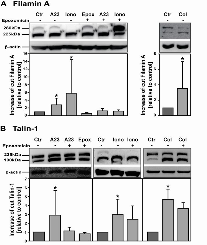
Für meine Großmutter
Christa Margarete Gerda Gründler
Katharina Gründler                                                            Vorwort

Vorwort

Vorab bedanke ich mich bei allen, die mich während meiner Promotion unterstützt
haben.

Ich bedanke mich ganz herzlich bei Herrn Prof. Dr. Zahler für die Vertretung meiner
Arbeit am Lehrstuhl Chemie und Pharmazie. Vielen Dank für die Bereitschaft und
Arbeit, die Sie auf sich genommen haben. Sie hatten stets ein offenes Ohr für mich
und haben meine Anliegen auch noch so groß oder klein schnellstens bearbeitet.
Bei Herrn PD Dr. Krämer bedanke ich mich für die Idee der Arbeit und die Betreuung
der Promotion. Danke für die Einarbeitung in das spannende Feld der kleinsten
Blutzellen. Ich freue mich, dass ich stets meine eigenen Ideen und Konzepte
einbringen und verwirklichen konnte. Vielen Dank für das Vertrauen, die
Zusammenarbeit und die daraus entstandenen Publikationen.
Herrn Prof. Dr. Pohl danke ich nicht nur für die Bereitschaft als Drittgutachter,
sondern auch insbesondere dafür, dass ich bei ihm am Lehrstuhl meinen Arbeitsplatz
hatte. Ich habe nicht nur die Laborarbeit am Walter-Brendel-Zentrum ausgeführt,
sondern wurde als vollwertiges Mitglied des Walter-Brendel-Zentrums behandelt.
Darüber hinaus vielen Dank für die regelmäßigen Treffen und daraus entstandenen
Diskussionen, die die Arbeit vorwärts gebracht haben.
Ein weiteres Dankeschön gilt den anderen Teilnehmern der Prüfungskommission,
namentlich Frau Prof. Dr. Vollmar, Herrn PD Dr. Michalakis, Herrn Prof. Dr. Biel.

Frau Dr. Mannell bin ich zu tiefst verbunden. Sie hat mich aufgenommen als
Arbeitsgruppenmitglied; Ich wurde als vollwertiges Mitglied behandelt und
angesehen. Vielen Dank für die nicht nur methodische, sondern auch
wissenschaftliche Unterstützung. Danke für das sehr schnelle Einarbeiten in das
fachfremde Thema und das daraus entstandene Voranschreiten meiner Dissertation.
Auch herzlichsten Dank für die wertvollen Diskussionen persönlicher und
wissenschaftlicher Art und die warme, liebevolle Umgangsweise, die in der
Arbeitsgruppe herrscht. Es ist Schade, dass die Zeit dem Ende zugeht. Herzlichsten
Dank für die Mitbetreuung von Anfang bis Ende. Unerwähnt soll hier auch nicht Prof.
Dr. Krötz bleiben, der diese Arbeitsgruppe aufgebaut und ins Leben gerufen hat.
Herrn Prof. Dr. Sohn danke ich für die Mitbetreuung meiner Arbeit und Herrn Prof. Dr.
Massberg für die Eingliederung in die Medizinischen Klinik und Poliklinik I. Die
Unterstützung der Dr. Kleist-Stiftung hat mir geholfen und an dieser Stelle sei
Matthias und Claudia Schulz herzlichst gedankt.
Im Rahmen dieser Arbeit hatte ich die Gelegenheit mit vielen Kooperationspartnern
zusammen zu arbeiten. Hier gilt mein Dank Herrn Dr. Drews für die Einführung in die
Katharina Gründler                                                            Vorwort

Proteasomaktivitätsassays und seiner Doktorandin Franziska Koser. Herrn Prof. Dr.
Stevanovic und Nico Trautwein danke ich für die Analyse der MHC I Peptide.
Darüber hinaus danke ich Herrn Prof. Dr. Spannagl für die schnelle Bereitschaft der
Hilfe bezüglich des Multiplate® Systems und der Thrombozytenkonzentrate.

Ebenso bedanke ich mich bei der ganzen Arbeitsgruppe Mannell herzlichst. Wir
haben gut miteinander gearbeitet und hatten viel Spaß zusammen. Namentlich
bedanke ich mich bei Ramona Mettler für die Einweisung in das Labor und die
täglichen persönlichen wie methodischen Diskussionen. Ein Dankeschön geht an
Joachim Pircher für das Beibringen der Grundtechniken der Thrombozyten. Riesig
danke ich Yvonn Stampnik, die mir immer mit Rat und Tat beiseite stand. Auch
Stefan, Franziska, Thomas, Philipp, Alex, Erik, Pascal und Georg sei an dieser Stelle
gedankt. Meiner Praktikantin Sloane und HiWi Raffaela danke ich herzlichst für die
wertvolle, gute und experimentelle Unterstützung.
Yvonn, du hast mir nicht nur mit Rat und Tat beiseite gestanden, wir haben
gemeinsam als Doktoranden die Zeit im Labor verbracht und viele weitere
persönliche und wissenschaftliche Unterhaltungen geführt, das Labor aufgeräumt
und Cafe-Pausen verbracht. Vielen Dank auch für das Korrekturlesen und deine
Freundschaft.
Allen Mitgliedern der Arbeitsgruppe Walzog (insbesondere Doris, Jennifer, Melanie
und Tanja) möchte ich zu tiefst für jegliche persönliche und wissenschaftliche
Unterstützung danken. Wir hatten eine schöne gemeinsame Zeit, nicht nur im Labor.
Ein weiteres Dankeschön möchte ich an Anna Bakovic richten, die nicht nur für eine
saubere Atmosphäre sorgt, sondern beigetragen hat zu dreieinhalb angenehmen
Jahren am Walter-Bendel-Zentrum.
Nicht zu verachten sind alle Blutspender, ohne deren Hilfe hätte kein einziges
Experiment durchgeführt werden können. Leider können nicht alle namentlich
genannt werden, aber regelmäßig neben Labormitgliedern kamen Claus, Chris,
Katha, Kitty und Stefan.

Ebenso danke ich meiner Familie und meinen Freunden.
Katha, dir kann ich gar nicht genug danken. Schön, dass es dich gibt. Ich bedanke
mich riesig für deine Freundschaft, die in der Studienzeit begann und hoffentlich
noch lange anhält. Hier möchte ich die fachlichen Diskussionen nicht unerwähnt
lassen, die mir immer weitergeholfen haben und großen Dank auch für das
Korrekturlesen.
Kitty, schön, dass es dich gibt und du mich auf andere Gedanken bringst. Danke für
die mentale Unterstützung.
Anja und Micha, ich danke euch für die Freundschaft, wir haben nicht nur eine
schöne Studienzeit verbracht, sondern sind darüber hinaus befreundet geblieben.
Katharina Gründler                                                         Vorwort

Meinen Berliner Freunden danke ich für die langjährige Freundschaft. Es ist schön,
dass wir trotz der Distanz befreundet sind.
Ein riesiges Dankeschön geht an Daniel. Du hast mir die Tür zu neuen Kulturen
geöffnet.
Meiner Mama, meinem Papa und meinem Bruder Alex danke ich für die
immerwährende, grenzenlose Unterstützung und Ermutigung. Danke, dass ich euch
habe und auf euch zählen kann. Ohne euch wäre diese Arbeit nicht so geworden wie
sie euch jetzt vorliegt.
Katharina Gründler                                                                                                              Table of contents

Table of contents
Table of contents ....................................................................................................... i
Abstract .................................................................................................................... IV
1 Introduction............................................................................................................ 1
  1.1 Blood platelet: Crucial part of vascular integrity ..................................................... 1
     1.1.1 Resting and activated platelets ............................................................................................... 1
     1.1.2 Receptor pathways of platelet activation ................................................................................ 2
       Fibrinogen receptor, integrin αIIbβ3 ............................................................................................... 2
       Thrombin (PAR) receptors ............................................................................................................ 3
       ADP receptors, P2Y receptors ..................................................................................................... 3
       Thromboxane receptors................................................................................................................ 3
       Collagen receptors, integrin α2β1 and GPVI ................................................................................. 4
     1.1.3 Aggregation ............................................................................................................................. 5
     1.1.4 Platelets in vascular inflammation and diseases .................................................................... 6

  1.2 Organization of cellular vitality and protein metabolism in platelets ..................... 6
     1.2.1 Migration and proliferation of platelets .................................................................................... 7
     1.2.2 Regulation of apoptosis ........................................................................................................... 7
     1.2.3 Platelets: Antigen presenting cells .......................................................................................... 8
     1.2.4 Transcription factors in human platelets affect platelet functions ........................................... 9
       NFκB signaling pathway ............................................................................................................. 10
       NFκB in platelets ........................................................................................................................ 10
     1.2.5 Protein de novo synthesis and protein degradation in platelets ........................................... 11

  1.3 The proteasome, a multicatalytic enzyme ...............................................................12
     1.3.1 Structure and complexity of the proteasome ........................................................................ 13
       The 20S core particle.................................................................................................................. 13
       Proteasome regulators and the formation of different complexes .............................................. 14
     1.3.2 Proteasome inhibitor classes ................................................................................................ 15
     1.3.3 Regulation of the proteasome ............................................................................................... 16
     1.3.4 Involvement of the proteasome in disease ........................................................................... 17
     1.3.5 The proteasome in anucleate platelets ................................................................................. 17

  1.4 Aim of the study ........................................................................................................18
2 Material and Methods .......................................................................................... 20
  2.1 Material ......................................................................................................................20
     2.1.1 Instruments ............................................................................................................................ 20
     2.1.2 Glas and platic labware ......................................................................................................... 21
     2.1.3 Inhibitors, chemicals and reagents........................................................................................ 22
     2.1.4 Bacteria strains ...................................................................................................................... 24
     2.1.5 Kits......................................................................................................................................... 24
     2.1.6 Gel preparations for SDS-PAGE home-made gels ............................................................... 24
     2.1.7 Primary antibodies used for western blotting ........................................................................ 25
     2.1.8 Secondary antibodies peroxidase conjugated used for western blotting .............................. 26
     2.1.9 Substrates for proteasome acitivity analysis ......................................................................... 26

                                                                                                                                                          i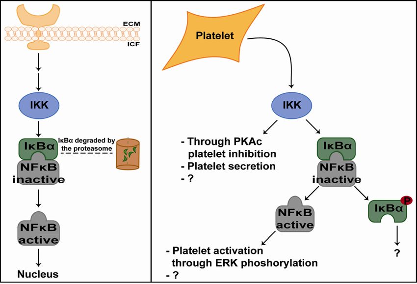
Katharina Gründler                                                                                                            Table of contents

  2.2 Methods .....................................................................................................................26
     2.2.1 Cultivation and handling of used cell lines ............................................................................ 26
     2.2.2 Thawing and freezing of cell lines ......................................................................................... 27
     2.2.3 Platelet isolation .................................................................................................................... 27
     2.2.4 Platelet stimulation ................................................................................................................ 28
     2.2.5 Patient studies ....................................................................................................................... 28
     2.2.6 Platelet and bacterial interaction studies .............................................................................. 29
     2.2.7 Protein solubilisation for western blot analysis ..................................................................... 29
     2.2.8 Protein solubilisation for proteasome activity measurements ............................................... 30
     2.2.9 Protein quantification ............................................................................................................. 31
     2.2.10 Gel electrophoresis and western blot analysis .................................................................... 32
     2.2.11 Proteasome activity measurements .................................................................................... 34
     2.2.12 Fluorescence activated cell sorting ..................................................................................... 36
     2.2.13 Aggregometry ...................................................................................................................... 37
     2.2.14 p65 transcription factor assay kit......................................................................................... 37
     2.2.15 MHC I peptide analysis ....................................................................................................... 38
     2.2.16 Statistical analysis ............................................................................................................... 38

3 Results.................................................................................................................. 40
  3.1 Characterization of the proteasome in human platelets.........................................40
     3.1.1 Human platelets express proteasome subunits .................................................................... 40
     3.1.2 Human platelets contain an active proteasome .................................................................... 41
       Detection and inhibition of the chymotrypsin-like activity of the 20S proteasome...................... 41
       Age dependency of 20S CT-L proteasome activity in platelets .................................................. 43
     3.1.3 26S and 20S proteasome complexes are active on all three catalytic activities .................. 43
       Comparison of all three catalytic 26S and 20S proteasome activities in human platelets and
       nucleated cells ............................................................................................................................ 44
       Specificity of the established proteasome activity assay............................................................ 45
     3.1.4 The proteasome is differentially regulated in human platelets .............................................. 46
       Effects of known proteasome inhibitors on all 26S and 20S activities ....................................... 47
       Calcium ionophores activate 26S proteasome activities ............................................................ 48
       The platelet agonist collagen enhances 26S CT-L proteasome activity .................................... 49
     3.1.5 Proteasome substrates ......................................................................................................... 50
       MHC I peptide analysis ............................................................................................................... 50
       Cytoskeletal proteins are cleaved by the proteasome................................................................ 51

  3.2 Role of the proteasome in platelet function ............................................................53
     3.2.1 Proteasome inhibition restrains platelet aggregation ............................................................ 53
     3.2.2 NFκB as a regulator of the proteasome in platelets .............................................................. 54
       NFκB influences aggregation in human platelets ....................................................................... 54
       NFκB inhibitors prevent collagen-stimulated proteasome activity enhancement ....................... 55
       NFκB activity is increased by collagen ....................................................................................... 56
       Collagen activates IκB kinase and promotes degradation of IκBα ............................................. 57

  3.3 Clinical investigation of mitochondrial function and proteolytic processes in
  platelets during sepsis ...................................................................................................58
     3.3.1 Markers of platelet apoptosis and mitochondrial control of platelet apoptosis ..................... 59
     3.3.2 Mitochondrial dysfunction of platelets correlates with clinical disease severity and outcome
           in sepsis ................................................................................................................................ 60
       Platelet mitochondrial membrane depolarization of sepsis patients correlates with clinical
       disease severity .......................................................................................................................... 60

                                                                                                                                                      ii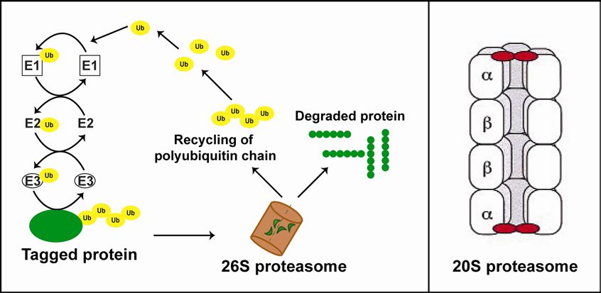
Katharina Gründler                                                                                                        Table of contents

       Reduction of platelet mitochondrial membrane potential and pro-apoptotic Bcl-xL in patients
       with severe sepsis ...................................................................................................................... 61
       Platelet mitochondrial membrane depolarization correlates with clinical disease outcome ....... 63
     3.3.3 Patients with severe sepsis show enhanced proteasome activity in human platelets .......... 64

4 Discussion ........................................................................................................... 66
  4.1 Analysis of the proteasome in human platelets ......................................................66
     4.1.1 Anucleate platelets contain an active proteasome................................................................ 66
     4.1.2 The proteasome is differentially regulated in human platelets .............................................. 67
     4.1.3 Proteasome substrates ......................................................................................................... 68

  4.2 Function of proteasomes in anucleate platelets .....................................................68
     4.2.1 The proteasome affects platelet aggregation ........................................................................ 69
     4.2.2 NFκB a regulator of the proteasome in platelets .................................................................. 69

  4.3 Sepsis as one of many clinical perspectives ..........................................................72
     4.3.1 The mitochondrial membrane potential in platelets as a marker of sepsis ........................... 72
     4.3.2 Proteasome activity is enhanced in platelets during sepsis .................................................. 73

  4.4 Conclusion and Outlook ...........................................................................................74

References .............................................................................................................. 75
Appendix ................................................................................................................. 83
  Abbreviations ..................................................................................................................83
  Publications .....................................................................................................................85

                                                                                                                                                iii
Katharina Gründler Abstract
Abstract
Even though platelets are the smallest cells in circulating blood, they play an integral
role in blood clotting where they are activated, adhere to the vessel wall, and
contribute to hemostasis. But over the years it was discovered that those anucleate
cells have more extended functions. They organize their cellular vitality similar to
nucleated cells and have an active protein metabolism performing protein de novo
synthesis as well as protein degradation. One of the main degradation systems in
cells is the proteasome. Besides protein quality control, the proteasome is involved in
important cellular processes like cell survival, transcription, development, selective
elimination of abnormal proteins and antigen processing.
A dysregulation of this multicatalytic protein complex leads to various disease
developments. Proteasome inhibitors, for instance, have been studied for treating
cancer. Platelets like nucleated cells contain a proteasome. However, the impact of
the proteasome on platelet functions remains poorly investigated until today. A better
knowledge of signaling pathways in platelets aids in understanding how alterations in
proteasome functions affect platelet-mediated processes and diseases.
This study confirms the existence of a functional proteasome in human platelets and
illustrates an important role in platelet biology, as well as sepsis.
With this study the role of the proteasome in anucleate platelets is demonstrated in
more detail and a signaling pathway regulating its activity was observed. Here, the
proteasome in platelets is linked to platelet aggregation. First, proteasome inhibitors
epoxomicin and bortezomib reduce ADP- and collagen-induced aggregation.
Furthermore, the 26S chymotrypsin-like activity of the proteasome is enhanced when
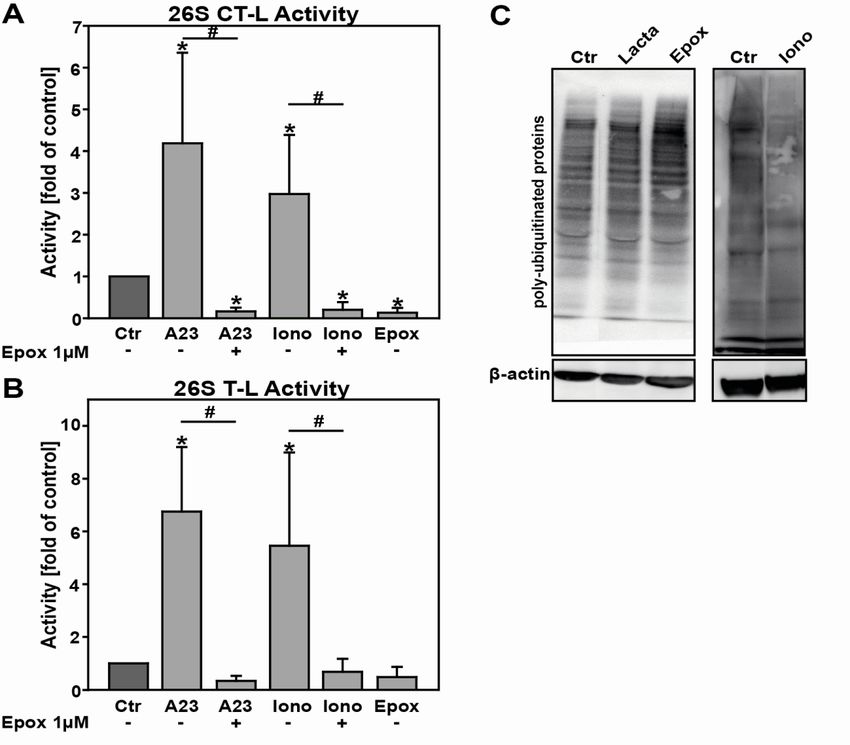
platelets are incubated with the platelet agonist collagen. Additionally, cytoskeletal
proteins Filamin A and Talin-1, which are crucial for platelet activation, were identified
as proteasome substrates and increased cleavage of these proteins occurs with
proteasome activation.
To investigate possible mechanisms of regulating the proteasome, the signaling
pathway related to NFκB was analyzed under platelet agonist treatment. The NFκB
pathway, that mediates aggregation, is initiated when platelets are treated with
collagen and the inhibitory protein of NFκB, IκBα, is degraded in collagen-stimulated
platelets. More interestingly, NFκB inhibitors prevent collagen-stimulated
enhancement of the proteasome activity. In return the connection of the proteasome
and the NFκB pathway is further demonstrated as NFκB inhibitors restrict cleavage
of the proteasome substrate Talin-1. These results propose a novel pathway that
involves the proteasome and that is in return connected with non-genomic functions
of NFκB in regulating platelet aggregation.
IVKatharina Gründler Abstract
In a second part this work shows for the first time that mitochondrial membrane
depolarization in platelets correlates with the disease course and disease severity in
patients with sepsis. Additionally, during these studies increased proteasome activity
was observed in sepsis patients compared to control patients and pathogenic
bacteria intensified the 26S trypsin-like activity of human platelets. Therefore,
molecular markers of platelet vitality may be valuable parameters to help evaluating
the clinical outcome of sepsis patients.
In summary, the study confirms the existence of a functional proteasome in human
platelets, contributes to our understanding how the proteasome affects platelet
functions such as aggregation and how this may be regulated on a molecular basis.
Furthermore, it allows for new insights in the disease course of sepsis and identifies
new molecular markers for assessing the disease severity and clinical outcome of
sepsis patients.
VKatharina Gründler 1 Introduction
1 Introduction
1.1 Blood platelet: Crucial part of vascular integrity
Platelets are the smallest cells in circulating blood with a diameter of 2-4µm [1, 2]. A
human platelet count lies between 150.000 and 300.000 platelets per µl blood. They
derive from megakaryocytes in the bone marrow and have a physiological lifetime of
about 7-10 days [3]. Platelets do not have a nucleus and are referred to as anucleate
cells. It is well studied that they play an important role in vessel and wound repair.
Platelets prevent blood loss by forming thrombi. Under physiological conditions
resting platelets circulate with the blood flow. They have a discoid shape and roll
along the intact endothelium. Traumatic action on the vessel wall, such as a cut
requires clot formation to stop bleeding. Platelets attach to the vascular lesion,
adhere, and form a primary hemostatic thrombus while attracting more platelets.
During adhesion platelets undergo shape change, spreading or rolling and activation.
In a last step platelets aggregate with each other.
1.1.1 Resting and activated platelets
As for other cells, those anucleate cells have a cytoplasmic membrane with
membrane proteins. The platelet plasma membrane expresses numerous integrated
proteins that are receptors for soluble agonist (such as ADP, thrombin or
thromboxane A2) or adhesion proteins (such as fibrinogen, collagen or von
Willebrand factor) [4, 5]. The plateletal cytoskeleton is important to maintain the
discoid shape of resting platelets and actively aids in the platelet shape change. The
cytoskeleton consists mainly of actin (15-20% of total protein mass), microtubuli, and
actin binding protein, myosin. Actin exists in a globular form, G-actin, and in a
filament form, F-actin [6]. Upon activation when the intracellular calcium
concentration reaches a specific threshold platelets undergo chape change and
pseudopods are formed [7]. The platelet looses its discoid shape. Microtubuli enrich
in the pseudopods and G-actin polymerizes to F-actin, which associates with other
structural proteins. F-actin filaments have a connection to the cell organelles and
reorganize them during this activation process. Well-known organelles of platelets
are mitochondria, glycogen stores and the storage granules (dense granules, α-
granules, and lysosomes) [8]. The lysosomes are similar to other cells and contain
hydrolytic enzymes. The granules are characteristic for platelets and store proteins
and other substances that are important for platelet function. The dense granules
contain compounds, such as ADP, ATP, Ca2+, and serotonin to promote aggregation
1Katharina Gründler 1 Introduction
and α-granules carry proteins, such as P-selectin or fibrinogen that play differing
roles in adhesion, aggregation, chemotaxis, proliferation and inflammation [9].
Figure 1: Morphology of resting and activated platelets. Upper panel shows electron micrographs
of a resting platelet and an activated platelet (x20000 and x10000 respectively). The lower panel
pictures transmission electron micrographs of cross-sections of a resting and an activated platelet
(x21000 and x30000 respectively) [4].
1.1.2 Receptor pathways of platelet activation
Platelets have a number of different transmembrane receptors that interact with its
physiological agonist to induce activation. Amongst others many integrins (such as
αIIbβ3, α2β1), G-protein-coupled seven transmembrane receptors (protease-
activated-receptor PAR-1 and PAR-4 thrombin receptors, P2Y1 and P2Y12 ADP
receptors, TPa and TPb TxA2 receptors), proteins of the immunoglobulin superfamily
(GPVI), and C-type lectin receptors (P-selectin) are found on platelets.
These platelet adhesion receptors are well understood and even though they differ in
their functions and signaling pathways they have some similarities. First, there is the
agonist/platelet receptor interaction followed by signaling pathways that promote
secretion. Then released substances induce various platelet responses and cause
further platelet activation as well as recruitment of other circulating platelets. The so-
called integrin activation and outside-in signaling is started. In the following the main
platelet receptors will be mentioned and Figure 2 gives a schematic representation of
platelet adhesion and agonist receptors with their cellular pathways.
Fibrinogen receptor, integrin αIIbβ3
The most abundant and best-studied integrin in platelets is the αIIbβ3 (GPIIb/IIIa)
integrin, the fibrinogen receptor. This receptor is inactive in resting platelets but upon
2Katharina Gründler 1 Introduction
platelet activation it undergoes conformational changes becoming able to bind
soluble plasma fibrinogen [10]. The outside-in signaling involves calcium mobilization,
phosphorylation of proteins, activation of the phosphoinositide metabolism and
cytoskeletal reorganization [11]. The αIIbβ3 integrin mediates the bridging between
two platelets where fibrinogen connects two αIIbβ3 integrins with eachother. Integrin
αIIbβ3 is the main adhesion molecule in platelet aggregation.
Thrombin (PAR) receptors
PAR receptors mediate platelet response to thrombin. PAR receptors are G-protein
coupled receptors and on human platelets PAR-1 and PAR-4 are found. Both trigger
PLC activation, Ca2+ mobilization, and PKC activation [12]. Thrombin stimulation
results in platelet activation, shape change and the release of granules. PAR-1 and
PAR-4 have different kinetics; to induce PAR-4 mediated signaling a higher thrombin
concentration is necessary [13].
Thrombin as a protease acts on PAR receptors by binding to the extracellular domain
and cleaving the receptor to form a new peptide ligand. Thrombin unmasks a specific
ligand for the PAR receptors that then activates the receptor and induces
transmembrane signaling [14].
ADP receptors, P2Y receptors
The agonist ADP binds to the ADP receptors P2Y1 and P2Y12, G-protein-coupled
receptors [15, 16]. One receptor is coupled to Gαq G protein (P2Y1) and the other is
coupled to Gαi (P2Y12). While P2Y1 stimulates PLCβ enhancing cytosolic Ca2+, that
activates PKC and leads to platelet shape change [15], P2Y12, which is coupled to
Gαi, goes another way. P2Y12 inhibits the adenylate cyclase and activates
phosphatidylinositol 3-kinase (PI3K) [17]. Activated PI3K promotes then AKT and
Rap1B activation [18]. This stimulation of Gαq and Gαi signaling pathways is
necessary for a fibrinogen receptor activation, thus for platelet-platelet adhesion.
Antiplatelet agents are effective in the treatment of arterial thrombosis. Those agents
target different critical steps in thrombogenesis. But important antiplatelet agents are
clopidogrel and prasugrel that target the ADP receptor P2Y12 [19].
Thromboxane receptors
Thromboxane A2 (TXA2) is a prostaglandin with potent platelet activating
characteristics. Thromboxane receptors (TPs) induce a cytosolic Ca2+ enhancement
and an influx of extracellular Ca2+, which activates the PLA2 resulting in arachidonic
acid (AA) hydrolysis from membrane phopholipids and converting it into TXA2 [20].
TXA2 then acts on its receptor again [21]. Because TPs are coupled to Gαq but not
Gαi family members they require a secretion of ADP for platelet aggregation. ADP
inhibits the adenylate cyclase mediated through the P2Y12 that is coupled to Gαi.
3Katharina Gründler 1 Introduction
Collagen receptors, integrin α2β 1 and GPVI
Following injury to the vascular wall, collagen is exposed on which amongst others
platelets adhere rapidly. Collagen I and III are considered the most important
collagens at the injured cite to induce platelet adhesion and platelets have many
collagen receptors. Most collagen receptors are known but there might exist more
due to the fact that there are receptors directly and indirectly binding collagen [22].
An indirect collagen receptor is the GPIb complex (CD42c) that interacts with von
Willebrand factor, which then binds to various collagens in the subendothelium. The
integrin α2β1 and the Ig superfamily receptor GPVI are the major direct collagen
receptors [22].
Integrin α2β1 is known as GPIa/IIa or CD49b/CD29. As all integrins, α2β1 is a large
glycoprotein with extracellular domains, transmembrane domains and cytoplasmic
domains. On resting platelets α2β1 is expressed in a low-affinity state (similar to the
inactive state of the fibrinogen receptor) and the affinity of α2β1 to soluble collagen
increases on platelet stimulation [23]. Thus integrin α2β1 seems to require an earlier
agonist-induced conformational change to bind to collagen [24]. Src and Syk family
tyrosine kinases are activated by α2β1 leading to the activation of PLCγ2 and the
formation of lamellipodia [25]. There is a complex crosstalk between α2β1 (collagen
receptor) and αIIbβb3 (fibrinogen receptor); α2β1 promotes the activation of αIIbβb3 and
as a consequence induces fibrinogen binding to adherent platelets [26].
The collagen receptor GPVI is connected to the immunoreceptor tyrosine-based
activation motif (ITAM) and its cytoplasmic domain is bound to the Src family kinases
Fyn and Lyn [27, 28]. When collagen binds to GPVI, ITAM is tyrosine phosphorylated
by those Src family kinases and initiates a complex signaling cascade activating a
series of adapter and effector proteins [29]. The main enzyme activated is PLCγ2 that
leads to the second messengers 1,2-diacylglycerol (DAG) and inositol 1,4,5-
triphosphate (IP3). IP3 in turn triggers intracellular Ca2+ mobilization, protein
phosphorylation and AA release. AA drives aggregation and secretion of TXA2 and
ADP [30].
4Katharina Gründler 1 Introduction
Figure 2: Schematic model of main platelet receptors and their cellular pathways [31]. The left
panel shows adhesion as well as agonist receptors and the right panel shows major cellular pathways
triggered by platelet agonists. The fibrinogen, thrombin, ADP, thromboxane A2 and collagen receptors
are discussed above.
1.1.3 Aggregation
While platelets adhere they not only undergo activation but also aggregate.
Aggregation is defined as the process of coadhesion of two platelets. Primary and
secondary aggregation are told apart. Primary aggregation is reversible and platelets
are only loosely connected by fibrinogen after activation. When they release their
granule components secondary aggregation starts, the binding of fibrinogen
becomes stronger and this process is irreversible [32]. Without fibrinogen or Ca2+
aggregation is not possible. Those two components are also stored in platelet
granules to achieve high concentrations of both of them in a thrombus.
The measurement of aggregation is the most common method for the diagnosis of
platelet function. Platelet aggregation can be detected amongst others by light
transmission aggregometry, LTA, also called Born aggregometry or through
impedance aggregometry, IA [33]. During IA electrical impedance in whole blood is
measured with two sensor electrodes. When platelets aggregate they adhere on the
metal wire and increase electrical resistance. During LTA light transmission is
detected by a photocell that sits behind a cuvette. While platelets aggregate, less
light is absorbed and the transmission increases. The aggregation curve measured
by LTA, Born aggregometry, can follow a monophasic or biphasic aggregation
depending on the agonist. After ADP stimulation platelets undergo a shape change
that is recognized by a decrease in light transmission, followed by the primary
aggregation (reversible) where the curve rises, and then the curve can either fall
(deaggregation) or goes through a plateau into the second phase of aggregation
(irreversible). Platelets activated with collagen show a delayed and longer decrease
in light transmission (shape change) and have a monophasic curve.
5Katharina Gründler 1 Introduction
1.1.4 Platelets in vascular inflammation and diseases
Next to inherited platelet disorders that affect, e.g., platelet adhesion, activation or
secretion, platelets play a crucial role in the involvement of various diseases.
Since platelets play a central role in stopping bleeding by clot formation they have a
major part in pathophysiological thrombus formation as well. During atherosclerosis
an interaction among platelets, endothelial cells, and leukocytes establishes a
localized inflammatory response that accelerates atherosclerosis, thrombus formation
and might result in a heart attack or stroke. Platelets normally interact with the
endothelium to maintain their physiological function and to enhance leukocyte
recruitment to sites of inflammation but when those physiological responses are
exaggerated at atherosclerosis sites pathogenesis of this disease is stimulated [34].
Moreover, growing evidence shows that platelets contribute to cancer progression.
Complex crosstalk between tumor cells and circulating platelets enhances tumor
growth and platelet receptors as well as platelet agonist play a role in cancer
metastasis [35].
Additionally, the exact role of platelets in sepsis with its underlying molecular
mechanisms is still to be analyzed. But it is suggested that platelets promote
inflammation during the early stages of infection thus helping prevent sepsis.
Nevertheless, thrombocytopenia is a common finding in severe sepsis and it might
result from platelets undergoing apoptosis [36, 37]. Although apoptotic proteolysis
might contribute to thrombocytopenia in sepsis other mechanisms are involved. It is
known that platelet activation and aggregation is regulated in sepsis through Toll-like
receptors expressed by platelets and bacterial exotoxins [38]. Moreover, the role of
patelet-leukocyte adhesion during sepsis coming from activated platelets needs to be
analyzed. While activated platelets secrete key components of the coagulation and
inflammatory cascade, there are only few studies on platelet function in sepsis.
Thrombocytopenia in general is found during many diseases. It might be drug-
induced however it needs to be considered during the process of healing.
1.2 Organization of cellular vitality and protein
metabolism in platelets
Platelets are known for their ability to stop bleeding and for many years that was
thought to be their only function. But over the years platelets have shown more
extended functions. They obsess many features that were believed, to be seen only
in nucleated cells. It was discovered that those anucleate cells are not as simple as
everyone believed. Those small particles are surprising. Platelets are able to migrate,
proliferate, go into apoptosis, and present antigens. They even contain transcription
factors and are able to perform protein de novo synthesis.
6Katharina Gründler 1 Introduction
1.2.1 Migration and proliferation of platelets
For a long time platelets were believed to be static cells that do not leave the site of
adhesion, even though there was some evidence that platelets in vitro are able to
move through a Boyden chamber [39]. Years later platelets were found to migrate in
vivo and more detailed analysis demonstrated that platelets are able to migrate in a
SDF-1-mediated fashion even through an endothelium [40-42]. Further evidence for
platelet migration is published continuously [43-45].
Yet, more astonishingly in 2010 Schwertz et al. discovered that platelets are able to
produce functional progeny. There seems to exist a cell division that does not require
a nucleus. Platelets form new cell bodies that contain mitochondria and α–granules.
Moreover the new fragments adhere and spread normally, express P-selectin and
annexin V in the typical way after stimulation [46].
1.2.2 Regulation of apoptosis
When apoptosis, programmed cell death, was observed it was exclusively assigned
to nucleated cells [47]. However, apoptotic signs were discovered in platelets [48,
49]. Numerous chemical agents such as the calcium ionophore A23187 trigger
platelet apoptosis but also the platelet activator thrombin is able to induce apoptotic
events in platelets [50, 51]. Moreover, the process of apoptosis might be induced
without stimulants in platelets, under pathological high shear stress or long-term
incubation of platelets under blood banking conditions [50, 52].
While the intrinsic mitochondria-dependent pathway is well studied in anucleate
platelets, the role of the extrinsic pathway remains unclear [53].
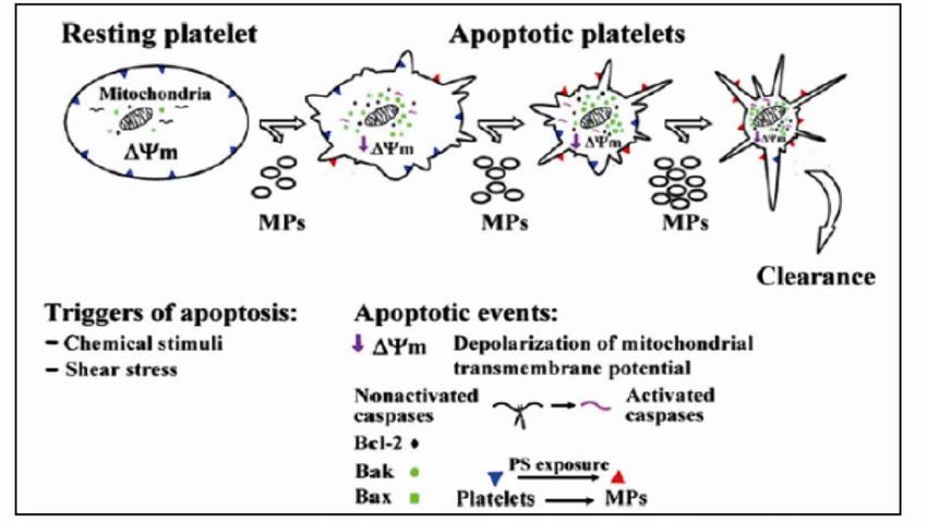
During apoptosis in platelets a depolarization of the mitochondrial membrane
potential has been demonstrated. The mitochondrial membrane potential decreases
in canine platelets after an estradiol treatment [54] or apoptotic stimulants A23187,
thrombin or high shear stress induce a reduction of the mitochondrial membrane
potential in human platelets [50, 51]. Degradation of the anti-apoptotic protein Bcl-xL
after treating platelets with bacteria was shown and pro-apoptotic proteins Bax and
Bak are higher expressed under, e.g., thrombin-induced plateletal apoptosis [36, 51].
Pro-apoptotic proteins also show higher expression in in vitro aged platelets [55].
Induction of activation of caspases -3, -8 and -9 was found in human platelets as well
[56]. Other apoptotic markers, such as phosphatidylserine translocation to the outer
leaflet of the plasmamembrane, and chytochrome c release were analyzed in
platelets [50, 57]. Furthermore, platelet shrinkage, membrane blebbing and
microparticle shedding, visible by microscopy, can be found in plateletal apoptosis
[50, 55]. All those apoptotic markers in platelets are summarized in a model for
apoptosis in platelets (Figure 3).
7Katharina Gründler 1 Introduction
Figure 3: Model of apoptosis in platelets [53]. Shown are platelets with their apoptotic markers
undergoing apoptosis when stimulated with chemical stimuli or high shear stress.
1.2.3 Platelets: Antigen presenting cells
A connection of platelets in processing and presenting antigens had long not been
made. However, in 2012 Chapman et al. stated that platelets express MHC I (major
histocompatibility complex class I) molecules and present antigens to naïve T cells.
They not only measured antigen presenting molecules on platelets in different in
vitro-incubations but also analyzed platelets isolated from infected and uninfected
mice [58]. Additionally, it is known that platelets express many proteins or RNA
transcripts, which are necessary for processing antigens and presenting them.
Platelets contain the endoplasmatic reticulum and a Golgi, where peptides to be
presented are processes and loaded on MHC I molecules. The proteasome that
helps by cleaving peptides that are to be presented is found in platelets, as well as
TAP (transporter associated with antigen processing), a protein that brings cytosolic
peptides to the ER [59, 60]. Many more proteins (such as calnexin, calreticulin and
Erp57) that facilitate correct folding of MHC I molecules and its association with β-
microglobulin, were detected in platelets [61, 62].
Proteomic analysis of platelets has brought further evidence for the existence of MHC
I molecules. The MHC I complex is found in α–granules of platelets [60] and the
global proteome analysis from Klockenbusch et al. identified the MHC I maschinery
as well [63]. Zufferey et al. provides a model of MHC I antigen-presenting pathway in
human platelets with its identified proteins (Figure 4).
8Katharina Gründler 1 Introduction
Figure 4: Schematic overview of the MHC I antigen-presenting pathway in human platelets.
MHC I is loaded in the ER and brought to the plasma membrane over the Golgi in a secretory granule.
Modified from Zufferey et al. [60].
1.2.4 Transcription factors in human platelets affect platelet
functions
Although platelets are anucleate, recent publications show that platelets express
transcription factors. Transcription factors including the steroid/nuclear receptors [64],
peroxisome proliferator activated receptor (PPAR) β/δ and γ [65, 66], the
glucocorticoid receptor (GR) [67], retinoid X receptors (RXR) [68] and nuclear factor
kappa-light-chain-enhancer of activated B cells (NFκB) were found in platelets [69,
70] and they indeed influence platelet functions. For instance, the nuclear receptor
estrogen β potentiates thrombin-stimulated platelet aggregation [71]. PPARγ prevents
the release of TXB2 and ATP after thrombin stimulation [66]. The glucocorticoid
receptor bound to its ligand prednisolone seems to inhibit platelet aggregation [67]
and the retinoid X receptors inhibit platelet aggregation through Rac inhibition and
prevention of Ca2+ release [68].
Taken together, this suggests non-genomic functions of transcription factors in
platelets. Also in erythrocytes, another anucleate cell type, transcription factors were
found [72]. Thus, anucleate cells like platelets and erythrocytes seem to be the ideal
human experimental model to study non-genomic functions of transcription factors.
Since some transcription factors like nuclear receptors are already known to possess
non-genomic functions in nucleated cells [73], it is not as surprising that they play a
non-genomic role in platelets. But that other transcription factors might also have
non-genomic functions is a whole new research field. Here I will focus on the
transcription factor NFκB that had mostly been studied in nucleated cells and its
genomic functions there are well known. Nevertheless, the recent identification of
NFκB in anucleate platelets is fascinating and promises a better understanding of
platelet biology.
9Katharina Gründler 1 Introduction
NFκB signaling pathway
The NFκB pathway has diverse functions and in accordance with this the NFκB
transcription factor family consists of 5 members (p50, p52, p65, RelB and c-Rel) [74,
75]. All of them contain a nuclear localization sequence (NLS) in their N-terminal
domain and they are normally found in the cytoplasm associated to an inhibitory
protein that masks their NLS [76]. The inhibitory proteins belong to the IκB family with
its most common member IκBα. The NFκB transcription factor proteins can form
homo- or heterodimers what again demonstrates the diverse function of this signaling
pathway. NFκB bound to the inhibitory protein might be considered inactive.
There are several NFκB activation pathways but the most frequent one is the
canonical. This pathway is activated in response to various inflammatory stimuli.
Hereby the IκB kinase (IKK) complex is activated and phosphorylates the inhibitory
protein IκBα at Ser32 and Ser36. This phosphorylation results in a rapid
ubiquitination and degradation of IκBα by the proteasome [76]. Then the so-called
active NFκB dimer with its liberated NLS sequence translocates to the nucleus where
it selectively activates the transcription of various genes mainly involved in
inflammation. The left panel of Figure 5 pictures this pathway.
As NFκB mainly activates inflammatory genes, it is found activated amongst others in
rheumatoid arthritis, an inflammatory disease. In the joint tissue NFκB of resident
macrophages provokes transcription of many pro-inflammatory cytokines and
chemokines leading to a subsequent invasion of a large number of immune cells to
the joint tissue. In an autocrine and paracrine manner cells are kept activated there
[77].
NFκB in platelets
Over the last years evidence was regularly published that NFκB exists in anucleate
platelets and exerts non-genomic functions. Already in 2002 Liu et al. showed the
existence of some NFκB/IκB family members in human platelets and demonstrated a
phosphorylation and degradation of IκB during platelet activation [69]. This was not
as absurd because thrombin, a platelet activator, is able to induce the NFκB pathway
in smooth muscle cells [78]. Malaver et al. analyzed further the functional significance
of NFκB in human platelets. They confirmed the expression of p65, IκBα and its
degradation. Moreover, they approached NFκB’s function, which seems to mediate
platelet aggregation [70]. NFκB inhibitors restricted platelet spreading, impaired
aggregation and reduced ATP release, TXB2 formation and P-selectin expression
[70]. Just a little later further publications stating functional NFκB in platelets were
published. Spinelli et al. used a different NFκB inhibitor and demonstrated reduced
platelet spreading, as well as lamellapodia formation [79]. Gambaryan et al.
described IKK activation after platelet activation in mice. However, they also showed
an induction of aggregation by IKK inhibitors [80]. This seems as the exact opposite
of findings from Malaver and Spinelli but it could just demonstrate the complexity of
10Katharina Gründler 1 Introduction
the IKK complex, which is known to have multiple substrates (not only NFκB) in
nucleated cells. The IKK complex might have multiple substrates in platelets as well
[81].
Furthermore, additional evidence of NFκB’s existence and non-genomic function in
platelets is given from year to year [82-86]. There was a study on a substance that
inhibits NFκB-mediated platelet aggregation [83] and the IKK seems to possess a
non-genomic function in platelet secretion [85].
Taken together, NFκB pathway members are present in platelets and seem to
function in a novel non-genomic way. The right panel of Figure 5 summarizes these
findings of NFκB in platelets.
Figure 5: NFκB pathway in eukaryotic cells and its non-genomic functions in platelets. Left
panel: Basic illustration of the canonical NFκB pathway in eukaryotic cells. ECM: extracellular matrix,
ICF: intracellular fluid. Right panel: NFκB and its family members in platelets with possible non-
genomic functions demonstrating the complexity of the NFκB pathway. This illustration is based on
publications until today [70, 79, 80, 85, 87].
1.2.5 Protein de novo synthesis and protein degradation in platelets
Essential for nucleated cells is their ability to transcripe DNA to RNA and translate it
in order to synthesize a protein. Platelets lack nuclei and as a consequence do not
possess cellular DNA, but they have mitochondria with the mitochondrial genome
[88, 89]. Nevertheless, they were considered incapable of regulating protein
11Katharina Gründler 1 Introduction
synthesis [90]. In the 1960s it was published that platelets can absorb amino acids
and might synthesize proteins but only in 1998 it was shown that platelets build a
specific protein upon stimulation [91, 92]. Since then many proteins found in the
platelet profile were found to be de novo synthesized in platelets [93]. Translation
does not require a nucleus and is encountered in platelets giving them an alternative
route for gene control [94]. Also an evolving area of research is the synthesis of
proteins during storage of platelet concentrates.
More evidence for protein de novo synthesis in platelets is published every year. For
instance, platelets indeed contain not only functional mRNA but also ribosomal RNAs
and other protein components (such as Dicer or the spliceosome) to perform
translation [93, 95].
Hand in hand with protein synthesis goes protein degradation. To maintain protein
homeostasis, cells balance protein synthesis with degradation. Protein degradation in
nucleated cells is well studied and is performed by two main pathways, the lysosomal
and the ubiquitin proteasome system. Our current understanding of degradation
pathways in platelets is not as detailed. Platelets contain the cysteine protease
calpain which regulates many cellular processes with its proteolytic activity. Known
substrates of calpain are cytoskeletal and membrane proteins. Therefore calpain
regulates amongst others granule secretion and cell spreading [96]. Furthermore,
platelets express many other proteases but essential here is that platelets possess
the special protease, the proteasome, which is one of the main systems to degrade
proteins [59, 97]. The proteasome is discussed in the following section.
All in all, anucleate platelets seem to have complex degradation systems [98].
1.3 The proteasome, a multicatalytic enzyme
Proteolysis is very important to regulate cellular protein levels and there exist two
main pathways to degrade proteins in eurkaryotes. The lysosomal and the ubiquitin
proteasome system are responsible for intracellular protein turnover [99, 100]. Since
its discovery the ubiquitin proteasome system came into focus expanding the role of
proteolysis from mere housekeeping to regulator of major cellular processes. It plays
a crucial role in regulating the cell cycle, division, survival, oncogenesis, transcription,
development, selective elimination of abnormal proteins, and antigen processing
[101-104]. The protein to be degraded, the substrate, is tagged by a polyubiquitin
chain to one of its lysine residues. This tagging is performed by a series of enzyme-
mediated reactions. First, ubiquitin is activated by ubiquitin-activating enzyme (E1),
the activated ubiquitin is brought to an ubiquitin-conjugating enzyme (E2) and finally
ubiquitin is ligated to the lysine residue of the substrate through the action of an E3
ligase. Further activated ubiquitins are attached to internal lysine residues within the
already attached ubiquitin to form polyubiquitin chains. The polyubiquitin tag is
recognized by the 26S proteasome. The proteasome, just one single protease, has
the central role of degrading the protein in the ubiquitin proteasome system [105,
12Katharina Gründler 1 Introduction
106]. Indeed regulation of the proteasome occurs on muliple levels, is extraordinarily
complex and not fully understood until today. The left panel of Figure 6 illustrates the
ubiquitin proteasome system. Degradation products are short peptides that can be
recycled to produce new proteins and the polyubiquitin tag can be hydrolyzed and
reused as well.
1.3.1 Structure and complexity of the proteasome
The proteolytic component of the ubiquitin proteasome system is the 26S
proteasome, which consists of two 19S (PA700) regulatory particles and a core
particle, the 20S proteasome.
The 20S core particle
The 20S proteasome is a 700kDa, cylinder-shaped protease with four stacked
heptameric rings. This cylinder contains 28 protein subunits that are arranged in a
specific way, two outer α rings and the two inner β rings (α7β7β7α7) [107, 108]. The
outer rings interact with the 19S particles and the inner rings harbor the proteolytic
activites. In eurkaryotes three β-type subunits contain proteolytically active centers
[107]. Their activity comes from their N-terminal threonine residue, which acts as a
nucleophile. Therefore proteasomes are classified as N-terminal nucleophilic
hydrolases/proteases [109]. The β5, β2, and β1 subunits contain the active centers
and according to their distinct cleavage preferences they are termed chymotrypsin-
like, trypsin-like, and caspase-like activities, respectively (CT-L, T-L, C-L) [110, 111].
The interior of the cylinder contains a cavity consisting of three contiguous chambers
joint by narrow constrictions [107, 108]. The right panel of Figure 6 clarifies the
structure of the 20S core particle of the proteasome. The structure indicates that
substrates enter through a gated-channel [112]. The unfolded amino acid chains are
brought through the particle in a continuous way and each active site can cleave the
chain after specific amino acid residues. The CT-L activity (β5 subunit) cuts after
hydrophobic amino acid residues [110], the T-L activity (β2 subunit) after basic amino
acid residues [110] and the C-L activity (β1 subunit) cleaves peptide bonds after
acidic and branched-chain amino acids [110, 111, 113, 114].
13Sie können auch lesen