Knochenumbaurate vier verschiedener Implantatoberflächen in regionalem Knochen mittels Polyfluorochrom-Markierung in Beagle Hunden
←
→
Transkription von Seiteninhalten
Wenn Ihr Browser die Seite nicht korrekt rendert, bitte, lesen Sie den Inhalt der Seite unten
238 ORIGINALARBEIT / ORIGINAL ARTICLE

                        1, 2              2                   2                4                       2, 3
      G. Weibrich           , W. Kleis , R.S.R. Buch , T. Hansen , Ph. Streckbein

      Knochenumbaurate vier verschiedener
      Implantatoberflächen in regionalem
      Knochen mittels Polyfluorochrom-
      Markierung in Beagle Hunden
      Bone remodeling around dental implant surfaces

             Diese in vivo Studie analysiert vier Implantatoberflä-                                This study analyzed the effect of four different implant sur-
             chen. Es wurden jeweils vier unterschiedliche Titan-                                  faces on bone regeneration in vivo. In twelve female beagle
      Schrauben-Implantate (Branemark MK III, Osseotite, Xive und                                  dogs, four different titanium screw implants (Branemark MK
      Compress) mit vier unterschiedlichen Oberflächen (TiUnite,                                   III, Osseotite, Xive and Compress) with four different surfaces
      Osseotite, Friadent Plus und maschiniert/sandgestrahlt) im                                   (TiUnite, Osseotite, Friadent Plus and machined/sandblasted,
      Unterkiefer von zwölf Beagle Hunden inseriert. Intravitale                                   respectively) were inserted in the mandible. Intravital fluoro-
      Fluorochrom-Färbung wurde in der 1. (Alizarin), 2.–3. (Cal-                                  chrome staining was performed in the 1st (alizarin), 2–3rd
      ceingrün) und 4.–5. (Xylenolorange) Woche durchgeführt.                                      (calcein green), and 4–5th (xylenol orange) weeks. Five ani-
      Fünf Tiere wurden nach sechs Wochen, die verbleibenden                                       mals were analyzed histologically and histomorphometrically
      sechs Tiere nach zwölf Wochen histologisch und histomor-                                     after six weeks and six were analyzed after twelve weeks. At
      phometrisch analysiert.                                                                      six weeks, the median peri-implant fluorochrome labeling
      Nach sechs Wochen Einheilzeit konnte kein statistisch signifi-                               (% of marked bone surface), a marker of bone regeneration,
      kanter Unterschied zwischen den vier untersuchten Implan-                                    was slightly greater for Xive (73.38 ± 10.3 %) and Brane-
      tatoberflächen sowohl hinsichtlich der analysierten peri-im-                                 mark (69.97 ± 25 %) than for Compress (54.47 ± 17.5 %)
      plantären (% der markierten Knochenoberfläche: Xive                                          and Osseotite (49.53 ± 9.5 %). At twelve weeks, the Xive im-
      (73,38 % ± 10, 3%), Branemark (69,97 % ± 25 %), Com-                                         plants had the highest median bone-implant contact rate
      press (54,47 % ± 17,5 %) und Osseotite (49,53 % ± 9,5 %))                                    (BIC) (72.36 ± 8.21 % vs. Branemark 53.37 ± 9.74 %, Os-
      Knochenregenerationsrate als auch bezüglich der resultieren-                                 seotite 47.47 ± 11.95 %, and Compress 42.7 ± 5.7 %). Peri-
      den BIC gezeigt werden. Der einzige signifikante Unter-                                      implant bone regeneration (fluorochrome staining) and the
      schied in der Knochenregenerationsrate konnte in der                                         resulting BIC did not differ significantly among the four im-
      12-Wochen-Gruppe für das Compress vs. Branemark Implan-                                      plant surfaces at six or twelve weeks (sign test, all p ≥
      tat gezeigt werden (sign test, p = 0,03; alle anderen sign                                   0.063). This study demonstrated that these four implant
      tests p > 0,063). Zu diesem Zeitpunkt fand sich in Analyse                                   surfaces did not differ significantly in their effect on peri-
      der Knochen-Implantat-Kontakt-Rate (BIC) (Xive 72,36 % ±                                     implant bone regeneration and the BIC.
      8,21 %, Branemark 53,37 % ± 9,74 %, Osseotite 47,47 % ±
      11,95 % und Compress 42,7 % ± 5,7%)) kein statistisch sig-                                   Keywords: Animal study, dental implant, implant surface, peri-
      nifikanter Unterschied (Vorzeichentest, alle p ≥ 0,063).                                     implant bone, polyfluorochrome, histomorphometry
      Diese Studie unterstützt die These, dass der Effekt der unter-
      suchten Implantatoberflächen auf die peri-implantäre Kno-
      chenregeneration und BIC sich nicht in statistisch signifikan-
      tem Umfang unterscheidet.

      Schlüsselwörter: Tierstudie, Implantat, Implantatoberfläche,
      Peri-implantärer Knochen, Polyfluorochrom, Histomorphometrie

      1                                                                                            1
          Poliklinik für zahnärztliche Prothetik (Direktor: Univ.-Prof. Dr. H. Scheller), Johan-     Department of Prosthetic Dentistry (Head: Scheller H., DDS, Professor), Johannes
          nes-Gutenberg-Universität, Augustusplatz 2, 55131 Mainz                                    Gutenberg-University, Augustusplatz 2, 55131 Mainz, Germany
      2                                                                                            2
          Klinik und Poliklinik für Mund-, Kiefer- und Gesichtschirurgie (Direktor: Univ.-           Department of Oral and Maxillo-Facial Surgery (Head: Wagner W., MD, DDS, Pro-
          Prof. Dr. Dr. W. Wagner), Johannes-Gutenberg-Universität, Augustusplatz 2,                 fessor), Johannes Gutenberg-University, Augustusplatz 2, 55131 Mainz, Germany
                                                                                                   3
          55131 Mainz                                                                                Department of Cranio-Maxillo-Facial Surgery (Head: Howaldt H.-P., MD, DDS,
      3
          Klinik für Mund-, Kiefer- und Gesichtschirurgie (Direktor: Univ.-Prof. Dr. Dr. H.-P.       Professor), Justus-Liebig-University, Klinikstrasse 29, 35385 Giessen, Germany
                                                                                                   4
          Howaldt), Justus-Liebig-Universität, Klinikstrasse 29, 35385 Gießen                        Department of Pathology, Mainz (Head: KirkpatrickC.J., MD, Professor), Johan-
      4
          Institut für Pathologie (Direktor: Univ.-Prof. Dr. C. J. Kirkpatrick), Johannes-Gu-        nes Gutenberg-University, Augustusplatz 2, 55131 Mainz, Germany
          tenberg-Universität, Augustusplatz 2, 55131 Mainz                                        Übersetzung: LinguaDent

  ■ © Deutscher Ärzte-Verlag | zzi | Z Zahnärztl Impl | 2009; 25 (3)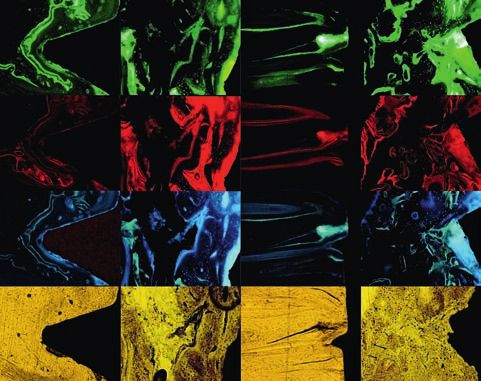
G. Weibrich et al.:
Knochenumbau an Zahnwurzelimplantaten
Bone remodeling around dental implant surfaces                                                                                                             239

Einleitung                                                            Introduction

In den letzten Jahren haben die meisten klinisch verfügbaren          In recent years, most of the clinically available implant systems
Implantatsysteme veränderte Implantatoberflächen einge-               have introduced modified implant surfaces, with each manu-
führt, wobei jeder Hersteller postuliert, dass seine Oberflächen-     facturer claiming that his surface structure optimizes osseoin-
struktur die Osseointegration optimiert. Bisher untersuchen           tegration. Most of the studies conducted hitherto have com-
die meisten vorliegenden Studien die jeweilige glatte (maschi-        pared the smooth (machined) surface with the corresponding
nierte) Oberfläche gegen die dazugehörige neu eingeführte             new surface modification from the same manufacturer. These
Oberflächenmodifikation des gleichen Herstellers. Diese vor-          histomorphometric studies show that a higher bone-implant
liegenden histomorphometrischen Studien konnten zeigen,               contact rate (BIC) results from increased and faster bone regen-
dass eine höhere Knochen-Implantat-Kontaktrate (BIC) auf              eration on the modified roughened implant surfaces [20, 22,
Grund einer erhöhten und beschleunigten Knochenneubil-                10]. Clinical use of one of these implant surface modifications,
dung an den modifizierten aufgerauten Implantatoberflächen            namely an anodized and oxidized surface (TiUnite), improved
resultiert [20, 22, 10]. Der klinische Einsatz einer dieser Implan-   the implant survival rate of dental implants inserted in the
tatoberflächenmodifikationen, einer anodisch-oxidierten               maxilla and mandible with immediate loading from 80 % to
Oberfläche (TiUnite), verbesserte die Implantat-Überlebens-           97 % compared with use of a machined implant surface from
rate unter Sofortbelastung der im Oberkiefer und Unterkiefer          the same manufacturer, using a similar study design [4, 5], in-
inserierten Zahnwurzelimplantate von 80 % auf 97 % vergli-            dicating the possible clinical relevance of such surface modifi-
chen mit der Anwendung der maschinierten Implantatoberflä-            cation. There have so far been very few studies that compare
che des gleichen Herstellers in analogem Studiendesign [4, 5],        the roughened surfaces of clinic implant systems available
was auf die mögliche klinische Relevanz einer solchen Oberflä-        from different manufacturers:
chenmodifikation hinweist. Es sind bisher nur sehr wenige Ar-             Initial comparative cell culture studies suggest that the co-
beiten vorhanden, welche die verfügbaren aufgerauten Im-              rundum-blasted and acid-etched titanium surface offers an ad-
plantatoberflächen der klinisch angewendeten Implantatsys-            vantage compared with plasma-sprayed and machined sur-
teme verschiedener Hersteller untereinander vergleichen:              faces [13]. In an animal model, the acid-etched surface demon-
    Erste vergleichende Zellkultur Untersuchungen lassen einen        strated an advantage compared with machined, plasma-
Vorteil der Korund-gestrahlten und Säure-geätzten Titanoberflä-       sprayed and corundum-blasted surfaces [1].
che gegenüber Plasma-gesprayten und maschinierten Oberflä-                The present study compares the histological and histomor-
chen vermuten [13]. Im Tiermodell zeigte die Säure-geätzte            phometric results of four clinically available titanium screw
Oberfläche einen Vorteil gegenüber den maschinierten, plasma-         implants with different surface structures with regard to the re-
gesprayten und den Korund-gestrahlten Oberflächen [1].                sulting peri-implant bone remodeling and BIC in the medium-
    Die vorliegende Studie vergleicht die histologischen und          term and late osseointegration period.
histomorphometrischen Ergebnisse von vier klinisch verfüg-
baren Titanschraubenimplantaten mit unterschiedlichen
Oberflächenstrukturen hinsichtlich des resultierenden peri-
implantären Knochenumbaues und der entstehenden BIC in
der mittleren und späten Osseointegrationsphase.

Material und Methoden                                                 Material and methods

Tiermodell                                                            Animal model

Nach Genehmigung durch die Tierversuchskommission wur-                Following approval by the animal experiment committee,
den zwölf weibliche Beagle Hunde im Alter von zwölf bis 15            twelve female beagle dogs aged twelve to 15 months and
Monaten und einem Gewicht von 12–16 kg in die Studie inte-            weighing 12–16 kg were included in the study. The animals
griert. Die Tiere wurden in Einzelkäfigen in der Tierversuchs-        were kept in individual cages in the Boehringer Ingelheim ani-
anstalt Boehringer Ingelheim gehalten und erhielten Wasser            mal experiment institute and were given water and standard
und Standard Futter. Drei Monate vor Beginn der Studie wur-           feed. Three months before the study, the mandibular premol-
den die Prämolaren im Unterkiefer entfernt. Alle Wunden wur-          ars were removed. All of the wounds were reviewed clinically
den klinisch nachuntersucht und zeigten unauffällige Hart-            and showed normal hard and soft tissue healing.
und Weichgewebeheilung.

Chirurgisches Vorgehen und Implantatdesign                            Surgical procedure and implant design

Nach Einleitung einer Anästhesie durch intramuskuläre Injek-          After induction of anesthesia by intramuscular injection of
tion von Dormitor (Orion Corporation, Espoo Finnland)                 Dormitor (Orion Corporation, Espoo Finland) (35 μg medeto-
(35 μg Medetomid pro kg Körpergewicht) und der intraven-              mide per kg body weight) and intravenous injection of Disopri-
ösen Injektion von Disoprivan (AstraZeneca AG, Grafenau 10,           van (AstraZeneca AG, Grafenau 10, CH-6301 Zug) 2 % (2 mg
CH-6301 Zug) 2 % (2 mg Propofol pro kg Körpergewicht) wur-            propofol per kg body weight) four different implants were in-

                                                                                      © Deutscher Ärzte-Verlag | zzi | Z Zahnärztl Impl | 2009; 25 (3) ■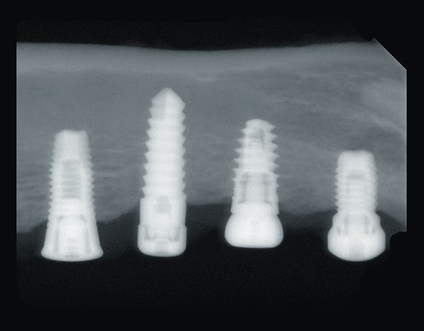
G. Weibrich et al.:
Knochenumbau an Zahnwurzelimplantaten
240 Bone remodeling around dental implant surfaces
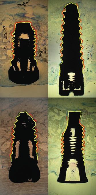
Abbildung 1 Röntgen Zahnfilm Aufnahme eines Präparates mit vier Abbildung 2 Fluorochrom-Markierung der Knochenumbauprozesse
verwendeten Implantaten im Unterkiefer. für die vier verschiedenen Implantate nach sechs Wochen, mittels
Figure 1 Radiograph of a preparation with four implants used in the Transmissions-Licht-Mikroskopie analysiert (von oben nach unten:
mandible. (Abb. 1-8: Ph. Streckbein) ohne Fluorochrom-Anregung, Alizarin, Xylenolorange, Calceingrün;
von links nach rechts: Branemark, Osseotite, Xive und Compress).
Figure 2 Fluorochrome labeling of the bone remodeling processes
for the four different implants after six weeks, analyzed using trans-
mission light microscopy (from above downward: without fluoro-
chrome stimulation, alizarin, xylenol orange, calcein green; from left to
right: Branemark, Osseotite, Xive and Compress).
den jeweils vier unterschiedliche Implantate randomisiert im serted randomized in the mandible according to the manufac-
Unterkiefer gemäß Hersteller-Empfehlungen inseriert. turer’s instructions.
Die folgenden Implantattypen mit unterschiedlichen Im- The following implant types with different implant surface
plantatoberflächenstrukturen wurden in randomisierter Positi- structures were inserted in randomized positions (Fig. 1):
on eingesetzt (Abb. 1): 1. Branemark (MK III): a self-tapping cylindrical titanium screw
1. Branemark (MK III): ein selbstschneidendes zylindrisches implant, Nobel Biocare (Göteburg, Sweden), length 7 mm,
Titanschraubenimplantat. Nobel Biocare (Göteburg, Schwe- diameter 3.75 mm. The TiUnite surface is produced by anod-
den), 7 mm Länge, 3,75 mm Durchmesser. Die TiUnite Ober- ized oxidation.
fläche wird durch anodische Oxidation hergestellt. 2. 3i Osseotite: a self-tapping cylindrical titanium screw im-
2. 3i Osseotite: ein selbstschneidendes zylindrisches Tit- plant, 3i (Miami, FL, USA), length 8.5 mm, diameter 4.0 mm.
anschraubenimplantat. 3i (Miami, FL, USA), 8,5 mm Länge, The Osseotite surface is acid-etched twice.
4,0 mm Durchmesser. Die Osseotite Oberfläche ist zweifach 3. Xive: a self-tapping conical titanium screw implant, Densply
Säure-geätzt. Friadent (Mannheim, Germany), length 8 mm, diameter 3.8
3. Xive: ein selbstschneidendes konisches Titanschraubenim- mm. The Friadent Plus surface is corundum-blasted and
plantat. Densply Friadent (Mannheim, Germany), 8 mm acid-etched.
Länge, 3,8 mm Durchmesser. Die Friadent Plus Oberfläche 4. Compress: a bone-compressing/condensing conical tita-
wird Korund-gestrahlt und Säure-geätzt. nium screw implant, IGZ eG (Diez, Germany), length 12
4. Compress: ein Knochen-verdrängendes/-kondensierendes mm, diameter 4.0 mm. The apical third of the surface is ma-
konisches Titanschraubenimplantat. IGZ eG (Diez, Germa- chined and sand-blasted.
ny), 12 mm Länge, 4,0 mm Durchmesser. Die Oberfläche ist
maschiniert und im apikalen Drittel sandgestrahlt. The wounds were closed with Vicryl 3.0 (Ethicon GmbH; Nor-
derstedt, Germany). The dogs were given soft feed for two
Der Wundverschluß erfolgte mit Vicryl 3.0 (Ethicon GmbH; weeks postoperatively.
Norderstedt, Deutschland). Die Hunde erhielten postoperativ
weiche Kost für zwei Wochen.
Antibiotische Abschirmung und Antibiotic protection and fluorochrome
Fluorochrom-Markierungs-Sequenz labeling sequence
Als postoperative antibiotische Abschirmung erhielten die Tie- As postoperative antibiotic cover, the animals were given an in-
re eine intramuskuläre Gabe von Tardomycel (BayerVital, Le- tramuscular injection of Tardomycel (BayerVital, Leverkusen,
■ © Deutscher Ärzte-Verlag | zzi | Z Zahnärztl Impl | 2009; 25 (3)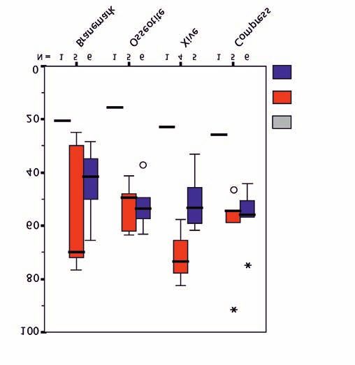
G. Weibrich et al.:
Knochenumbau an Zahnwurzelimplantaten
Bone remodeling around dental implant surfaces                                                                                                                                           241

                                                                                                                   Alizarinrot (1. Woche) /
                                                                                                                   Alizarin (week 1)
                                          Abbildung 3 Ver-
                                          kleinerte Darstellung

                                                                    Alizarinrot-markierte Knochenfläche in (%) /
                                          der histologischen Prä-
                                          parate von vier ver-
                                          schiedenen Implantat-

                                                                    Alizarin-labeled bone surface in (%)
                                          typen/-oberflächen,
                                          markiert für die Mess-
                                          ung der Knochen-Im-
                                          plantat-Kontaktrate
                                          nach sechs Wochen
                                          (oben links: Brane-                                                                                                      Lebenszeit /
                                                                                                                                                                   Lifetime
                                          mark, oben rechts: Os-                                                                                                     0 Wochen /
                                          seotite, unten links:                                                                                                        weeks
                                                                                                                                                                     6 Wochen /
                                          Xive und unten rechts:                                                                                                       weeks
                                          Compress).                                                                                                                 12 Wochen /
                                                                                                                                                                        weeks
                                          Figure 3 Reduced
                                          image of the histologi-
                                          cal preparations of
                                          four different implant
                                          types/surfaces, labeled    Abbildung 4 Alizarin-markierte Knochenfläche als Marker für die
                                          to measure the bone-       Knochenregeneration während der ersten Woche postoperativ für die
                                                                                                                              1,2
                                          implant contact rate       vier Implantatoberflächen.
                                          after six weeks (top       Figure 4 Alizarin-labeled bone surface as a marker of bone regener-
                                          left: Branemark, top       ation during postoperative week one. 1,2
                                          right: Osseotite, bot-
                                          tom left: Xive and bot-
                                          tom right: Compress).

verkusen, Deutschland) (Benzylpenicillin-Procain) 0,1 ml/kg          Germany) (procaine benzyl penicillin) 0.1 ml/kg body weight
Körpergewicht direkt präoperativ und dann alle 48 Stunden für        directly preoperatively and then every 48 hours for an overall
eine Gesamtdauer von zehn Tagen.                                     period of ten days.
    Die sequentielle Intravital-Markierung des regenerieren-             Sequential intravital labeling of the regenerating bone was
den Knochens wurde postoperativ mit 3 % Alizarin (0,83 ml/kg         performed postoperatively with 3 % alizarin (0.83 ml/kg body
Körpergewicht) in der ersten Woche (Applikation am OP Tag),          weight) in week one (given on the day of surgery), 1 % calcein
1 % Calceingrün (5 ml/kg Körpergewicht) in der zweiten und           green (5 ml/kg body weight) in weeks two and three (7 and 14
dritten Woche (7. und 14. post OP Tag), und 6 % Xylenoloran-         days postoperatively), and 6 % xylenol orange (1.5 ml/kg body
ge (1,5 ml/kg Körpergewicht) in der vierten und fünften Woche        weight) in weeks four and five (21 and 28 days postoper-
(21. und 28. post OP Tag) durchgeführt.                              atively).

Präparationstechnik                                                  Preparation technique

Eines der zwölf Tiere verstarb fünf Tage postoperativ und wurde      One of the twelve animals died five days postoperatively and
als zusätzliche Information über die Reparationsvorgänge fünf        was shown descriptively as additional information about the
Tage postoperativ deskriptiv dargestellt. Fünf Tiere wurden          repair processes five days postoperatively. Five animals were
sechs Wochen post OP, sechs Tiere zwölf Wochen post OP his-          analyzed histologically after six weeks and six animals after
tologisch aufgearbeitet. Die Präparat-Vorbereitung erfolgte          twelve weeks. The specimens were prepared using Donath and
mittels der Trenn-Dünnschlifftechnik nach Donath und Breu-           Breuner’s cutting and grinding technique [2, 3]. The specimens
ner [2, 3]. Die Präparate wurden entlang der Implantatachse in       were prepared along the implant axis in 40–60 μm sections.
40–60 μm dünnen Schnitten aufbereitet. Nach einer histomor-          Following histomorphometric analysis of the fluorochrome
phometrischen Analyse der Fluorochrom-Markierungen wur-              labeling, the specimens were stained with toluidine blue and
den die Präparate mit Toluidinblau gefärbt und histologisch          examined histologically.
untersucht. Die BIC wurde histomorphometrisch gemessen.

Histomorphometrische Auswertung                                      Histomorphometric analysis

Um die peri-implantäre Knochenregeneration zu analysieren            In order to analyze peri-implant bone regeneration, the fluoro-
wurde die Fluorochrom-Markierungen des peri-implantären              chrome labeling of the peri-implant bone was always measured
Knochens an immer den selben drei Stellen pro Implantat ge-          at the same three sites per implant: at implant threads 1 and 3

                                                                                                                    © Deutscher Ärzte-Verlag | zzi | Z Zahnärztl Impl | 2009; 25 (3) ■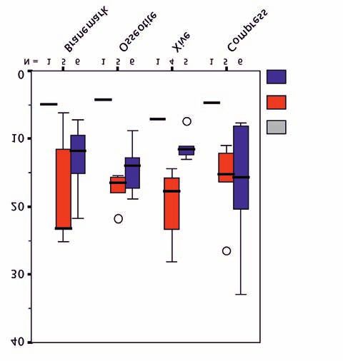
G. Weibrich et al.:
          Knochenumbau an Zahnwurzelimplantaten
242       Bone remodeling around dental implant surfaces

                                                       Summe (%)                          Perzentile /         Alizarin (%)                   Perzentile /
                                                          sum                              percentile             alizarin                     percentile

                             Zeitpunkt -
                              Wochen /                                         Median /                                           Median /
                                                  n        MW          SD                 25%/75%          n      MW      SD                   25%/75%
                               time -                                          median                                             median
                               weeks

            Branemark               0              1       20,6            –     20,6          –           1      10,5        –     10,5            –

                                    6              5       54,8        25         70        27,6/74,4      5      17,8    9,6       18,3        8,8/26,6

                                    12             6       43,7       13,1       41,6       33,2/54        6      14,6    7,9       12,9         10/20,7

             Osseotite              0              1       15,5            –     15,5          –           1       6,5        –      6,5            –

                                    6              5       52,9        9,5       49,5      44,7/62,8       5      16,6    6,2       14,5        11,7/22,5

                                    12             6       52,4        8,8       53,7      46,5/58,7       6      18,1        9     16,2        11,6/23,7

                XiVe                0              1        23             –      23           –           1       5,1        –      5,1            –

                                    6              4       71,7       10,3       73,4      61,5/80,2       4      21,4    2,1       20,9        19,7/23,6

                                    12             5       50,5       11,6       53,3      39,3/60,3       5      16,1    8,1       16,8          8/24

            Compress                0              1       25,9            –     25,9          –           1       9,4        –      9,4            –

                                    6              5       61,1       17,5       54,5       50,4/75        5       20         8     15,4         14/28,3

                                    12             6       56,3       10,2       55,8       49,1/61,2      6      17,5    5,4       16,4         14/20,2

          messen: an jedem Implantat von crestal gezählt an den Im-                         counted from the crestal end and at the last thread in the apical
          plantatwindungen Nr. 1 und Nr. 3 sowie an der letzten Win-                        region (caudolateral corner) of the implants, with the areas to
          dung im Apikalbereich (caudolaterale Ecke) der Implantate,                        be measured directly next to the implant surface (Fig. 2). Four
          wobei die zu messenden Areale direkt der Implantatoberfläche                      photos were taken of each of these three areas (Leica DMRX,
          anlagen (Abb. 2). Vier Fotoaufnahmen wurden jeweils von die-                      Leica; CCD color video camera, Sony; 100x magnification): the
          sen drei Arealen angefertigt (Leica DMRX, Leica; CCD Farb-                        first photo was taken under transmitted light microscopy with-
          videokamera, Sony; 100-fache Vergrößerung): zuerst eine Auf-                      out a specific filter and this was followed by three fluorescence
          nahme unter Durchlicht-Mikroskopie ohne einen spezifischen                        photos to analyze the fluorochromes alizarin, calcein green
          Filter, danach drei Fluoreszens-Aufnahmen um die Fluorochro-                      and xylenol orange (Fig. 2). These four photos were stored di-
          me Alizarin, Calceingrün und Xylenolorange zu analysieren                         gitally and analyzed histomorphometrically using an image
          (Abb. 2). Diese vier Aufnahmen wurden digital abgespeichert                       analysis system (Image Tool for MS Windows, University of
          und histomorphometrisch mittels Bild-Analyse-System (Image                        Texas Health Science Center, San Antonio, TX, USA). With this
          Tool für MS-Windows, University of Texas Health Science Cen-                      system the number of fluorochrome-labeled pixels (as % of the
          ter, San Antonio, TX, USA) ausgewertet. Mit Hilfe dieses Sys-                     total area) was determined separately for alizarin, calcein green
          tems wurde die Anzahl der Fluorochrom-markierten Pixel                            and xylenol orange at three regions per implant. The mean al-
          (in % der Gesamtfläche) getrennt für Alizarin, Calceingrün und                    izarin staining of the corresponding three implant regions was
          Xylenolorange an jeweils drei Regionen pro Implantat be-                          analyzed as a measure of bone regeneration in the first post-
          stimmt. Der Mittelwert der Alizarin-Anfärbung der jeweils zu-                     operative week, the mean calcein green as a measure of regen-
          sammengehörenden drei Implantatregionen wurde als Maß                             erative processes in weeks two and three and the mean xylenol
          für die Knochenregeneration in der ersten postoperativen Wo-                      orange fluorescence as a measure for weeks four and five.
          che, der Mittelwert von Calceingrün als Maß für regenerative                           After staining the specimens with toluidine blue, a further
          Prozesse der zweiten und dritten Woche und der Mittelwert                         digital photo of the entire implant together with the surround-
          von Xylenolorange als Maßzahl für die vierte und fünfte Wo-                       ing bone was taken at 16x magnification. The bone-implant
          che gewertet.                                                                     contact rate (BIC) was determined as the percentage of the im-
               Nach Anfärbung der Präparate mit Toluidine blau wurde ei-                    plant surface covered with bone, using an image analysis pro-
          ne zusätzliche digitale Aufnahme des ganzen Implantates zu-                       gram (Adobe Photoshop CS and Paint Shop Pro 7 for MS
          sammen mit dem umgebenden Knochengewebe mit 16facher                              Windows) (Fig. 3).
          Vergrößerung angefertigt. Die Knochen-Implantat-Kontakt-
          rate (BIC) wurde als knochenbedeckter Prozentsatz der Implan-
          tatoberfläche mittels Bild-Analyse-Programm (Adobe Photo-
          shop CS und Paint Shop Pro 7 für MS-Windows) bestimmt
          (Abb. 3).

      ■ © Deutscher Ärzte-Verlag | zzi | Z Zahnärztl Impl | 2009; 25 (3)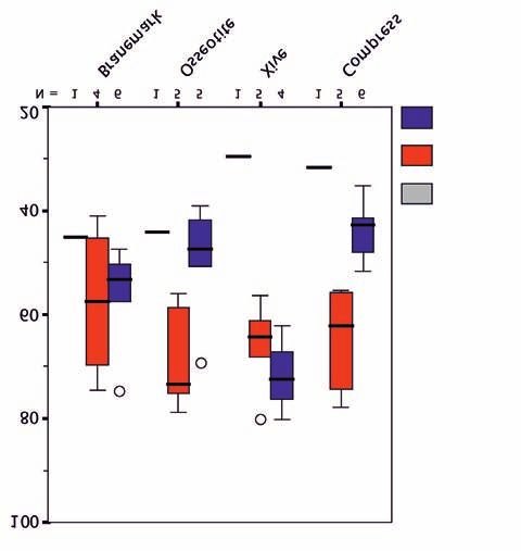
G. Weibrich et al.:
Knochenumbau an Zahnwurzelimplantaten
Bone remodeling around dental implant surfaces                                                                                                              243

    Calceingrün (%)                          Perzentile /    Xylenolorange (%)                        Perzentile /          Tabelle 1 Mittel-
     calcein green                            percentile       xylenol orange                          percentile           wert, Median, Per-
                                                                                                                            zentile und Stan-
                              Median /                                                Median /                              dardabweichung
   n      MW        SD                       25%/75%           n      MW      SD                       25%/75%
                              median                                                  median                                der Fluoreszenzmar-
                                                                                                                            kierten Knocheno-
 1 4,9     –        4,9            –             1 5,3         –      5,3       -                                           berfläche aufgeteilt
                                                                                                                            nach Implantattyp
   5      17,9      8,5          23,2          8,9/24,2        5      19,1    9,9       19,4             9,9/28,2
                                                                                                                            und Untersuchungs-
                                                                             11,1/2
   6      12,9      5,1      11,9 9/16,8 6       16,2         4,5     17                                                    zeitpunkt.
                                                                               0,5
   1      4,2        –            4,2              –           1      4,9      –         4,9                  -             Table 1 Mean,
                                                                                                                            median, percentile
   5      17,5      2,6          16,5          15,5/19,9       5      18,9     5        19,2              14/23,5           and standard devi-
   6      14,3      3,6          13,9          11,8/17,7       6      20,1    3,8       19,3             17,1/23,6          ation of the fluor-
                                                                                                                            escence-labeled
   1       7,1       –            7,1              –           1      10,8     –        10,8                  -
                                                                                                                            bone surface classi-
   4      19,5       6           17,8          15,1/25,7       4      30,8    7,7       33,6              22,7/36           fied according to im-
                                                                                                                            plant type and time
   5      11,1      2,2          11,6          9,3/12,7        5      23,3    5,5       24,7              18,8/27
                                                                                                                            of examination.
   1      4,6        –            4,6              –           1      11,8     –         11,8                 -             (Tab. 1-2: Ph. Streckbein)

   5      16,3      6,1          15,3          11,6/21,4       5      24,9    5,7       24,5             19,8/30,1

   6      16,7      9,3          15,7          8,1/23,5        6      22,1    4,6       19,9              19/26,5

Statistische Methoden                                                 Statistical methods

Alle quantitativen Messwerte wurden mittels deskriptiver Statis-      All quantitative measurements were described using descrip-
tik beschrieben (n, MW, Standardabweichung, Median, Mini-             tive statistics (n, mean, standard deviation, median, mini-
mum, Maximum, und andere Quartile). Zur Überprüfung, ob               mum, maximum and other quartiles). To examine whether the
die Oberflächenmodifikationen der Implantate die Knochen-             surface modifications of the implants influence bone regener-
regeneration beeinflusst, wurden die Tiere der 6- und 12-Wo-          ation, the animals in the 6-week and 12-week groups were ana-
chen-Gruppen getrennt für die einzelnen Implantattypen ana-           lyzed separately: 1. Branemark, 2. Osseotite, 3. Xive and
lysiert: 1. Branemark, 2. Osseotite, 3. Xive und 4. Compress.         4. Compress.
    Der Mittelwert der drei Fluorochrom-Messungen pro Im-                 The mean of the three fluorochrome measurements per im-
                                                                                                                                        st
plantat (Messung der markierten Knochenoberfläche in % an             plant (measurement of the labeled bone surface in % at the 1
                                                                                 rd
1. Windung, 3. Windung, letzter Windung) wurde als Kombina-           thread, 3 thread and last thread) was interpreted as a com-
tionsparameter für die Knochenregeneration im peri-implantä-          bined parameter for bone regeneration in the peri-implant re-
ren Bereich interpretiert. Die vier verbundenen Mittelwerte der       gion. The four associated bone regeneration means (four im-
Knochenregeneration (vier Implantate pro Tier) wurden gra-            plants per animal) were shown graphically for each of the four
fisch für jedes der vier Implantattypen mittels Boxplots dar-         implant types using box plots.
gestellt.                                                                 To examine the temporal sequence of possible implant sur-
    Um die zeitliche Abfolge möglicher Implantatoberflächen-          face effects on bone regeneration, each fluorochrome labeling
effekte auf die Knochenregeneration zu überprüfen wurde jede          was analyzed separately and shown side by side for the four im-
Fluorochrom-Markierung getrennt analysiert und für die vier           plant types: the alizarin measurements represent bone
Implantattypen nebeneinander dargestellt: Die Messwerte von           formation during week one, calcein green in weeks two and
Alizarin repräsentieren dabei die Knochenbildung während              three and xylenol orange in weeks four and five.
der ersten Woche, Calceingrün die Wochen zwei und drei und                In order to compare overall bone activity during the os-
Xylenolorange die Wochen vier bis fünf.                               seointegration process, the total fluorochrome labeling of each
    Um die gesamte Knochenaktivität während des Osseointe-            implant was calculated (alizarin + calcein green + xylenol or-
grationsprozesses zu vergleichen, wurde die Summe der Fluoro-         ange) and shown in a box plot for the four implant surfaces.
chrom-Markierungen eines jeden Implantates berechnet (Ali-                Box plots of bone regeneration and BIC were plotted separ-
zarin + Calceingrün + Xylenolorange) und in einem Boxplot             ately for the different implant types after a healing period of six
für die vier Implantatoberflächen dargestellt.                        weeks (n = 5 animals) and twelve weeks (n = 6 animals) (for pro-
    Boxplots der Knochenregeneration und der BIC wurden für           duction reasons, number of specimens n = 4–6 implants/time-
die unterschiedlichen Implantattypen getrennt nach sechs (n = 5       group), although the median was also used for the groups,
Tiere) bzw. zwölf (n = 6 Tiere) Wochen Einheilzeit (herstellungs-     though it has only limited validity. For the four implant types,
bedingte Präparateanzahl n = 4–6 Implantate/Zeit-Gruppe) ge-          the sign test for non-normally distributed linked data was cal-
zeichnet, obwohl der Median auch für die Gruppen verwendet            culated and the p values were shown.
wurde, für welche er nur begrenzte statistische Validität aufweist.

                                                                                       © Deutscher Ärzte-Verlag | zzi | Z Zahnärztl Impl | 2009; 25 (3) ■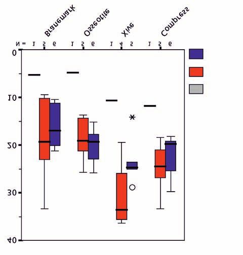
G. Weibrich et al.:
                                   Knochenumbau an Zahnwurzelimplantaten
244                                Bone remodeling around dental implant surfaces

                                                     Calceingrün (2.-3. Woche) /                                                                             Xylenolorange (4.-5. Woche) /
                                                     Calcein green (week 2-3)                                                                                Xylenol orange (week 4-5)

                                                                                                            Xylenolorange-markierte Knochenfläche in (%) /
  Calceingrün-markierte Knochenfläche in (%) /

                                                                                                            Xylenol orange-labeled bone surface in (%)
  Calcein green-labeled bone surface in (%)

                                                                                            Lebenszeit /                                                                                     Lebenszeit /
                                                                                            Lifetime                                                                                         Lifetime
                                                                                              0 Wochen /                                                                                      0 Wochen /
                                                                                                weeks                                                                                           weeks
                                                                                              6 Wochen /                                                                                      6 Wochen /
                                                                                                weeks                                                                                           weeks
                                                                                              12 Wochen /                                                                                     12 Wochen /
                                                                                                 weeks                                                                                           weeks

                                   Abbildung 5 Calceingrün markierte Knochenfläche als Marker für                             Abbildung 6 Xylenolorange markierte Knochenfläche als Marker für
                                   die Knochenregeneration während der zweiten und dritten Woche                              die Knochenregeneration während der vierten und fünften Woche
                                   postoperativ für die vier Implantatoberflächen. 1,2                                        postoperativ für die vier Implantatoberflächen. 1,2
                                   Figure 5 Calcein green-labeled bone surface as a marker of bone re-                        Figure 6 Xylenol orange-labeled bone surface as a marker of bone re-
                                   generation during postoperative weeks two and three for the four im-                       generation during postoperative weeks four and five for the four im-
                                   plant surfaces. 1,2                                                                        plant surfaces. 1,2

                                   Für die vier Implantattypen wurde der Vorzeichentest für nicht                             Results
                                   normal verteilte verbundene Daten berechnet und p-Werte dar-
                                   gestellt.                                                                                  Intravital fluorochrome labeling

                                                                                                                              In the 6-week (n = 5 animals) and 12-week (n = 6 animals)
                                   Ergebnisse                                                                                 specimens, the box plots showed only small differences in the
                                                                                                                              fluorochrome-labeled bone surface between the four implant
                                   Intravitale Fluorochrom-Markierung                                                         types for alizarin, calcein green and xylenol orange (Fig. 4–6).
                                                                                                                              In the 6-week group, median alizarin and calcein green labe-
                                   Bei den 6- (n = 5 Tiere) und 12- (n = 6 Tiere) Wochen Präparaten                           ling was higher with the Branemark and Xive implants than
                                   zeigten die Boxplots nur geringe Unterschiede in der Fluoro-                               with the other two implant types, whereas the Xive and Com-
                                   chrom-markierten Knochenfläche zwischen den vier Implan-                                   press implants demonstrated higher fluorochrome staining for
                                   tattypen für Alizarin, Calceingrün und Xylenolorange (Abb.                                 xylenol orange. In most cases, the median fluorochrome labe-
                                   4–6). In der 6-Wochen-Gruppe lag der Median der Alizarin-                                  ling was the same or slightly lower in the 12-week specimens
                                   und Calceingrün-Markierungen höher bei den Branemark und                                   than in the 6-week specimens. The means and standard devi-
                                   Xive Implantaten als bei den beiden anderen Implantattypen,                                ation of the fluorescence-labeled bone surfaces are shown in
                                   wohingegen die Xive- und Compress-Implantate höhere Fluo-                                  Table 1.
                                   rochromeinfärbungen für Xylenolorange aufwiesen. In den                                         Analysis of the three individual fluorochromes did not
                                   meisten Fällen lag der Median der Fluorochrom-Markierungen                                 show any statistically significant difference between the im-
                                   bei den 12-Wochen-Präparaten gleich oder leicht niedriger als                              plant surfaces (sign test, six weeks: all p ≥ 0.125; twelve weeks:
                                   bei den 6-Wochen-Präparaten. Die Mittelwerte und Standard-                                 all p ≥ 0.063).
                                   abweichung der Fluoreszenzmarkierten Knochenoberflächen                                         As a marker of bone regeneration, the median of the entire
                                   sind in Tabelle 1 dargestellt.                                                             fluorochrome-labeled bone surface (sum of alizarin + calcein
                                       Eine Analyse der drei individuellen Fluorochrome zeigte                                green + xylenol orange labeling) did not differ between the four
                                   keinen statistisch signifikanten Unterschied zwischen den Im-                              implant types in the 6-week specimens. Based on the sign test,
                                   plantatoberflächen (Vorzeichentest, sechs Wochen: alle p ≥                                 there was only a single statistically significant difference in
                                   0,125; zwölf Wochen: alle p ≥ 0,063).                                                      overall fluorochrome labeling, namely, between Compress and
                                       Als Marker der Knochenregeneration unterschied sich der                                Branemark (p = 0.03) in the 12-week group (Fig. 7), (12 weeks:
                                   Median der gesamten Fluorochrom-markierten Knochenflä-                                     all others p ≥ 0.063; six weeks: all others p ≥ 0.125).
                                   che (Summe von Alizarin + Calceingrün + Xylenolorange Mar-
                                   kierung), zwischen den vier Implantattypen bei den 6-Wo-

      ■ © Deutscher Ärzte-Verlag | zzi | Z Zahnärztl Impl | 2009; 25 (3)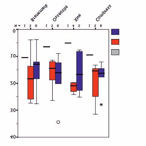
G. Weibrich et al.:
Knochenumbau an Zahnwurzelimplantaten
Bone remodeling around dental implant surfaces                                                                                                          245

                                                                                            Perzentile /         Tabelle 2 Mittelwert,
                                                     BIC (%)
                                                                                             percentile          Median, Perzentile und
                                                                                                                 Standardabweichung der
                      Zeitpunkt -
                      Wochen /                                               Median /                            Knochen-Implantat-Kon-
                                           n           MW          SD                        25%/75%
                        time -                                               median                              taktrate (BIC) aufgeteilt
                        weeks
                                                                                                                 nach Implantattyp und Un-
     Branemark              0              1            45          –             45                 -           tersuchungszeitpunkt.
                                                                                                                 Table 2 Mean, median,
                            6              4           59,4        13,8          65,4          45,2/70,7
                                                                                                                 percentile and standard
                            12             6           56,1        9,7           53,4          49,6/61,8         deviation of the bone-im-

      Osseotite             0              1           44,1         –            44,1                -           plant contact rate (BIC)
                                                                                                                 classified according to im-
                            6              5           68,4        10,4          73,3          57,3/77,1
                                                                                                                 plant type and time of
                            12             5           49,6        12            47,5           40,3/60          examination.

        XiVe                0              1           29,6         –            29,6                -

                            6              5           66,1         9            64,4          58,8/74,2

                            12             4           69,5        8,2           72,4          61,2/76,3

      Compress              0              1           31,8         –            31,8                -

                            6              5           65,1        10,6          62,2          55,5/76,2

                            12             6           43,6        5,7           42,7           39,8/49

chen-Präparaten nicht. Für die 12-Wochen-Gruppe fand sich          Bone-implant contact rate (BIC)
(Abb. 7), basierend auf dem Vorzeichentest nur ein einziger sta-
tistisch signifikanter Unterschied in der gesamten Fluoro-         There were no statistically significant morphological or histo-
chrom-Markierung, nämlich der zwischen Compress und Bra-           morphometric differences in the BIC between the four implant
nemark (p = 0,03) in der 12-Wochen-Gruppe (zwölf Wochen:           types in either the 6-week or 12-week specimens (Fig. 8) (sign
alle anderen p ≥ 0,063; sechs Wochen: alle anderen p ≥ 0,125).     test, six weeks: all p ≥ 0.063; twelve weeks: all p ≥ 0.125). After
                                                                   twelve weeks, the median BIC was higher for the Xive implant
Knochen-Implantat-Kontaktrate (BIC)                                (69.5 ± 8.2%) than for the other three implant types (Brane-
                                                                   mark 56 ± 9.7 %, Osseotite 49.6 ± 11.9 %, and Compress: 43.6
Es fanden sich keine statistisch signifikanten morphologischen     ± 5.7 %). The means are shown in Table 2.
oder histomorphometrischen Unterschiede in der BIC zwi-
schen den vier Implantattypen, weder in den 6- noch in den
12-Wochen-Präparaten (Abb. 8) (Vorzeichentest, sechs Wo-
chen: alle p ≥ 0,063; zwölf Wochen: alle p ≥ 0,125). Nach zwölf
Wochen war der Median der BIC höher für das Xive Implantat
(69,5 ± 8,2%) als für die anderen drei Implantattypen (Brane-
mark 56 ± 9,7 %, Osseotite 49,6 ± 11,9 %, und Compress: 43,6
± 5,7 %). Die Mittelwerte sind in Tabelle 2 dargestellt.

Diskussion                                                         Discussion

Die in der vorliegenden Studie verwendete Fluorochrom-Mar-         The fluorochrome labeling employed in this study is an estab-
kierung ist eine etablierte Methode [11, 12]. Die histhomor-       lished method [11, 12]. The bone surface as determined histo-
phometrisch bestimmte Knochenoberfläche kann als Korrelat          morphometrically can be used as a correlative for bone regen-
für die Knochenregeneration verwendet werden [8, 21, 17, 14].      eration [8, 21, 17, 14]. The median of most of the fluoro-
Der Median der meisten Fluorochrom-markierten Präparate in         chrome-labeled specimens in the 12-week group was lower
der 12-Wochen-Gruppe war niedriger als der entsprechende           than the corresponding median in the 6-week group. Osseoin-
Median in der 6-Wochen-Gruppe. Die Osseointegration der            tegration of the implants therefore appears to be largely com-
Implantate scheint also bereits nach sechs Wochen weitest-         plete after just six weeks and the fluorochromes are leached out
gehend abgeschlossen und die Fluorochrome im weiteren Hei-         during subsequent healing. Overall, the fluorochrome labeling
lungsverlauf bereits ausgewaschen. Insgesamt konnte die Fluo-      sequence can be used as a valid indicator for the time course of
rochrom-Markierungs-Sequenz als valider Indikator für den          bone regeneration.
zeitlichen Verlauf der Knochenregeneration verwendet wer-              During the first postoperative week (alizarin labeling), re-
den.                                                               sorption of the peri-implant bone, which has been damaged

                                                                                   © Deutscher Ärzte-Verlag | zzi | Z Zahnärztl Impl | 2009; 25 (3) ■
G. Weibrich et al.:
Knochenumbau an Zahnwurzelimplantaten
246 Bone remodeling around dental implant surfaces
Alizarin + Calcein + Xylenol(1.-5. Woche) /
Alizarin + Calcein + Xylenol (week 1-5)
Fluorochrom-markierte Knochenfläche in (%) /
Knochen-Implantat-Kontaktrate (BIC)in (%) /
Fluorochrom-labeled bone surface in (%)
Bone-implant contact rate in (%)
Lebenszeit / Lebenszeit /
Lifetime Lifetime
0 Wochen / 0 Wochen /
weeks weeks
6 Wochen / 6 Wochen /
weeks weeks
12 Wochen / 12 Wochen /
weeks weeks
Abbildung 7 Gesamte Fluorochrom-markierte (Alizarin + Calcein- Abbildung 8 Boxplot der Knochen-Implantat-Kontaktrate nach sechs
1,2
grün + Xylenolorange) Knochenfläche als Marker für die Knochenre- und zwölf Wochen für die vier verschiedenen Implantatoberflächen.
generation während der ersten bis fünften Woche postoperativ für die Figure 8 Box plot of the bone-implant contact rate after six and
vier Implantatoberflächen. 1, 2 twelve weeks for the four different implant surfaces. 1,2
Figure 7 Total fluorochrome-labeled bone surface (alizarin + calcein
green + xylenol orange) as a marker of bone regeneration during post-
operative weeks one to five for the four implant surfaces. 1, 2
1 1
Herstellungsbedingt Artefakt belastete Präparate wurden nicht mit einbezogen. Preparations with artifacts due to production were not included.
2 2
Datenpunkte mit einem Abstand von mehr als drei Interquartilsabständen wer- Data points with a gap of more than three interquartile intervals are identified
den als Extremwerte mit einem Stern gekennzeichnet, der Median wird als hori- with a star as extreme values, the median is shown as a horizontal line across the
zontaler Strich der Box dargestellt, die dünnen horizontalen Linien zeigen Daten- box, the thin horizontal lines show data points at an interquartile interval of up to
punkte bis zu einem Abstand von 1,5 Interquartielabstand, Daten mit größerem 1.5, and data with a greater interval are shown as a circle.
Abstand werden als Kreis ausgewiesen.
Während der ersten postoperativen Woche (Alizarin-Mar- by the implantation, predominates [19]. Bone remodeling be-
kierung) überwiegt die Resorption des durch die Implantation gins in the second postoperative week and can be visualized
geschädigten peri-implantären Knochens [19]. Das knöcher- using fluorochrome labeling. The greater dye uptake with
ne Remodelling beginnt in der zweiten postoperativen Wo- Branemark and Xive after six weeks indicates increased bone
che und kann mittels der durchgeführten Fluorochrom-Mar- regeneration compared with Osseotite and Compress. After
kierung visualisiert werden. Die höhere Farbstoffeinlagerung these six weeks, the fluorochrome labeling fell by more than
bei Branemark und Xive nach sechs Wochen weist auf eine one third in the case of the Branemark and Xive specimens.
vermehrte Knochenregeneration im Vergleich zu Osseotite This indicates persisting remodeling processes, whereas the
und Compress hin. Nach den sechs Wochen reduziert sich die labeling remained largely stable in the case of Osseotite and
Fluorochrom-Markierung bei Branemark und Xive Präpara- Compress implant surfaces, which might indicate reduced
ten um mehr als ein Drittel. Dies deutet auf anhaltende Remo- bone remodeling during weeks seven to twelve (Fig. 7). How-
dellingprozesse hin, wohingegen die Markierungen bei Os- ever, the clinical relevance of these histomorphometric differ-
seotite- und Compress-Implantatoberflächen weitestgehend ences remains unclear.
stabil bleiben, was auf einen verminderten Knochenumbau The Xive surface appears remarkable as it seems to show a
während der siebten bis zwölften Woche hinweisen könnte tendency to increased bony regeneration in the first week (al-
(Abb. 7). Die klinische Relevanz dieser histomorphometri- izarin) and in weeks four to five (xylenol orange). The highest
schen Unterschiede bleibt jedoch unklar. median of all the sums of fluorochrome labeling is found in
Bemerkenswert erscheint die Xive Oberfläche, da sie eine the 6-week specimens of the Xive implant (Fig. 4, 6). The Bra-
tendenziell erhöhte knöcherne Regeneration in der ersten nemark implant surface has the second highest value in the
Woche (Alizarin) und in der vierten bis fünften Woche (Xyle- 6-week group. The limited number of specimens is possibly
nolorange) aufzuweisen scheint. Der höchste Median aller the cause of the lack of significance of the differences that ap-
summierten Flourochrom-Markierungen wird in den 6-Wo- pear to be present. After twelve weeks, however, the Compress
chen-Präparaten des Xive-Implantates gefunden (Abb. 4, 6). implant had the highest median total fluorochrome labeling,
Die Branemark Implantatoberfläche weist den zweithöchs-
■ © Deutscher Ärzte-Verlag | zzi | Z Zahnärztl Impl | 2009; 25 (3)G. Weibrich et al.:
Knochenumbau an Zahnwurzelimplantaten
Bone remodeling around dental implant surfaces 247
ten Wert in der 6-Wochen Gruppe auf. Die limitierte Anzahl showing a statistically significant difference from the Bra-
der Präparate ist möglicherweise die Ursache für eine fehlen- nemark implant.
de Signifikanz der scheinbar vorhandenen Unterschiede. However, these higher values for the Compress implant do
Nach zwölf Wochen zeigte das Compress-Implantat jedoch not necessarily confirm a higher bone regeneration rate but could
mit dem höchsten Median-Wert der gesamten Flourochrom- also be an expression of as yet incomplete and therefore more pro-
Markierung einen statistisch signifikanten Unterschied zum longed and thus greater incorporation of fluorochromes, indicat-
Branemark-Implantat. ing that increased bone remodeling is still ongoing.
Diese höheren Werte für das Compress-Implantat belegen After six weeks the BIC of the conical implants Osseotite,
aber nicht unbedingt eine höhere Knochenregenerationsrate Xive and Compress was slightly higher than that of the cylin-
sondern könnten genauso Ausdruck einer immer noch nicht ab- drical Branemark implant. The cause might be the lateral con-
geschlossenen und damit länger dauernden und somit auch stär- densation of the bone due to the conical implant design.
keren Einbausituation von Fluorochromen als Ausdruck eines After twelve weeks, the Xive implant achieved the highest
immer noch anhaltenden vermehrten Knochenumbaus sein. BIC, followed by the Branemark, Osseotite and Compress im-
Nach sechs Wochen war die BIC der konischen Implantate plants. Compared with the BIC of the 6-week group, there was
Osseotite, Xive und Compress leicht höher als bei dem zylin- a slight increase with the Xive implant in the 12-week group,
drischen Branemark-Implantat. Die Ursache könnte in der late- the value remained constant with the Branemark implant and
ralen Kondensation des Knochens bedingt durch die koni- the BIC was slightly reduced with Osseotite and Compress im-
schen Implantatdesigns liegen. plants.
Nach zwölf Wochen erreichte das Xive-Implantat die The lower BIC of the conical Osseotite and Compress im-
höchste BIC, gefolgt von Branemark-, Osseotite- und Com- plants after twelve weeks might possibly be caused by increased
press-Implantaten. Verglichen mit den BIC der 6-Wochen bone resorption during the late osseointegration phase. This
Gruppe ergibt sich in der 12-Wochen-Gruppe eine leichte Stei- coincides with the results of a study on the effect of bone site
gerung beim Xive-Implantat sowie ein gleichbleibender Wert condensation prior to implant insertion, which also showed
beim Branemark- und eine gering verminderter BIC für Osseo- increased bone remodeling processes [9]. Furthermore, avail-
tite- und Compress-Implantate. able studies show the lowest BIC values for machined surfaces,
Die niedrigere BIC der konischen Osseotite- und Com- which might also explain the reduced values found here, at
press-Implantate nach zwölf Wochen könnte möglicherweise least for the two-thirds machined Compress implant.
durch eine erhöhte Knochenresorption während der späten However, the difference in implant length might also be an
Osseointegrationsphase verursacht sein. Dies deckt sich mit influence, as a greater proportion of the longer implants was
den Ergebnissen einer Studie zur Auswirkung einer Knochen- inserted in the medullary space of the bone. On the other
lagerkondensation vor Implantatinsertion, welche ebenfalls hand, such differences may be suspected after only six weeks,
resultierend erhöhte Knochenumbauprozesse nachweisen but these were not evaluated here.
konnte [9]. Des weiteren zeigen vorhandene Studien die nied- BIC rates between 40–50 % appear valid compared with
rigsten BIC Werte für maschinierte Oberflächen, was eben- data from other studies on different implant surfaces: Trisi et al.
falls die hier ermittelten verminderten Werte zumindest für found a BIC of 47 % for the Osseotite implant in the human
das zu zwei Drittel maschinierte Compress-Implantat erklä- maxilla [18]; Kim et al. report a BIC of 47.7 ± 13.4 % for 36 im-
ren könnte. plants with TiUnite surface in twelve dogs [7]; Shibli et al. docu-
Allerdings könnte auch die unterschiedliche Implantatlän- mented a BIC of 32.19 ± 15.68 % for seven implants with a TiU-
ge einen Einfluss haben, da dann ein größerer Anteil der länge- nite surface in a human study (n = 7) [15]; Grassi et al. also ob-
ren Implantate im Markraum des Knochens eingesetzt wurde. served a BIC of 42.83 ± 9.80 % in a human study of 14 implants
Dagegen spricht, dass solche Unterschiede dann auch schon with a sand-blasted and acid-etched surface [6]. Sul et al.
nach sechs Wochen zu vermuten sind, diese hier aber nicht showed an increased loosening torque for the TiUnite surface
evaluiert wurden. compared with the Osseotite surface [16].
BIC-Werte zwischen 40–50 % erscheinen valide verglichen Analysis of the BIC showed no significant differences be-
mit Daten aus anderen Studien an verschiedenen Implantatober- tween the four different implant surfaces inserted in local
flächen: Trisi et al. fanden eine BIC von 47 % für das Osseotite Im- bone, possibly because of the limited number of specimens (n
plantat in der humanen Maxilla [18]; Kim et al. berichten über ei- = 4–6 per group and time point).
ne BIC von 47,7 ± 13,4 % für 36 Implantate mit TiUnite Oberflä- The clinical relevance of the differences evaluated here
che in zwölf Hunden [7]; Shibli et al. konnten eine BIC von 32,19 under other general conditions such as human use or aug-
± 15,68 % für sieben Implantate mit TiUnite Oberfläche in einer mented implant site is currently still unclear.
Humanstudie dokumentieren (n = 7) [15]; Grassi et al. beobachte-
ten ebenfalle in einer Humanstudie eine BIC von 42,83 ± 9,80 %
bei 14 Implantaten mit sandgestrahlter und säuregeätzter Ober- Conclusion
fläche [6]. Sul et al. zeigten einen erhöhtes Ausdrehmoment der
TiUnite Oberfläche im Vergleich zur Osseotite Oberfläche [16]. After a healing period of six weeks, no statistically significant
Eine Analyse der BIC zeigte keine signifikanten Unterschie- difference between the four investigated implant surfaces was
de zwischen den vier unterschiedlichen im ortsständigen Kno- demonstrated with regard to either the peri-implant bone re-
chen inserierten Implantatoberflächen, möglicherweise auf generation rate or to the resulting BIC.
Grund der limitierten Anzahl an Präparaten (n = 4–6 pro Grup- The only significant difference in the bone regeneration
pe und Zeitpunkt). rate was shown in the 12-week group for the Compress vs. Bra-
© Deutscher Ärzte-Verlag | zzi | Z Zahnärztl Impl | 2009; 25 (3) ■G. Weibrich et al.:
Knochenumbau an Zahnwurzelimplantaten
248 Bone remodeling around dental implant surfaces
Die klinische Relevanz der hier evaluierten Unterschiede nemark implant (sign test, p = 0.03). At this time, there was no
bei anderen Rahmenbedingungen wie Humananwendung statistically significant difference when the bone-implant con-
oder augmentiertem Implantatlager ist derzeit noch ungeklärt. tact rate (BIC) was analyzed.
Schlussfolgerung Acknowledgement
Nach sechs Wochen Einheilzeit konnte kein statistisch signifi- The implants used in this study were provided free of charge by
kanter Unterschied zwischen den vier untersuchten Implantat- Nobel Biocare Deutschland GmbH, Cologne, Germany, 3i,
oberflächen sowohl hinsichtlich der analysierten peri-implan- Miami, USA, Densply Friadent GmbH, Mannheim, Germany,
tären Knochenregenerationsrate als auch bezüglich der resul- and IGfZ eG Diez, Germany.
tierenden BIC gezeigt werden.
Der einzige signifikante Unterschied in der Knochenrege-
nerationsrate konnte in der 12-Wochen-Gruppe für das Com-
press vs. Branemark Implantat gezeigt werden (sign test, p =
0,03). Zu diesem Zeitpunkt fand sich in Analyse der Knochen-
Implantat-Kontakt-Rate (BIC) kein statistisch signifikanter Un-
terschied.
Korrespondenzadresse
Danksagung
Dr. Dr. Philipp Streckbein
Klinik und Poliklinik für Mund-, Kiefer- und Gesichtschirurgie
Die in dieser Studie verwendeten Implantate wurden von No-
- Plastische Operationen, Justus-Liebig-Universität Giessen
bel Biocare Deutschland GmbH, Köln/Deutschland, 3i, Miami/ Klinikstrasse 29, 35385 Giessen
USA, Densply Friadent GmbH, Mannheim/Deutschland, und Tel.: 06 41 / 99-46271, Fax: 06 41 / 99-46279
der IGfZ eG Diez/Deutschland ohne Berechnung zur Ver- E-Mail: philipp.streckbein@uniklinikum-giessen.de
fügung gestellt.
Literatur
1. Cordioli G, Majzoub Z, Piattelli A, Sca- 6. Grassi S, Piattelli A, de Figueiredo LC, 11. Rahn BA, Perren SM: Xylenol Orange, a
rano A: Removal torque and histomor- Feres M, de Melo L, Iezzi G, et al.: Histo- fluorochrome useful in polychrome se-
phometric investigation of 4 different logic evaluation of early human bone quential labeling of calcifying tissues.
titanium surfaces: an experimental stu- response to different implant surfaces. J Stain Technology 1971;46:125-129
dy in the rabbit tibia. Int J Oral Maxillo- Periodontol 2006;77:1736-1743 12. Rahn BA: Die polychrome Sequenz-
fac Implants 2000;15:668-674 7. Kim SK, Lee HN, Choi YC, Heo SJ, Lee markierung des Knochenanbaus. Zeiss
2. Donath K, Breuner G: A method for the CW, Choie MK: Effects of anodized oxi- Information 1976;22:36-39
study of undecalcified bones and teeth dation or turned implants on bone he- 13. Sammons RL, Lumbikanonda N, Gross
with attached soft tissues. The Säge- aling after using conventional drilling or M, Cantzler P: Comparison of osteo-
Schliff (sawing and grinding) tech- trabecular compaction technique: his- blast spreading on microstructured
nique. J Oral Pathol 1982;11:318-326. tomorphometric analysis and RFA. Clin dental implant surfaces and cell beha-
3. Donath K: Die Trenn-Dünnschliff- Oral Implants Res 2006; 17:644-650 viour in an explant model of osseointe-
Technik zur Herstellung histologischer 8. Knöfler W, Graf H, Gröschel T, Löwicke gration. A scanning electron microsco-
Präparate von nicht schneidbaren Ge- G: Zur Knochenreaktion auf Biomateria- pic study. Clin Oral Implants Res
weben und Materialien. Der Präparator lien. II. Ergebnisse der fluoreszenzmi- 2005;16:657-666
1988;34:197-206 kroskopischen Untersuchung zur Beob- 14. Schlegel KA, Kloss FR, Schultze-Mosgau
4. Glauser R, Ree A, Lundgren A, Gottlow achtung der initialen Knochenbildung. S, Neukam FW, Wiltfang J: Healing pat-
J, Hammerle CH, Scharer P: Immediate Z Zahnärztl Impl 1990; VI:145-152 terns at the bone-implant-interface
occlusal loading of Branemark im- 9. Nkenke E, Kloss F, Wiltfang J, Schultze- using different topical measurements.
plants applied in various jawbone regi- Mosgau S, Radespiel-Troger M, Loos K, Deutsche Zahnärztliche Zeitschrift
ons: a prospective, 1-year clinical study. et al.: Histomorphometric and fluores- 2002;57:194-199
Clin Implant Dent Relat Res 2001; cence microscopic analysis of bone re- 15. Shibli JA, Grassi S, de Figueiredo LC,
3:204-213 modelling after installation of implants Feres M, Iezzi G, Piattelli A: Human
5. Glauser R, Lundgren AK, Gottlow J, using an osteotome technique. Clin Peri-Implant Bone Response to Turned
Sennerby L, Portmann M, Ruhstaller P, Oral Implants Res 2002;13:595-602 and Oxidized Titanium Implants Inser-
et al.: Immediate occlusal loading of 10. Oyonarte R, Pilliar RM, Deporter D, ted and Retrieved After 2 Months. Im-
Branemark TiUnite implants placed Woodside DG: Peri-implant bone re- plant Dent 2007;16:252-259
predominantly in soft bone: 1-year re- sponse to orthodontic loading: Part 2. 16. Sul YT, Johansson C, Albrektsson T:
sults of a prospective clinical study. Implant surface geometry and its effect Which surface properties enhance bo-
Clin Implant Dent Relat Res 2003;5 on regional bone remodeling. Am J Or- ne response to implants? Comparison
Suppl 1:47-56 thod Dentofacial Orthop 2005; of oxidized magnesium, TiUnite, and
128:182-189 Osseotite implant surfaces. Int J Pro-
sthodont 2006;19:319-328
■ © Deutscher Ärzte-Verlag | zzi | Z Zahnärztl Impl | 2009; 25 (3)G. Weibrich et al.:
Knochenumbau an Zahnwurzelimplantaten
Bone remodeling around dental implant surfaces 249
17. Terheyden H, Jepsen S, Moller B, Tucker MM, Rueger DC: Sinus
floor augmentation with simultaneous placement of dental
implants using a combination of deproteinized bone xeno-
grafts and recombinant human osteogenic protein-1. A his-
tometric study in miniature pigs. Clin Oral Implants Res 1999;
10:510-521
18. Trisi P, Lazzara R, Rebaudi A, Rao W, Testori T, Porter SS: Bone-
implant contact on machined and dual acid-etched surfaces
after 2 months of healing in the human maxilla. J Periodontol
2003; 74:945-956
19. Weibrich G, Hansen T, Kleis W, Buch R, Hitzler WE: Effect of
platelet concentration in platelet-rich plasma on peri-implant
bone regeneration. Bone 2004; 34:665-671
20. Weng D, Hoffmeyer M, Hurzeler MB, Richter EJ: Osseotite vs. ma-
chined surface in poor bone quality. A study in dogs. Clin Oral
Implants Res 2003; 14:703-708
21. Wetzel AC, Stich H, Caffesse RG: Bone apposition onto oral
implants in the sinus area filled with different grafting materi-
als. A histological study in beagle dogs. Clin Oral Implants Res
1995; 6:155-163
22. Zechner W, Tangl S, Furst G, Tepper G, Thams U, Mailath G, et
al.: Osseous healing characteristics of three different implant
types. Clin Oral Implants Res 2003; 14:150-157
© Deutscher Ärzte-Verlag | zzi | Z Zahnärztl Impl | 2009; 25 (3) ■Sie können auch lesen