MÜNCHEN 2021 - IDENTIFIZIERUNG MOLEKULARGENETISCHER URSACHEN VON SELTENEN ANGEBORENEN IMMUNDEFEKTEN AM BEISPIEL DER HYPER-IGE SYNDROME
←
→
Transkription von Seiteninhalten
Wenn Ihr Browser die Seite nicht korrekt rendert, bitte, lesen Sie den Inhalt der Seite unten
Identifizierung molekulargenetischer Ursachen von

seltenen angeborenen Immundefekten am Beispiel der

Hyper-IgE Syndrome

 Benedikt D. Spielberger

 München 2021
Aus der Kinderklinik und Kinderpoliklinik

 im Dr. von Haunerschen Kinderspital

 der Ludwig-Maximilians-Universität München

Identifizierung molekulargenetischer Ursachen von seltenen angeborenen Immundefekten

 am Beispiel der Hyper-IgE Syndrome

 Dissertation

 zum Erwerb des Doktorgrades der Medizin

 an der Medizinischen Fakultät

 der Ludwig-Maximilians-Universität zu München

 vorgelegt von

 Benedikt Daniel Spielberger

 aus

 München

 im Jahr

 2021

 2
Mit Genehmigung der Medizinischen Fakultät

 der Universität München

Berichterstatter: Prof. Dr. med. Ellen D. Renner

Mitberichterstatter: Prof. Dr. K. Spiekermann

 Prof. Dr. H. Schulze-Koops

 PD Dr. Dr. E. Strobel

Mitbetreuung durch die

promovierte Mitarbeiterin: Dr. Beate Hagl

Dekan: Prof. Dr. med. dent. Reinhard Hickel

Tag der mündlichen Prüfung: 18.02.2021

 3
Einleitende Zusammenfassung

 der schriftlichen kumulativen Promotion

 gemäß §4a der Promotionsordnung für die Fakultät

 der Ludwigs-Maximilians-Universität München

 vom 1. Juni 1983

in der konsolidierten Fassung der elften Änderungssatzung vom 15. September 2016

 4
Eidesstattliche Versicherung

Benedikt Daniel Spielberger

Ich erkläre hiermit an Eides statt, dass ich die vorliegende Dissertation mit dem Thema

Identifizierung molekulargenetischer Ursachen von seltenen angeborenen Immundefekten

am Beispiel der Hyper-IgE Syndrome

selbstständig verfasst, mich außer der angegebenen keiner weiteren Hilfsmittel bedient und

alle Erkenntnisse, die aus dem Schrifttum ganz oder annähernd übernommen sind, als solche

kenntlich gemacht und nach ihrer Herkunft unter Bezeichnung der Fundstelle einzeln

nachgewiesen habe.

Ich erkläre des Weiteren, dass die hier vorgelegte Dissertation nicht in gleicher oder in

ähnlicher Form bei einer anderen Stelle zur Erlangung eines akademischen Grades eingereicht

wurde.

Freiburg, 10.03.2021 Benedikt Spielberger

 Unterschrift Doktorand

 5

Inhaltsverzeichnis

1 Abkürzungsverzeichnis 7

2 Beitrag zu den Publikationen 8

 2.1 Beitrag zur Publikation „ Challenges of genetic counseling in patients with autosomal

 dominant diseases, such as the Hyper-IgE syndrome (STAT3-HIES)” 8

 2.2 Beitrag zur Publikation „Somatic alterations compromised molecular diagnosis of

 DOCK8 hyper-IgE syndrome caused by a novel intronic splice site mutation" 9

3 Einleitung 12

4 Zusammenfassung 18

5 Summary 20

6 Schriftenverzeichnis 22

 6.1 Originalarbeit: Challenges of genetic counseling in patients with autosomal

 dominant diseases, such as the hyper-IgE syndrome (STAT3-HIES) 22

 6.2 Originalarbeit: Somatic alterations compromised molecular diagnosis of DOCK8

 hyper-IgE syndrome caused by a novel intronic splice site mutation 26

7 Literaturverzeichnis 49

8 Danksagung 51

9 Publikationsliste 53

 6

1 Abkürzungsverzeichnis

AD-HIES Autosomal-dominantes Hyper-IgE Syndrom
AR-HIES Autosomal-rezessives Hyper-IgE Syndrom
DNA Desoxyribonukleinsäure
DOCK8 Dedicator of cytokinesis 8
ESID European society for immunodeficiencies
GEF G-protein exchange factor
HHV3 Humanes Herpesvirus 3
HIES Hyper-IgE Syndrom
HSV1/2 Herpes simplex-Virus 1/2
HSZT Hämatopoetische Stammzelltransplantation
IL6 Interleukin 6
IL10 Interleukin 10
NGS Next Generation Sequencing
NK-Zellen natürliche Killerzellen
PBMC periphere mononukleäre Zellen
PID Primärer Immundefekt
STAT1 Signal transducer and activator of transcription 1
STAT3 Signal transducer and activator of transcription 3
STAT5 Signal transducer and activator of transcription 5
Tem effector memory T-Zellen
Temra exhausted effector memory T-Zellen
Tyk2 Tyrosinkinase 2
WES Whole-exome sequencing
WGS Whole-genome sequencing

 7
2 Beitrag zu den Publikationen

Nach methodischer Einarbeitung in die Durchführung von Polymerase Kettenreaktion (PCR)

und deren Sequenzierung, sowie Planung, Durchführung und Analyse von intrazellulären

Färbungen mittels Durchflusszytometrie (FACS) begann ich mit der Bearbeitung eines

eigenständigen Projekts.

2.1 Beitrag zur Publikation „ Challenges of genetic counseling in patients with

 autosomal dominant diseases, such as the Hyper-IgE syndrome (STAT3-HIES)”

Zur Publikation „Challenges of genetic counseling in patients with autosomal dominant

diseases, such as the hyper-IgE syndrome (STAT3-HIES)“ erstellte ich gemeinsam mit Frau Prof.

Ellen Renner und Frau Dr. Beate Hagl das Studienprotokoll und –design. In dieser Publikation

deckte ich die Ursache für eine überproportionale Häufung an STAT3-HIES bei den

Nachkommen gesunder Eltern auf. Mein Beitrag bestand darin, die Hypothese einer

Keimbahnmutation im Sperma des Vaters zu untersuchen. Hierfür warb ich bei den

betroffenen Familien um Zustimmung an der wissenschaftlichen Studie teilzunehmen. Ich

etablierte die Sequenzierung von STAT3 aus Spermien in Kooperation mit Frau Prof. Katja

Anslinger und Frau Birgit Bayer aus dem Institut für Rechtsmedizin der LMU München. Ich

führte selbstständig die genetische Untersuchung von STAT3 aus Spermien im Labor von Frau

Prof. Ellen Renner durch. Des Weiteren bestand mein Beitrag in der Erhebung und Analyse der

klinischen Daten, weiterer laborchemischer Parameter, Erstellung von HIES-Scores und der

Interpretation der Ergebnisse sowie der Korrespondenz mit Koautoren zur Gewinnung der

Daten und des Untersuchungsmaterials. Dr. Kathrin Siepermann, Dr. Gregor Dückers und Prof.

Tim Niehues betreuten Patienten. Dr. Christina Wöllner führte die Mutationsanalyse bei

einem Patienten durch und Prof. Bodo Grimbacher diagnostizierte und betreute Patienten,

die in der Publikation enthalten sind. Julie Sawalle-Belohradsky führte die STAT3 PCR aus
 8
genomischer DNA der betroffenen Kinder durch. Prof. Bernd Belohradsky betreute Patienten

der Publikation vor der Übergabe an Prof. Ellen Renner. Gemeinsam mit Frau Prof. Ellen

Renner schrieb ich hauptverantwortlich unter Beteiligung aller Autoren das Manuskript und

brachte es zur erfolgreichen Publikation im Journal of Allergy and Clinical Immunology. Alle

Autoren beteiligten an der Manuskripterstellung und gaben ihr Einverständnis zur

Veröffentlichung der finalen Manuskriptversion.

2015 konnte ich die Ergebnisse dieses Forschungsprojektes im Rahmen der Summer School

der European Society for Immunodeficiencies (ESID) vorstellen.

2.2 Beitrag zur Publikation „Somatic alterations compromised molecular diagnosis

 of DOCK8 hyper-IgE syndrome caused by a novel intronic splice site mutation"

Für die Publikation „Somatic alterations compromised molecular diagnosis of DOCK8 hyper-

IgE syndrome caused by a novel intronic splice site mutation" erarbeitete ich zusammen mit

Frau Prof. Ellen Renner und Frau Dr. Beate Hagl das Studienprotokoll und –design, und

unterstützte Frau Prof. Ellen Renner erfolgreich beim Einwerben von Forschungsgeldern der

Wilhelm Sander-Stiftung. Für diese Publikation erhob und analysierte ich zunächst klinische

Daten einer Patientenfamilie mit zwei Kindern mit dem klinischen Bild eines HIES und unklarer

krankheitsverursachender Mutation. Im Rahmen der experimentellen Arbeit führte ich

genetische und immunologische Untersuchungen der Patienten, ihren Familienmitgliedern

und von gesunden Kontrollpersonen durch. Hierfür etablierte ich Verfahrensprotokolle für

Durchflusszytometrische Untersuchungen wie die Stimulation und Analyse der

Phosphorylierung von STAT1 (pSTAT1) und STAT5 (pSTAT5) in PBMCs und expandierten T-

Zellen (nicht in der finalen Publikation enthalten) und von STAT3 (pSTAT3) in Fibroblasten

 9
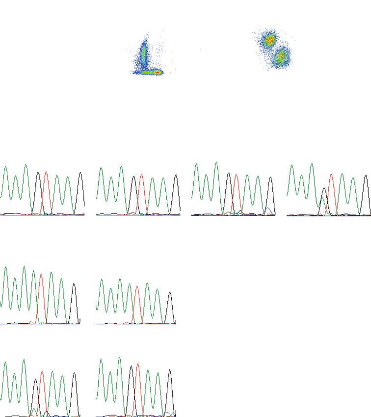
sowie die intrazelluläre Färbung für DOCK8 mit insgesamt acht Oberflächenmarkern (Fig. 4

und 5). Darüber hinaus etablierte ich das Einzelzell-Sorting für die anschließende

Sequenzierung von DOCK8 DNA und RNA in verschiedenen Zellsubpopulationen (Fig. 4 und 5).

Zusätzlich etablierte ich die in der Publikation im Text erwähnte Luminex-basierten Analysen

sowie das Serum-Inkubations-Assay (Supplementary Fig. S1), um Auto-Antikörper als

mögliche Ursache der fehlenden Stimulierbarkeit von pSTAT3 auf Interleukin-6

auszuschließen. Die Etablierung der Auto-Antikörper Untersuchung wurde von Dr. Anne Puel

durch ihre Vorerfahrungen bei der Etablierung einer ähnlichen Methode unterstützt. Hierbei

gelang es mir IL6, IL6R und gp130 an fluoreszierende beads zu koppeln und mittels Luminex-

Assay die Bindung von Proteinen aus Patientenserum darzustellen. Zusammen mit Prof.

Jordan Orange und Prof. Ellen Renner analysierte ich die Daten des whole-exome Sequencing.

Gemeinsam mit Shira Verduin, Dr. Isaac Njman, Dr. Beate Hagl und Prof. Mirjam von Gjin

analysierte ich die Daten des whole-genome sequencing. Hierbei zeigte sich ARHGAP32 als

vielversprechendes Kandidatengen, sodass ich hierfür Transfektionsexperimente mit

Patientenzellen etablierte und erfolgreich durchführte (Supplementary Fig. S2). Daten aus den

Abbildungen 1, 2, 4 und 5 sowie Supplementary Figures S1, S2 und S4 basieren auf von mir

etablierten und durchgeführten Experimenten. Dr. Silvia Thöne, Dr. Beate Hagl, Dr. Christof

Winter und Prof. Jürgen Ruland führten die ddPCR und deren Analyse durch und

interpretierten die Ergebnisse. Sophie Bonnal und Dr. Beate Hagl klonierten die Minigene-

Konstrukte und führten die Minigene-Experimente durch (Fig. 3). Dr. Christian Mertes, Dr.

Beate Hagl, Prof. Julien Gagneur und Prof. Thomas Meitinger führten die in silico Analysen zur

kryptischen und kanonischen Splice-Site durch (Fig. 3). Prof. Detlev Schindler führte

Radiosensitivitätsversuche mit Fibroblasten der Patienten durch und konnte dadurch die

 10
Pathogenität einer Veränderung im Gen ATM ausschließen. Andreas Eberherr führte die

Experimente mit den HAP1 ARHGAP32 Knock-out-Zellen durch.

Nach Abschluss aller experimentellen Arbeiten waren Frau Prof. Ellen Renner, Frau Dr. Beate

Hagl und ich die Hauptautoren des publizierten Manuskripts. Die geteilte Erstautorenschaft

ist durch Frau Dr. Beate Hagls Einsatz bei der Etablierung und Durchführung der ddPCR, der

Minigene-Experimente, der Sequenzierung von Immunzellsubpopulationen und die in silico

Analyse der kryptischen Splice-Site, sowie das gemeinsame Schreiben der finalen Publikation

begründet. Alle Autoren beteiligten sich an der Manuskripterstellung und gaben ihr

Einverständnis zur Veröffentlichung der finalen Manuskriptversion.

 11
3 Einleitung

Primäre Immundefekte und Gendiagnostik

Die Gruppe der primären Immundefekte (PID) umfasst aktuell über 350 verschiedene,

genetisch definierte Erkrankungen (1-5), die mit einer eingeschränkten Entwicklung oder

Aktivität des Immunsystems einhergehen. Wie aus Abbildung 1 ersichtlich wird, konnten

insbesondere in den vergangenen zehn Jahren durch verbesserte genetische

Untersuchungsmöglichkeiten wie dem Next Generation Sequencing (NGS) und die gestiegene

Wahrnehmung für diese seltenen Erkrankungen eine Vielzahl von Gendefekten die zu PID

führen identifiziert werden. Im Gegensatz zur Sanger-Sequenzierung, bei der in einem

Versuchsansatz ein definierter Basenabschnitt der DNA amplifiziert wird, werden unter dem

Begriff Next Generation Sequencing neue Genomanalyse-Verfahren verstanden, bei denen

eine sehr große Anzahl von DNA-Molekülen parallel sequenziert wird. Es kommen

verschiedene Techniken zum Einsatz, die eine massive, parallele Amplifikation von DNA-

Fragmenten in einem Ansatz ermöglichen. Ein Vorteil von NGS liegt somit in der verkürzten

Amplifikationsdauer. Es besteht dabei die Möglichkeit einige gezielt ausgewählte Gene, ein

gesamtes Exom (whole exome sequencing, WES) oder Genom (whole genome sequencing,

WGS) zu amplifizieren (6).

 12
Abb. 1.: Anstieg der mit angeborenen Immundefekten assoziierten Gene (y-Achse) im Verlauf der Jahre (x-

Achse). Abkürzungen: AR: autosomal-rezessiv, AD: autosomal-dominant, X: X-chromosomal vererbt. Abb

modifiziert nach (6).

Hyper-IgE Syndrome

Hyper-IgE Syndrome (HIES) stellen eine Untergruppe der PID dar und zeichnen sich durch

Ekzem, erhöhtes Serum-IgE und rezidivierende Infektionen aus (4, 7-12).

Die Erstbeschreibung 1966 – damals als Job Syndrom – geht auf zwei Patienten mit

ungewöhnlich häufigem Auftreten von Abszessen durch Staphylokokkus aureus und

ekzematöser Haut zurück (13). Die Assoziation mit erhöhtem Serum-IgE prägte 1972 den

Begriff „Hyper-IgE Syndrom“ (14). 1999 konnte das Hyper-IgE Syndrom (HIES) als

Multisystemerkrankung mit Ekzem, erhöhtem Serum-IgE und rezidivierenden Haut- und

Atemwegsinfektionen und einem autosomal-dominantem Erbgang beschrieben werden (15).

Klinische Hauptcharakteristika des autosomal-dominanten HIES (AD-HIES) sind, neben

erhöhtem Serum-IgE, eine variabel ausgeprägte Eosinophilie sowie ein frühzeitig auftretendes

Ekzem, welches sich bereits als ausgeprägtes Neugeborenenekzem präsentieren kann.

 13
Häufiges Zeichen der Immunschwäche sind Superinfektionen der ekzematösen Haut mit

Staphylokokkus aureus oder Candida albicans. Darüber hinaus kommt es zu häufigen Infekten

der Atemwege wie Sinusitiden oder Pneumonien durch Staphylokokkus aureus oder

Streptokokkus pneumoniae. In den unteren Atemwegen ist die Ausbildung von

Pneumatozelen aufgrund schwerer rezidivierender Atemwegsinfektionen, die ein Reservoir

für Erreger sein können, beim AD-HIES beschrieben (16, 17). Ein charakteristisches Aussehen

des Gesichts mit rauer Haut, prominenter Stirn, tiefliegenden Augen und breitem

Nasenrücken ist bekannt. Zusätzlich kommt es bei einer Vielzahl an Patienten zum Auftreten

von pathologischen Frakturen der langen Röhrenknochen, und verlängerter Retention der

Milchzähne. Auf Basis dieser distinkten Unterscheidungsmerkmale wurde im Jahr 1999 ein

Score, basierend auf klinischen Merkmalen wie Milchzahnpersistenz, Gesamt-IgE und Anzahl

der Pneumonien oder Hautabszesse, für die Wahrscheinlichkeit des Vorliegens eines

autosomal-dominanten Hyper-IgE Syndroms erarbeitet, der die Diagnose bestätigen sollte

und somit frühere Therapien ermöglichen sollte (18).

2004 erfolgte die klinische Erstbeschreibung einer Kohorte mit autosomal-rezessivem

Vererbungsmuster, welches als autosomal-rezessives HIES (AR-HIES) definiert wurde (19). Das

AR-HIES lässt sich klinisch vom AD-HIES durch andere klinische und laborchemische Merkmale

abgrenzen. So sind beim AR-HIES das Auftreten von Virusinfektionen durch Herpesviren

(HSV1/2, HHV3) mit generalisiertem Ekzema herpeticatum oder Molluscum contagiosum

Infektionen charakteristisch. Laborchemisch zeigen AR-HIES-Patienten häufig eine

Lymphopenie, erniedrigtes Immunglobulin M und eine ausgeprägte Eosinophilie (12). Des

Weiteren zeigen Patienten mit AR-HIES keine skeletalen Auffälligkeiten, Milchzahnpersistenz

oder überstreckbare Gelenke. AR-HIES Patienten leiden dagegen häufig unter Allergien und

anaphylaktischen Reaktionen gegen Nahrungsbestandteile wie Kuhmilchprotein,

 14
Hühnereiweiß oder Hülsenfrüchte (20). Aufgrund der rezidivierenden und persistierenden

Virusinfektionen ist bei AR-HIES das Auftreten von malignen Tumoren häufig (10).

Im Rahmen der Ursachenforschung zu Hyper-IgE Syndromen gelang 2006, ausgehend von

einem Patienten mit HIES-Phänotyp mit erhöhtem IgE, Ekzem und Immundefekt die

Identifikation von Mutationen im Gen Tyrosin Kinase 2 (Tyk2) (21). Kurze Zeit später konnten

durch Untersuchungen des JAK-STAT-Signalweges in größeren Kohorten und in einem der

erstbeschriebenen Patienten dominant-negative Veränderungen im Gen STAT3 (Signal

transducer and activator of transcription 3) als Ursache für das autosomal-dominante Hyper-

IgE Syndrom (AD-HIES) aufgezeigt werden (7-9, 22). Dieses mutierte Protein hat einen

dominant-negativen Effekt auf den STAT3-Signalweg und inhibiert die Aktivität des Wildtyp-

Allels durch Bildung von Heterodimeren. In der Folge wird die Signaltransduktion von

proinflammatorischen Zytokinen, wie Interleukin 6 oder Interleukin 1b durch STAT3

vermindert und die Differenzierung von TH17-Zellen erschwert, weshalb bei AD-HIES

Patienten die TH17-Zellzahlen erniedrigt sind (9, 23). TH17-Zellen und Interleukin-17 spielen

eine wichtige Rolle bei der Immunabwehr von extrazellulären Bakterien und Pilzen

(insbesondere Candida albicans) und somit konnte die Infektanfälligkeit bei AD-HIES gut

erklärt werden (24-26).

In einer Patientenkohorte mit dem klinischen Bild eines autosomal-rezessivem Hyper-IgE

Syndroms konnten durch gezielte Genuntersuchungen compound-heterozygote und

homozygote Mutationen sowie Deletionen im Gen DOCK8 (Dedicator of Cytokinesis 8) als

Ursache für AR-HIES identifiziert werden (12, 27). Durch die Mutationen kommt es entweder

zum Expressions- oder zum Funktionsverlust des DOCK8-Proteins. DOCK8 wirkt, als Mitglied

der DOCK180-Proteinfamilie, als ein Steuerelement des Aktin-Zytoskeletts und wird

hauptsächlich in Zellen des blutbildenden Gewebes exprimiert, wurde aber auch in Gewebe

 15
von Lunge und Pankreas gefunden (11). DOCK8 trägt als GTP-Austauschfaktor (guanine

exchange factor, GEF) zur Aktivierung von Guanintriphosphat-bindenden Proteinen (G-

Proteine) wie CDC42 bei und moduliert so Aktin-Polymerisation und das Zytoskelett-

Rearrangement. Klinisch kommt es hierdurch zu verringerter T-Zellzahl mit geringem Anteil

naiver T-Zellen, unzureichender Formation der immunologischen Synapse und verringerter

Antikörperbildung (28).

Therapeutisch stehen für AD-HIES und AR-HIES unterschiedliche Konzepte zur Verfügung. Die

Therapie für das AD-HIES besteht derzeit vor allem in konsequenter prophylaktischer

antiinfektiver und supportiver Therapie, um Überleben und Lebensqualität möglichst zu

verbessern (10, 29). Hämatopoetische Stammzelltransplantationen (HSZT) in AD-HIES sind als

Einzelbeschreibungen bekannt (30, 31) und bleiben bisher eine individuelle Entscheidung.

Aufgrund der Multisystemerkrankung ist bislang nicht klar ist, ob eine HSZT zum Beispiel auch

für die Lungenstrukturveränderungen wie Pneumatozelenbildung hilfreich ist. Wohingegen,

aufgrund des schwer ausgeprägten Erkrankungsbildes mit Tendenz zur Bildung maligner

Tumore, die möglichst frühe HSZT Therapie der Wahl für DOCK8-HIES ist (10, 29).

Ausblick:

Trotz der Fortschritte in der genetischen Diagnostik ist es derzeit noch nicht gelungen für alle

Patienten mit einem Hyper-IgE Syndrom eine genetische Diagnose zu sichern. Diagnostische

Schwierigkeiten sind beispielsweise darin begründet, dass mit den gängigen Methoden der

Sequenzierung vorrangig das Exom mit kleinen Teilbereichen der angrenzenden Introne, also

die codierende DNA untersucht wird. Somit kann zwar eine rasche Aussage über das Vorliegen

von Mutationen in diesen Bereichen getroffen werden, jedoch keinerlei Aussage zu den weiter

im Intron liegenden, nicht-kodierenden Sequenzen. Diese nicht-kodierenden Sequenzen üben

 16
jedoch in der Funktion als Promotoren, Enhancer oder auch Silencer zum Teil großen Einfluss

auf die Regulation der DNA Transkription aus. Neben technisch bedingten Herausforderungen

aufgrund großer Datenmenge können auch beispielsweise ein genetisches Mosaik, also

Mutationen in nur einem Gewebe, oder Reversionen von Mutationen in schnell teilenden

Zellreihen, die molekulargenetische Diagnose und damit die individuelle

Therapieentscheidung erschweren.

Zusammenfassend kann festgehalten werden, dass trotz verbesserter genetischer Diagnostik,

für die Versorgung der Patienten die Aufmerksamkeit bezüglich der klinischen, PID

spezifischen Warnsignale, wie erhöhte Infektanfälligkeit und Infektionen mit protrahiertem

oder therapierefraktärem Verlauf, weiterhin entscheidend ist. Neben der Basisdiagnostik von

Differentialblutbild, Immunglobulinspiegeln im Serum und spezifischer Antikörperbildung

kann gegebenenfalls eine weiterführende immunologische Diagnostik hilfreich sein. Nur

durch frühzeitige und effektive Therapie sowie Infektionsprophylaxe kann die Entstehung

irreversibler Schäden begrenzt werden.

 17
4 Zusammenfassung

Ziel der vorliegenden Arbeit ist es genauere Einblicke in die Vererbung und Pathogenese von

Immundefekten aus dem Spektrum der Hyper-IgE Syndrome zu erlangen und so frühzeitig

eine molekulargenetische Diagnose zu sichern um eine supportive oder kurative Therapie

ermöglichen zu können bevor irreversible Krankheitskomplikationen entstehen.

In der Veröffentlichung „Challenges of genetic counseling in patients with autosomal

dominant diseases, such as the hyper-IgE syndrome (STAT3-HIES)“ beschreiben wir drei Kinder

mit AD-HIES, die einen gemeinsamen Vater, jedoch zwei unterschiedliche Mütter haben. Als

Ursache konnten wir erstmalig die Vererbung einer heterozygoten, dominanten STAT3-

Variante als paternales Keimzellmosaik belegen. Autosomal-dominante Veränderungen in

STAT3 sind vorwiegend zufällige Keimbahnveränderungen, sogenannte Spontanmutationen,

sodass die Wiederholungswahrscheinlichkeit für gesunde Eltern als gering eingeschätzt wird.

Sollte es aber zu gehäuftem Auftreten einer identischen Mutation bei Nachkommen gesunder

Eltern kommen zeigt unsere Veröffentlichung auf, dass ein Keimzellmosaik abgeklärt werden

sollte, um gezielt genetisch beraten zu können.

In der Arbeit „Somatic alterations compromised molecular diagnosis of DOCK8 hyper-IgE

syndrome caused by a novel intronic splice site mutation“ untersuchten wir eine Patientin mit

Verdacht auf ein HIES eingehend. Klinisch präsentierte sich die Patientin mit ausgeprägtem

Ekzem, rezidivierenden kutanen Superinfektionen, Gedeihstörung und multiplen

Nahrungsmittelallergien. Bereits im ersten Lebensjahr litt die Patientin unter einer schweren

Pneumonie, die zu Intubation und invasiver Beatmung führte. Auf der Basis der aus der

Beatmung folgenden Bronchiolitis obliterans entwickelte sich durch eine Pseudomonas

aeruginosa Besiedlung ein fibrotischer Umbau der Lunge. Trotz normaler Th17-Zellzahl zeigte

sich eine verminderte Stimulierbarkeit von STAT3 auf Interleukin 6 (IL6), nicht aber auf
 18
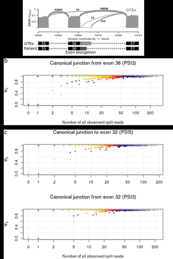
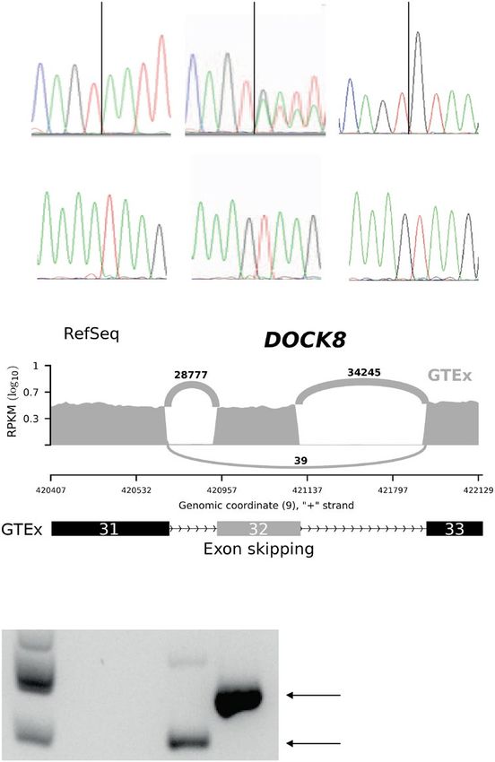
Interleukin 10 (IL10), hinweisend für einen Defekt in der IL6-STAT3-Achse. DOCK8-Protein war

in der Durchflusszytometrie- und Western-Blot-Analyse darstellbar. Ein PID-Genpanel und ein

whole exome sequencing (WES) blieben ohne Ergebnis zur Klärung der Krankheitsursache. Erst

die Untersuchung des gesamten Genoms (whole genome sequencing, WGS) und dezidierte

Sequenzierung der cDNA von DOCK8 zeigte eine bislang nicht beschriebene Intronvariante,

die zu fehlerhaftem Spleißen von DOCK8 führte. Zusätzlich zeigte sich, dass es in Teilen der T-

Zellen und nahezu allen natürlichen Killerzellen (NK-Zellen) zu spontanen Reversionen der

Intronmutation gekommen ist. Die Zellen mit Reversion hatten somit vermutlich einen

relativen Überlebensvorteil und konnten übermäßig expandieren, weshalb es zur Expression

von DOCK8-Protein und dem positiven Nachweis in der Durchflusszytometrie und Western-

Blot kam. Das defekte STAT3-Signalling ließ sich, nach Ausschluss von Autoantikörpern, durch

eine Verschiebung von T-Zelluntergruppen zu, bei der Patientin vorherrschenden, effector

memory T-Zellen (Tem) und exhausted effector memory T-Zellen (Temra) erklären, für welche

bekannt ist, dass diese eine verminderte IL6-STAT3 Stimulierbarkeit zeigen. Zeitlich parallel zu

den Untersuchungen in der Patientin wurde ihre jüngere Schwester geboren, bei der die

identische genetische Veränderung gefunden wurde. Für beide Kinder konnte erfolgreich eine

Stammzelltransplantation erfolgen, so dass insbesondere die jüngere Schwester vor

schwerwiegenden Infektionskomplikationen bewahrt werden konnte.

Zusammenfassend zeigen diese Familien einige Herausforderungen der genetischen und

klinischen Aufarbeitung bei Verdacht auf Immundefektsyndrome deutlich auf.

Hoffnung für die noch nicht diagnostizierten Patientinnen und Patienten gibt die Entwicklung

hin zur Sequenzierung des gesamten Genoms gekoppelt mit funktionellen

Proteinuntersuchungen (Multi-Omics-Verfahren), sodass eine weitere Verbesserung der

individuellen Prognose durch frühzeitige Therapien greifbar wird.

 19
5 Summary

The aim of the present study is to obtain detailed insights into the inheritance and

pathogenesis of immunodeficiencies like the hyper-IgE syndromes and to ensure a genetic

diagnosis in order to enable a supportive and curative therapy before irreversible disease

complications arise.

In the publication "Challenges of genetic counseling in patients with autosomal dominant

diseases, such as the hyper-IgE syndrome (STAT3-HIES)", we describe three children with

AD-HIES, who have one father but two mothers. We were able to show for the first time the

inheritance of a heterozygous, dominant-negative STAT3 variant as a paternal germ cell

mosaic. Autosomal dominant changes in STAT3 are predominantly random germline changes,

so-called spontaneous mutations, so that the probability of recurrence for healthy parents is

considered low. However, frequent occurrence of an identical mutation in offspring of healthy

parents might be due to a germ cell mosaicism and should be clarified in order to be able to

give good genetic counseling as shown in our publication.

In "Somatic alterations compromised molecular diagnosis of DOCK8 hyper-IgE syndrome

caused by a novel intronic splice site mutation" we assessed a patient with a suspected HIES

in detail. Clinically, the patient presented with pronounced eczema, recurrent cutaneous

superinfections, failure to thrive, and multiple food allergies. Already in the first year of life,

the patient suffered from severe pneumonia, which led to intubation and invasive ventilation.

On the basis of the bronchiolitis obliterans resulting from the respiration, a fibrotic remodeling

of the lung developed through a Pseudomonas aeruginosa colonization. Despite normal Th17

cell count, decreased stimulation of STAT3 to interleukin 6, but not to interleukin 10, indicated

a defect in the IL6-STAT3 axis. DOCK8 protein was present in flow cytometry and western blot

analysis. A PID gene panel and whole exome sequencing (WES) analysis identified no cause of
 20
the disease. Only the investigation of the entire genome and the dedicated sequencing of the

cDNA of DOCK8 revealed a previously unknown pathogenic intron variant, which led to

defective splicing of DOCK8. In addition, it has been shown that parts of the T cells and almost

all natural killer cells (NK cells) have spontaneously reversed the intron mutation. The cells

with reversion presumably showed a relative survival advantage and were able to expand

excessively, resulting in expression of DOCK8 protein and detection in flow cytometry and

Western blot. Defective STAT3 signaling, after exclusion of autoantibodies, was shown to be

due to a shift of T cell compartments with patient-dominated effector memory T cells (Tem)

and exhausted effector memory T cells (Temra) which are known to be less responsive to IL6

stimulation and subsequent STAT3 activation. During the patient`s assessment, her younger

sister was born, in whom the identical genetic change was found. Both children successfully

received a hematopoietic stem cell transplantation (HSCT), with the result that especially in

the younger sister serious infectious complications could be prevented.

In summary, these families clearly show some challenges of genetic and clinical workup in

suspected immunodeficiency syndromes.

Hope for yet undiagnosed patients arises from the movement towards sequencing of the

entire genome coupled with functional protein studies (multi-omics methods), enabling

further improvement of the individual prognosis by early therapies.

 21
6 Schriftenverzeichnis

6.1 Originalarbeit: Challenges of genetic counseling in patients with autosomal

 dominant diseases, such as the hyper-IgE syndrome (STAT3-HIES)

Spielberger BD, Woellner C, Dueckers G, Sawalle-Belohradsky J, Hagl B, Anslinger K, Bayer B,

Siepermann K, Niehues T, Grimbacher B, Belohradsky BH, Renner ED. Challenges of genetic

counseling in patients with autosomal dominant diseases, such as the hyper-IgE syndrome

(STAT3-HIES). J Allergy Clin Immunol. 2012 2012 Dec;130(6):1426-8.

 22
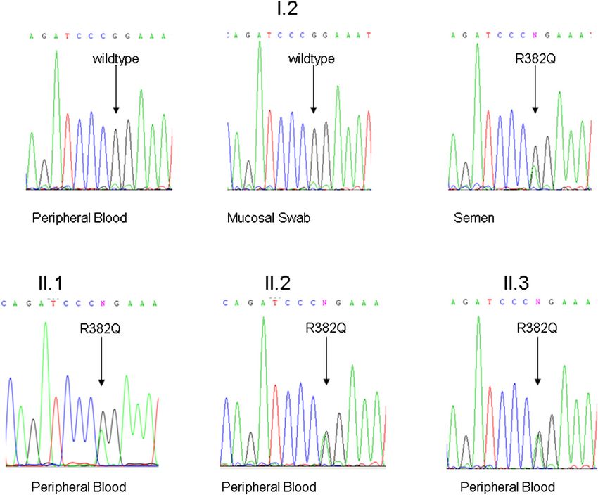
1426 LETTERS TO THE EDITOR J ALLERGY CLIN IMMUNOL
 DECEMBER 2012

This study was supported by the PCIRN (Public Health Agency of Canada–Canadian
 Institutes for Health Research Influenza Research Network).
Disclosure of potential conflict of interest: G. De Serres received the vaccines used in
 this study from GlaxoSmithKline and has received grants from GlaxoSmithKline
 and Sanofi. J. P. Drolet has received payment for lectures/service on speakers’ bureaus
 from Merck and Pfizer. D. Banerjee receives payment from Schering-Plough for short
 articles that are published online about topics related to allergies. C. Lemire has re-
 ceived payment for lectures/service on speakers’ bureaus from Abbott, Merck, and
 FIG 1. Pedigree of the family. The index patient (subject II.1), as well as her
 Nestl!e. A. Moore has consultant arrangements with Nycomed and has received pay-
 2 half brothers (individuals II.2 and II.3), are shown as solid symbols, which
 ment for lectures/service on speakers’ bureaus from King Pharmaceuticals Canada
 indicate affected subjects. Open symbols present unaffected subjects with
 and Pfizer. E. S. Chan has received grants and travel support from the Public Health
 the wild-type sequence (individuals I.1 and I.3), whereas the striped symbol
 Agency of Canada. D. Stark has received travel support from the University of Laval
 reflects the mosaicism of individual II.2. Squares indicate male individuals,
 and the government of Quebec. M. Benoit was employed by Janssen-Ortho and
 and circles indicate female individuals.
 owned stock in Janssen-Ortho and Johnson & Johnson. The rest of the authors declare
 that they have no relevant conflicts of interest.

 unremarkable pregnancy. At 5 months of age, she started
REFERENCES experiencing eczematous dermatitis, which spread from the
1. Lafleche J, Ahmandipour N, Anyoti H, Pless R, Law B. The safety profile of pan-
 demic H1N1 vaccines: reports of adverse events following immunization (AEFI) re-
 head to the shoulders. She had recurrent oral and diaper
 ceived by the Public Health Agency of Canada (PHAC), October 2009 through dermatitis, otitis media, urinary tract infections, bronchitis, and
 March 2010. Can J Infect Dis Med Microbiol 2010;21:223. pneumonia, some of which required intravenous antibiotic
2. Kurz X, Domergue F, Slattery J, Segec A, Szmigiel A, Hidalgo-Simon A. Safety treatment. At 4 years of age, she was started on prophylactic
 monitoring of influenza A/H1N1 pandemic vaccines in EudraVigilance. Vaccine
 antibiotic treatment with cefuroxime. Failure to shed primary
 2011;29:4378-87.
3. Groupe Central ESPRI. Surveillance des manifestations cliniques inhabituelles sur- teeth, increased serum IgE levels (9.716 IU/mL; value for age,
 venues apr"es la vaccination contre la grippe A(H1N1) lors de la campagne de masse
J ALLERGY CLIN IMMUNOL LETTERS TO THE EDITOR 1427
VOLUME 130, NUMBER 6

 FIG 2. Mutation analysis of affected family members. Mutation analysis of individual II.2 shows the
 wild-type sequence of gDNA isolated from peripheral blood and mucosal cells. The gDNA of individual II.2
 isolated from semen carries the heterozygous STAT3 mutation c.1145G>A, R382Q, which has been
 identified in all his clinically affected offspring (individuals II.1-II.3).

mutation of the gene STAT3 was identified in the genetic material Mosaicism is present if 1 individual has 2 or more cell lines
extracted from the semen (Fig 2). with a different genotype that developed from a single fertilized
 To show the functional consequence of the identified STAT3 homogeneous zygote.7 Because only the germ cells of the semen
mutation, peripheral mononuclear cells were stimulated with and not the somatic cells (oral mucosa and PBMCs) are mutated,
phorbol 12-myristate 13-acetate/ionomycin to assess the TH17 subject I.2 is healthy but can pass down the mutation to his off-
cell numbers, as previously described.4 TH17 cell counts are spring (individuals II.1, II.2, and II.3) with variable penetration.
known to be significantly decreased in patients with STAT3- The cause is usually a mutation that occurred in an early stem
HIES,4,6 and indeed, they were diminished in subjects II.1, II.2, cell that gave rise to the gonadal tissue. The maximum amount
and II.3 (0.03%, 0.07%, and 0.15% TH17 cells of CD41 cells; of affected germ cells is 50% and is dependent on the develop-
value in healthy control individuals, >0.2% CD41 cells), whereas mental stage at which the mutation occurred.7
subject I.2, with the wild-type STAT3 sequence in peripheral This family illustrates the complexity of genetic counseling
blood, had normal values (1.40% CD41 cells). in cases of autosomal dominant inherited diseases. Sequenc-
 As with other autosomal dominant diseases, STAT3-HIES in ing the gDNA of the parents’ peripheral blood might not
children with unaffected parents is suggested to be a result of be sufficient in patients with STAT3-HIES to rule out an
spontaneous in utero mutations. Thus the risk of subsequent off- increased risk for additional affected children. An increased
spring being affected with the same condition is thought to be risk of penetrance caused by an unknown mosaicism in germ
negligible. However, the fact that individual I.2 has healthy par- cells has to be considered. Zlotogora et al7 stated in 1998 that
ents and 3 children with the complete clinical spectrum of there are too few data on carriers of germline mosaicism and
STAT3-HIES caused by the identical heterozygous STAT3 muta- its recognition in genetic diseases. Only a few germline mosa-
tion (R382Q) involving subjects I.1 and I.3 suggests a paternal icisms have been observed in patients with primary immuno-
germline mosaicism. deficiencies, such as Wiskott-Aldrich syndrome, X-linked

 24
1428 LETTERS TO THE EDITOR J ALLERGY CLIN IMMUNOL
 DECEMBER 2012

severe combined immunodeficiency, DiGeorge syndrome, and 8. Arveiler B, de Saint-Basile G, Fischer A, Griscelli C, Mandel JL. Germ-line mo-
severe congenital neutropenia.8-11 This report of a germline saicism simulates genetic heterogeneity in Wiskott-Aldrich syndrome. Am J Hum
 Genet 1990;46:906-11.
mosaicism in an additional autosomal dominant primary im- 9. Puck JM, Pepper AE, Bedard PM, Laframboise R. Female germ line mosaicism as
munodeficiency should further raise awareness of this the origin of a unique IL-2 receptor gamma-chain mutation causing X-linked se-
condition. vere combined immunodeficiency. J Clin Invest 1995;95:895-9.
 Overall, we recommend informing family members of patients 10. Sandrin-Garcia P, Macedo C, Martelli LR, Ramos ES, Guion-Almeida ML, Ri-
 chieri-Costa A, et al. Recurrent 22q11.2 deletion in a sibship suggestive of parental
with STAT3-HIES about the possibility of mosaicism. Further- germline mosaicism in velocardiofacial syndrome. Clin Genet 2002;61:380-3.
more, genetic testing of every newborn in families with known 11. Newburger PE, Pindyck TN, Zhu Z, Bolyard AA, Aprikyan AA, Dale DC, et al.
members carrying STAT3 mutations is suggested to ensure the Cyclic neutropenia and severe congenital neutropenia in patients with a shared
diagnosis of STAT3-HIES early in life. Only an early diagnosis of ELANE mutation and paternal haplotype: evidence for phenotype determination
 by modifying genes. Pediatr Blood Cancer 2010;55:314-7.
STAT3-HIES allows initiation of the right treatment necessary for
limiting complications caused by infections and to benefit the Available online September 13, 2012.
quality of life of the individual patient. http://dx.doi.org/10.1016/j.jaci.2012.07.030

 We thank the patients and their family for their participation, primary care LRBA gene deletion in a patient presenting with
provider Dr M€ oller for patient care, Dr Sarah Andrews for critical review of autoimmunity without hypogammaglobulinemia
the manuscript, and Irmgard Eckerlein and Mayumi Hoffmann for technical
assistance and performing flow cytometry. To the Editor:
 Benedikt D. Spielbergera Primary immunodeficiencies are a highly heterogeneous group
 Cristina Woellner, MScb of genetic disorders caused by Mendelian mutations in >150
 Gregor Dueckers, MDc
 immune-related genes.1 Primary immunodeficiencies manifest as
 Julie Sawalle-Belohradskya
 Beate Hagl, PhDa
 severe and/or disseminated recurrent infections and may also
 Katja Anslinger, MDd have autoimmune manifestations.
 Birgit Bayerd We studied a female patient P1 of Pakistani origin who
 Kathrin Siepermann, MDc presented at the age of 4 years with a generalized lymphadenop-
 Tim Niehues, MDc athy, splenomegaly, neutropenia (range, 0.05-0.28 3109/L), and
 Bodo Grimbacher, MDb thrombocytopenia (platelet count, 20-40 3109/L). A lymph
 Bernd H. Belohradsky, MDa node biopsy showed reactive changes, bone marrow aspirate
 Ellen D. Renner, MDa was unremarkable, and antineutrophil antibodies were present.
 She also had chronic diarrhea associated with an autoimmune en-
From athe University Children’s Hospital and dthe Institute of Legal Medicine, Ludwig- teropathy, characterized by duodenal villous atrophy and large
 Maximilians-Universit€ at, Munich, Germany; bthe Centre of Chronic Immunodefi- bowel lymphocytic infiltration on biopsy. Her initial immunology
 ciency (CCI), University Medical Center Freiburg and University of Freiburg,
 Freiburg, Germany; and cChildren’s Hospital, Helios Kliniken, Krefeld, Germany.
 workup found only raised IgG levels (22.6 g/L), raised inflamma-
 E-mail: Ellen.Renner@med.lmu.de. tory markers, and a low number of natural killer cells (0.00-0.02).
Supported by the Kindness for Kids Foundation grant (to J.S.-B.), the German Research Lymphocyte subsets, double-negative T cells, T-cell proliferation
 Foundation (DFG RE2799/3-1), the Fritz-Thyssen research foundation grant (Az. assays, IgA (1.33 g/L), IgM (1.43 g/L), tetanus vaccine responses,
 10.07.1.159), the LMU Munich F€ oFoLe grant #680/658 (to E.D.R.), and the German
 and a nitroblue tetrazolium test were normal (Table I). P1 had no
 Federal Ministry of Education and Research (BMBF 01 EO 0803) (to B.G.). Data
 included in this publication are part of a medical thesis at the School of Medicine, significant history of infections except for a psoas abscess associ-
 Ludwig Maximilian University Munich (BDS). ated with chronic neutropenia. Over time she manifested growth
Disclosure of potential conflict of interest: B. Grimbacher has received payment for failure and developed new autoimmune features, including an ep-
 a lecture from the American Academy of Allergy, Asthma & Immunology and has isode of erythema nodosum, transient arthritis of both feet, and re-
 received research support from the German Federal Ministry of Education and Re-
 search. The rest of the authors declare that they have no relevant conflicts of
 current hemolytic anemia. As a result, she received several
 interest. courses of steroids, rituximab (with prophylactic immunoglobu-
 lin replacement), and mycophenolate mofetil. Five years after ini-
 tial presentation, after multiple courses of rituximab, she
REFERENCES
 developed recurrent infections (Streptococcus pneumoniae facial
 1. Grimbacher B, Holland SM, Gallin JI, Greenberg F, Hill SC, Malech HL, et al. Hy-
 per-IgE syndrome with recurrent infections—an autosomal dominant multisystem cellulitis, Streptococcus pneumoniae sepsis, and Haemophilus in-
 disorder. N Engl J Med 1999;340:692-702. fluenzae empyema) after withdrawal of immunoglobulin therapy.
 2. Minegishi Y, Saito M, Tsuchiya S, Tsuge I, Takada H, Hara T, et al. Dominant-neg- Although both her CD191 B cells and IgG level were normal, fur-
 ative mutations in the DNA-binding domain of STAT3 cause hyper-IgE syndrome. ther investigation found a new-onset antibody deficiency with ab-
 Nature 2007;448:1058-62.
 3. Holland SM, DeLeo FR, Elloumi HZ, Hsu AP, Uzel G, Brodsky N, et al.
 sent vaccine responses (Table I). Because of further chest
 STAT3 mutations in the hyper-IgE syndrome. N Engl J Med 2007;357: symptoms, despite recommencing immunoglobulin replacement,
 1608-19. a chest computed tomographic scan was performed that showed
 4. Schimke LF, Sawalle-Belohradsky J, Roesler J, Wollenberg A, Rack A, Borte M, extensive lung infiltration. Lung biopsy showed a florid diffuse
 et al. Diagnostic approach to the hyper-IgE syndromes: immunologic and clinical
 lymphoid interstitial infiltrate that consisted of a mixture of
 key findings to differentiate hyper-IgE syndromes from atopic dermatitis. J Allergy
 Clin Immunol 2010;126:611-7, e1. CD31 T and CD201 B cells, with scattered lymphoid follicles,
 5. Anslinger K, Bayer B, Rolf B, Keil W, Eisenmenger W. Application of the BioRo- particularly around airways. No granulomata were seen, and
 bot EZ1 in a forensic laboratory. Leg Med (Tokyo) 2005;7:164-8. stains for bacteria, fungi, and mycobacteria, as well as in situ hy-
 6. Milner JD, Brenchley JM, Laurence A, Freeman AF, Hill BJ, Elias KM, et al. Im- bridisation for EBV, were negative.
 paired T(H)17 cell differentiation in subjects with autosomal dominant hyper-IgE
 syndrome. Nature 2008;452:773-6.
 Patient P1 was born to a consanguineous marriage of first
 7. Zlotogora J. Germ line mosaicism. Hum Genet 1998;102:381-6. cousins. Therefore, we hypothesized that her disease was caused

 25
6.2 Originalarbeit: Somatic alterations compromised molecular diagnosis of DOCK8

 hyper-IgE syndrome caused by a novel intronic splice site mutation

Beate Hagl*, Benedikt D Spielberger*, Silvia Thoene, Sophie Bonnal, Christian Mertes,

Christof Winter, Isaac J. Nijman, Shira Verduin, Andreas C. Eberherr, Anne Puel, Detlev

Schindler, Jürgen Ruland, Thomas Meitinger, Julien Gagneur, Jordan S. Orange, Marielle E. van

Gijn, Ellen D. Renner. Somatic alterations compromised molecular diagnosis of DOCK8 hyper-

IgE syndrome caused by a novel intronic splice site mutation. Sci Rep. 2018 Nov 13;8(1):16719.

doi: 10.1038/s41598-018-34953-z.

*shared first author, contributed equally.

 26
OPEN Somatic alterations compromised

 hyper-IgE syndrome caused by a
Received: 25 June 2018
Accepted: 24 October 2018 novel intronic splice site mutation
Published: xx xx xxxx
 Beate Hagl Spielberger Thoene Bonnal
 Christian Mertes Winter Nijman Verduin
 Eberherr Puel Schindler Ruland
 Thomas Meitinger Gagneur Orange van Gijn &
 Renner

 and and corresponding

 intronic homozygous + >

 Chair and Institute of Environmental Medicine, UNIKA-T, Technical University of Munich and Helmholtz Zentrum
 Munich, Munich/Augsburg, Munich, Germany. University Children’s Hospital, Dr. von Haunersches Kinderspital,
 Ludwig Maximilian University, Munich, Germany. Institute of Clinical Chemistry and Pathobiochemistry, Klinikum
 rechts der Isar, Technical University of Munich, Munich, Germany. German Cancer Consortium (DKTK), partner site
 Munich, Munich, Germany. German Cancer Research Center (DKFZ), Heidelberg, Germany. Centre for Genomic

 Universitat Pompeu Fabra (UPF), Barcelona, Spain. Department of Informatics, Technical University of Munich,
 Garching, Germany. Department of Genetics, University Medical Center Utrecht, Utrecht, The Netherlands.
 Laboratory of Human Genetics of Infectious Diseases, Necker Branch, Necker Medical School, Paris, France. Paris
 Descartes University, Sorbonne Paris Cité, Institut Imagine, Paris, France. St Giles Laboratory of Human Genetics
 of Infectious Diseases, Rockefeller Branch, Rockefeller University, New York, NY, USA. Department of Human
 Genetics, University of Würzburg, Würzburg, Germany. German Center for Infection Research (DZIF), partner site
 Munich, Munich, Germany. Institute of Human Genetics, Technical University of Munich and Helmholtz Zentrum
 Munich, Neuherberg, Germany. Quantitative Biosciences Munich, Gene Center, Department of Biochemistry,
 Ludwig Maximilian University, Munich, Germany. Center for Human Immunobiology of Texas Children’s Hospital/
 Department of Pediatrics, Baylor College of Medicine, Houston, TX, USA. Department of Pediatrics, Division of
 Immunology, Allergy, and Rheumatology, Baylor College of Medicine, and Texas Children’s Hospital, Houston, TX,
 USA. Department of Pediatrics, Baylor College of Medicine, and Texas Children’s Hospital, Houston, TX, USA.
 Hochgebirgsklinik and Christine-Kühne-Center for Allergy Research and Education (CK-Care), Davos, Switzerland.
 Beate Hagl and Benedikt D. Spielberger contributed equally. Correspondence and requests for materials should be
 addressed to E.D.R. (email: ellen.renner@tum.de)

 SCIENTIFIC REPORTS | (2018) 8:16719 1

 27
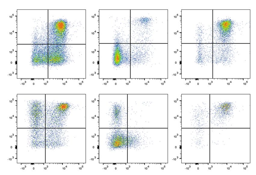
www.nature.com/scientificreports/

 Patients with primary immunodeficiencies (PIDs), such as hyper-IgE syndromes (HIES), have benefited tremen-
 dously from clinical classifications and the discovery of underlying gene defects and corresponding molecular
 testing. HIES are rare immunodeficiencies characterized by eczema, elevated serum IgE levels, eosinophilia and
 recurrent infections; and depending on the underlying genetic defect, additionally persistent primary teeth, aller-
 gic findings, lymphopenia or low Th17 cell counts1–7. All HIES entities overlap significantly with more common
 diseases, particularly severe forms of atopic dermatitis. Hence, prior to the possibility of molecular testing and
 due to low awareness of HIES, diagnosis was often delayed until severe complications, particularly irreversible
 lung changes, have impacted patients’ quality of life.
 The identification of genes causing HIES enabled diagnostic blood testing of low Th17 cell counts and
 reduced STAT3 phosphorylation in STAT3-HIES8,9, or lack of DOCK8 protein expression in DOCK8-HIES10.
 The improved understanding of the immunopathology resulted in treatment optimization1–4, such as the ben-
 efit of immunoglobulin substitution therapy in addition to rigorous antibiotic treatments due to the discovered
 impaired adaptive immunity5,11 and the consensus of early hematopoietic stem cell transplantation (HSCT) as
 treatment of choice in HIES caused by DOCK8 deficiency (DOCK8-HIES)11–13.
 With the knowledge that early diagnosis often determines the disease outcome, targeted next-generation
 sequencing (NGS) has become a cost-efficient tool in PID diagnostics14,15. Due to the rapidly increasing number
 of newly defined monogenic PIDs molecular PID diagnostics already needs to cover over 350 genes and targeted
 NGS approaches are limited to a pre-defined set of disease-causing genes15,16. Therefore, whole exome sequencing
 (WES) and whole genome sequencing (WGS) analyses are starting to replace targeted approaches15.
 The experience of the following family shows how somatic alterations complicated molecular diagnosis and
 how the close interplay of clinical, immunological, molecular, and bioinformatic diagnostic approaches identified
 a genetically determined disease.

 Results
 The index patient (patient II.2) is the second child of healthy
 first-degree cousins (Fig. 1a); term born with 2860 g (7th percentile) and 34 cm head circumference (25th percen-
 tile) after uneventful pregnancy. Recurrent upper and lower respiratory infections started at 2 months, including
 a fulminant pneumonia, requiring intubation and ventilation resulting in bronchiolitis obliterans at 6 months of
 age. Recurrent infections led to additional chronic lung changes with bronchiectasis formation and Pseudomonas
 aeruginosa positive lung specimen following frequent exacerbations (Fig. 1b). She developed eczema at 4 months
 with generalized eczema herpeticatum at 12 months of age. Several flares of eczema herpeticatum followed,
 including a herpes simplex blepharitis at around 8 years of age. There were repeated Molluscum contagiosum
 and mucocutaneous Candida infections as well as recurrent onychomycosis. She developed multiple specific IgE
 positive food allergies to milk, eggs, soy, and peanuts; at 12 months of age she suffered an anaphylactic reaction
 to lentils. Her growth started to slow to the 3rd percentile at 6 months of age and dropped further with significant
 growth retardation, malnutrition, and iron deficient anemia.
 At 3.5 years of age she was referred for PID evaluation. HIES was suspected due to elevated serum IgE (max.
 value 30434 IU/ml), eosinophilia (max. value 9100 cells/µl), chronic eczema, and recurrent bacterial, fungal, and
 viral infections, particularly of skin and lungs. Except for hyperextensible joints, she had no other skeletal findings
 associated with STAT3-HIES. The overall clinical presentation was in-between STAT3- and DOCK8-HIES with
 48 points in the NIH-HIES score17, which sums up findings of STAT3-HIES and is considered predictive for HIES
 above 40 points.
 Immunological work-up revealed high IgE, IgG, and IgA levels, and low to normal IgM levels (Fig. 1c). Mainly
 normal or high total lymphocyte and NK cell counts with high eosinophil counts were measured (Fig. 1d).
 Memory B cell counts affecting total, switched and unswitched memory B cells were low. Absolute numbers
 of total T cells, T helper cells, and cytotoxic T cells were within normal range, except for individual low and
 high values. Considering the patient’s young age, her T cell subsets showed a shift towards a memory pheno-
 type with low percentages of naïve T cells (CCR7+CD45RA+) and high percentages of effector memory T cells
 (CCR7−CD45RA−) in CD4+ and CD8+ T cells and CD8+ TEMRA cells (CCR7−CD45RA+) (Fig. 1e). Th17 cell
 counts were normal on two time-points with 0.32% and 0.23% of CD4+ cells (normal value: >0.20% of CD4+
 cells). Lymphocyte stimulation tests assessing proliferation by [ 3H]thymidine uptake had partially reduced
 response to mitogens such as Phytohaemagglutinin, OKT3, Concanavalin A and Pokeweed mitogen and mark-
 edly reduced reactions to an antigen mixture of Tetanus and Diphteria toxoid antigen (data not shown).
 Since recurrent infections and severe failure to thrive were not controlled by intensive antibacterial, -viral
 and -fungal treatment, the patient was referred again at 8 years of age. When the family ultimately agreed to
 HSCT, she received donor cells of her mother at 8 years of age. After an uneventful HSCT, her general health
 condition progressed over the next months with significant improvement of eczema and skin infections and
 restart of weight- and height-gain. Lymphocyte stimulation test to mitogens as well as B and T cell compartments,
 including naïve T cells, normalized within the first year after HSCT except for a shift towards cytotoxic T cells.
 Initially recurrent lung infections remained unchanged, most likely due to the bronchiolitis obliterans resulting
 from the severe infection at 6 months of age. Three years after HSCT, the frequency of respiratory infections has
 improved while continuous antibiotic and inhalation treatment are still required. Food allergies to milk, egg, and
 nuts were still present.
 Shortly after HSCT of patient II.2, a third child (patient II.3) was born (3790 g of weight; 77th percentile;
 36.5 cm head circumference; 89th percentile). Within the first months of age, she developed eczema, hard to
 control skin infections, and chronic otitis externa. Except for slightly elevated serum IgE (initially 18.2 IU/ml;
 max. value 1532 IU/ml at 7 months) and eosinophilia, she presented with lymphocyte counts and proliferation
 unremarkable for age (data not shown). Also NK, T and B cell counts, including subpopulations and IgG, IgA,
 and IgM serum were unremarkable for age (Fig. 1c–e). At 8 months of age, she received HSCT from a healthy

SCIENTIFIC REPORTS | (2018) 8:16719 2

 28
www.nature.com/scientificreports/

 Figure 1. Pedigree, clinical and immunologic presentation of the two affected siblings. (a) Pedigree of the
 investigated consanguineous family of the two affected siblings: II.2: index patient; II.3: second affected
 child. (b) Chest CT scan of patient II.2 at 3.5 years of age with areas of ground glass opacities, air trapping,
 multiple irregular nodular opacities (tree-in-bud sign), and bilateral bronchiectasis representing lung
 parenchyma destruction. (c) Serum immunoglobulin levels, (d) absolute numbers of eosinophil, lymphocyte
 and lymphocyte subsets of patient II.2 (triangles) and patient II.3 (circles) before (open symbols) and after
 HSCT (black filled symbols) compared to age-related normal range (gray filled area). (e) Representative
 flow cytometric plots showing CD4+ and CD8+ T cell subsets assessed by CCR7 and CD45RA expression in
 patient II.2 and patient II.3 compared to a healthy control. Percentages of naïve T cells (CCR7+CD45RA+),
 central memory T cells (CCR7+CD45RA−), effector memory T cells (CCR7−CD45RA−) and TEMRA cells
 (CCR7−CD45RA+) of CD4+ or CD8+ T cells are indicated by numbers in the respective quadrant.

SCIENTIFIC REPORTS | (2018) 8:16719 3

 29
www.nature.com/scientificreports/

 Figure 2. STAT3 phosphorylation analysis after stimulation. (a) Western blot analysis of whole cell lysates of
 PBMCs, unstimulated or 20 min. stimulated with 200 ng/ml IL6 or IL10. Expression of STAT3 phosphorylated
 at Y705 (pSTAT3) and total STAT3 (STAT3) of the two affected siblings and a healthy control was assessed;
 Actin as loading control. (b) Representative flow cytometric analysis showing diminished Y705-STAT3
 phosphorylation after 20 min. stimulation with 200 ng/ml IL6 (solid line) versus unremarkable results after
 stimulation with 20 ng/ml IL10 (dotted line) and 10 ng/ml IL21 (dashed line) in lymphocytes of patient
 II.2 compared to unremarkable results in patient II.3 and a healthy control; filled gray area: unstimulated
 lymphocytes. (c) Flow cytometric analysis showing Y705-STAT3 phosphorylation after 20 min. stimulation with
 20 ng/ml IL6 (solid line) or IL10 (dotted line) and 10 ng/ml IL21 (dashed line) comparable to healthy control in
 lymphocytes of one (representative of four) DOCK8-HIES patient. (d) Restored STAT3 phosphorylation after
 IL6 stimulation (solid line) in patient II.2 15 months after HSCT compared to unstimulated (filled gray area)
 and IL10-stimulated (dotted line) lymphocytes.

 unrelated donor. So far, she has developed completely normal without eczema, skin or unusual respiratory infec-
 tions more than two years after HSCT.

 Patient II.2 showed diminished Y705-STAT3 phosphorylation in PBMCs after
 IL6 stimulation, yet not after IL10 or IL21 stimulation by western blot and flow cytometric analysis (Fig. 2a,b).
 There was no altered STAT3 phosphorylation in lymphocytes of patient II.3 and four molecularly-defined
 DOCK8-HIES patients compared to healthy controls (Fig. 2a–c). Slightly decreased STAT3 phosphorylation in
 lymphocytes of patient II.3 compared to healthy controls was seen in some flow cytometric experiments (Fig. 2b)
 but overall similar to the range observed in healthy controls and not as clearly reduced as seen in patient II.3.
 The STAT3 signaling defect in patient II.2 was restricted to immune cells, since STAT3 phosphorylation after IL6
 stimulation in patient’s fibroblasts was comparable to control fibroblasts. We ruled out IL6-, IL6-receptor (IL6R)
 and gp130-autoantibodies as cause of the STAT3 signaling defect by multiplex analyses with serum antibody
 concentrations below or comparable to levels found in control sera. Furthermore, pre-incubation of PBMCs
 of a healthy control with serum of patient II.2 had no effect on IL6- and IL10-induced STAT3 phosphorylation
 (Supplementary Fig. S1). Similarly to other immunological alterations, STAT3 phosphorylation normalized in
 lymphocytes of patient II.2 within one year after HSCT (Fig. 2d).

 In search of the genetic cause of the underlying PID of patient II.2, we performed a pre-
 viously described PID targeted NGS approach14, including STAT3 and DOCK8. A homozygous single nucleotide
 variant in the ATM gene (c.1010 G > A, p.R337H) was detected and ruled-out to be disease-causing by normal
 radiation sensitivity testing of fibroblasts of patient II.2 and later a normal ATM sequence of patient II.3. Next,
 we completed WES analysis in patient II.2 and identified a homozygous alteration in ARHGAP32 (c.6038 G > A,
 p.R2013H). This alteration was considered disease-causing because ARHGAP32 is a GTPase-activating protein
 (GAP)18 and another GAP, called MgcRacGAP, has been associated with IL6-induced STAT3 phosphorylation
 in human and murine in vitro models19,20. However, STAT3 phosphorylation was not restored by overexpressing
 wildtype ARHGAP32 in patient cells and IL6-induced STAT3 phosphorylation in an ARHGAP32 knock-out
 HAP1 cell line was intact (Supplementary Fig. S2).
 WGS of patient II.2 and the healthy parents and a recessive model approach analysis did not lead to any addi-
 tional candidates. De novo variants analysis of the WGS data revealed four putative candidate variants, which
 were excluded due to their intergenic location or their location of more than 9 kb up- or downstream of adjacent
 exons.

SCIENTIFIC REPORTS | (2018) 8:16719 4

 30
Sie können auch lesen