ZELLBIOLOGIE & ANGEWANDTE VIROLOGIE CELL BIOLOGY & APPLIED VIROLOGY
←
→
Transkription von Seiteninhalten
Wenn Ihr Browser die Seite nicht korrekt rendert, bitte, lesen Sie den Inhalt der Seite unten
I n s t i t u t f ü r B i o m e d i z i n i s c h e T e c h n i k ( S t. I n g b e r t, S u l z b a c h , M ü n s t e r / W o l b e c k )

Zellbiologie & Angewandte Virologie
CELL BIOLOGY & APPLIED VIROLOGY
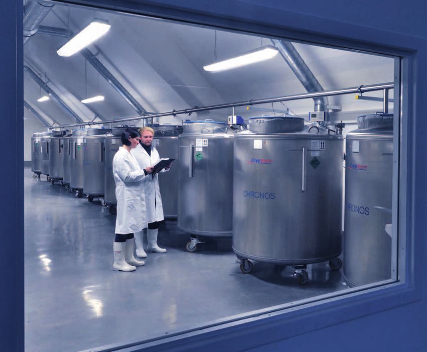
Titelbild: Lager Wolbeck: Einlagerung von Humanproben der Cover picture: Wolbeck repository: Storage of human samples of the
Umweltprobenbank des Bundes in Kryotanks zur späteren retro German Environmental Specimen Bank (Umweltprobenbank des Bundes,
spektiven Analyse (Foto: Bernd Müller). UPB) in cryotanks for later, retrospective analysis (Photo: Bernd Müller).
Prof. Dr. Hagen von Briesen
+49 (0) 6894/980-286
hagen.briesen@ibmt.fraunhofer.de
Biobanken & Kryolager (mit Außenstelle Münster/Wolbeck) Bioprozesstechniken & Nanotechnologie
Biobanks & Cryorepositories (including Münster/Wolbeck branch) Bioprocess Technologies & Nanotechnology
Prof. Dr. Hagen von Briesen Prof. Dr. Hagen von Briesen
+49 (0) 6894/980-286 +49 (0) 6894/980-286
hagen.briesen@ibmt.fraunhofer.de hagen.briesen@ibmt.fraunhofer.de
Umweltprobenbank des Bundes – Humanproben Präklinische Nanobiotechnologie
German Environmental Specimen Bank – Human Samples Preclinical Nanobiotechnology
Dr. Dominik Lermen Dr. Sylvia Wagner
+49 (0) 6894/980-251 +49 (0) 6894/980-274
dominik.lermen@ibmt.fraunhofer.de sylvia.wagner@ibmt.fraunhofer.de
HIV Specimen Cryorepository (Bill & Melinda Gates Foundation) In-vitro-Kulturtechniken
HIV Specimen Cryorepository (Bill & Melinda Gates Foundation) In Vitro Culture Technologies
Dr. Anja Germann Dr. Erwin Gorjup
+49 (0) 6897/9071-73 +49 (0) 6894/980-274
anja.germann@ibmt.fraunhofer.de erwin.gorjup@ibmt.fraunhofer.de
CRYO-BREHM – Zellbank für Wildtiere Prüflaboratorien & Qualitätssicherung
CRYO-BREHM – Cellbank for Wildlife Test Laboratories & Quality Assurance
Dr. Dominik Lermen Prof. Dr. Hagen von Briesen
+49 (0) 6894/980-251 +49 (0) 6894/980-286
dominik.lermen@ibmt.fraunhofer.de hagen.briesen@ibmt.fraunhofer.de
2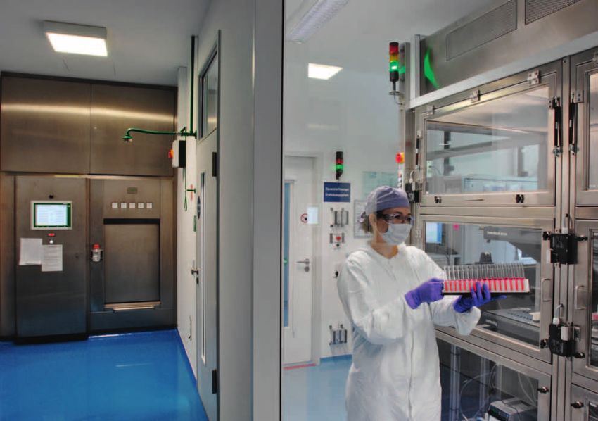
Zellbiologie &
Angewandte Virologie
CELL BIOLOGY &
APPLIED VIROLOGY

Angebote, Ergebnisse und Produkte der Abteilung        Offers, results and products of the Biobanks &
Biobanken & Kryolager                                  Cryorepositories Department
Umweltprobenbank des Bundes – Humanproben              Environmental Specimen Bank – Human Samples
HIV Specimen Cryorepository (B & M Gates Foundation)   HIV Specimen Cryorepository (B & M Gates Foundation)
CRYO-BREHM – Zellbank für Wildtiere                    CRYO-BREHM – German Cell Bank for Wildlife

Angebote, Ergebnisse und Produkte der Abteilung        Offers, results and products of the Bioprocess
Bioprozesstechniken & Nanotechnologie                  Technologies & Nanotechnology Department
Präklinische Nanobiotechnologie                        Preclinical Nanobiotechnology
In-vitro-Kulturtechniken                               In vitro Culture Technologies

Prüflaboratorien & Qualitätssicherung                  Test Laboratories & Quality Assurance

Projektbeispiel: Biokompatible Hybridkeramiken als     Project example: Biocompatible hybrid ceramics as
Knochenersatz                                          bone substitute

Ausstattung                                            Equipment

                                                                                                              3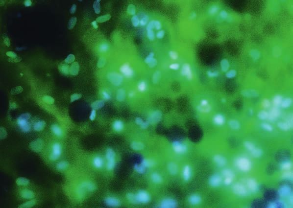
Z e l l bi o l o g i e & A n g e w a n d t e V i r o l o g i e

Verbesserte und neuartige Zellkulturtechniken und die darauf       Weiterhin lassen sich diese Nanopartikel als Genfähren einset-
aufbauenden analytischen Messverfahren müssen bei der              zen, um gezielt DNA in Zellen zu transportieren, die die
rasanten biotechnologischen Entwicklung von zukunftsori-           Expression bestimmter Proteine ermöglicht. Üblicherweise
entierten therapeutischen Konzepten Schritt halten. Hierbei        erfolgen gezielte genetische Veränderungen von Zellen durch
gewinnen Standardisierung und Optimierung im Bereich der           virale Vektoren. Die Nutzung dieser bereitet aber erhebliche
präklinischen und klinischen Testungen von Wirk- und Impf-         Probleme wie die Gefahr der Ausbildung von Krebs durch die
stoffen zunehmend an Bedeutung. Die Hauptabteilung Zell-           Aktivierung von Proto-Onkogenen. Die virusfreie genetische
biologie & Angewandte Virologie entwickelt hierfür alternative     Veränderung von Zellen mittels DNA-beladener Nanopartikel
Zellkultursysteme und Testverfahren für die verschiedensten        stellt sowohl für die Stammzellforschung wie auch für die
Bereiche der Stammzellforschung und Nanobiotechnologie.            zukünftige Zelltherapie eine vielversprechende Alternative
Die Wirkstoffentwicklung wird durch den Einsatz entspre-           dazu dar. Auch dieses ist ein vom BMBF unterstütztes Ver-
chender Technologieplattformen unterstützt. Hierzu zählen          bundprojekt (»NanoGene«) im Rahmen des europäischen For-
Transport- und Freisetzungsuntersuchungen therapeutischer          schungsnetzes EuroNanoMed.
Applikationssysteme über zelluläre Barrieren wie z. B. der Blut-
Hirn-Schranke, wofür wir der menschlichen Blut-Hirn-Schranke       Darüber hinaus kommen die in der Hauptabteilung neu entwi-
physiologisch sehr nahestehende Systeme entwickelt haben.          ckelten Zellkultursysteme und Testverfahren im Bereich der
Hierin können zum Beispiel Nanopartikel getestet werden,           Nanotoxikologie zum Einsatz: In einem Marie Curie Initial Trai-
die zuvor mit Alzheimer-Medikamenten beladen wurden                ning Network (ITN)-Projekt des 7. Rahmenprogramms der EU
und auf deren Oberfläche sich Ankermoleküle befinden, die          (»NanoTOES«) entwickelt die Abteilung hochsensitive chipba-
bestimmte Strukturen der Blut-Hirn-Schranke erkennen. So           sierte Testverfahren, um physikalische, chemische und biologi-
sollen die Nanopartikel ein Alzheimer-Medikament gezielt           sche Faktoren von Nanopartikeln und deren Einflüsse auf bio-
ins Gehirn transportieren. Die Entwicklung dieser neuartigen       logische Zellen zu untersuchen. Des Weiteren werden
Transportmethode von Medikamenten und die Untersuchung             zukunftsweisende Plattformen zum Sammeln, Präparieren,
von Chancen und Risiken werden vom Bundesministerium               Konservieren und zur Verteilung von Bioreagenzien und klini-
für Bildung und Forschung (BMBF) im Rahmen des Verbund-            schen Proben für weltweite Netzwerke entwickelt. Hierzu zäh-
projekts »NanoBrain« unterstützt, das Teil des europäischen        len optimierte Prozesse der Probenaufarbeitung und deren
Forschungsnetzes Era-NET »NEURON« ist. Eine weitere                Kryokonservierung. In diesem Zusammenhang wurde am
zelluläre Barriere, die es bei oraler Applikation zu überwinden    Fraunhofer IBMT im Rahmen der globalen Initiative zur Ent-
gilt, ist die Darmbarriere. In speziell hierfür ausgelegten        wicklung eines HIV-Impfstoffes (»Collaboration for AIDS Vac-
Darmbarriere-Modellen testen wir Formulierungen von                cine Discovery – CAVD«) eine globale HIV-Kryobank der
Wirkstoffen für die Photodynamische Therapie, die ansonsten        Sicherheitsstufe S3 aufgebaut, die von der Bill & Melinda
oral nicht bioverfügbar sind. Hier besteht die Chance, diese       Gates Foundation und der saarländischen Landesregierung
Wirkstoffgruppe in Tablettenform zu verabreichen und somit         finanziell getragen wird. Die Kryobank für Bioreagenzien
die mit Nachteilen behaftete intravenöse Gabe zu umgehen.          untersteht einem zertifizierten Qualitätssicherungsprogramm,
Dieses Projekt wird im Rahmen des Verbunds »BioTraP for            welches das Arbeiten nach den Richtlinien der »Good Clinical
CCC« durch das BMBF gefördert.                                     Laboratory Practice« (GCLP) kontrolliert. Hier werden Proben
                                                                   in einer vollautomatisierten Anlage zur Kultivierung eukaryoti-

4
CELL B I OLOG Y & A P P L I ED V I ROLOG Y
Improved and innovative cell culture techniques and the ana- These nanoparticles can also be used as gene ferries to trans-
lytical measurement methods based on these have to keep up port DNA in a targeted manner to cells to allow the expression
with the rapid biotechnological development of future-ori- of certain proteins. Targeted genetic alterations of cells are
ented therapeutic concepts. Standardization and optimization usually achieved using viral vectors. This, however, gives rise to
in the area of preclinical and clinical testing of active ingredi- substantial problems such as the risk of the development of
ents and vaccines are becoming increasingly important in this cancer due to the activation of proto-oncogenes. The virus-
context. For this purpose the Divison of Cell Biology & Applied free genetic alteration of cells using DNA-loaded nanoparticles
Virology Department is developing alternative cell culture sys- represents a promising alternative in this regard both for stem
tems and test methods for the various areas of stem cell cell research and for cell therapy in the future. This is also a
research and nanobiotechnology. The development of drugs is joint project funded by the BMBF ("NanoGene") within the
supported by the use of the corresponding technology plat- framework of the European research network EuroNanoMed.
forms. This includes transport and release investigations of
therapeutic application systems via cellular barriers such as the In addition to this, the cell culture systems and test methods
blood-brain barrier, for which we have developed systems newly developed in the division are being used in the area of
which are physiologically very close to the human blood-brain nanotoxicology: In a Marie Curie Initial Training Network (ITN)
barrier. These can be used, for example, to test nanoparticles project of the 7th framework program of the EU ("Nano-
which are loaded with Alzheimer drugs and which are surface- TOES"), the department is developing highly sensitive, chip-
modified with anchor molecules that recognize corresponding based test methods in order to investigate physical, chemical
structures on the blood-brain barrier. In this way, the nanopar- and biological factors of nanoparticles and their influences on
ticles can transport Alzheimer drugs in a targeted manner to biological cells.
the brain. The development of this innovative transport
method of drugs and the examination of chances and risks are Future-oriented platforms for the collection, preparation, con-
supported by the Federal Ministry for Education and Research servation and distribution of bioreagents and clinical samples
(BMBF) within the framework of the joint project "Nano- for worldwide networks are also being developed. This
Brain", which is part of the European research network Era- includes optimized processes for the preparation and cryo-
NET "NEURON". Another cellular barrier which has to be over- preservation of samples. In this context a global HIV cryobank
come in the case of oral application is the intestinal barrier. of safety level S3 was developed at the Fraunhofer IBMT
With the aid of intestinal barrier models especially designed within the framework of the global initiative for the develop-
for this purpose, we test formulations of drugs for photody- ment of an HIV vaccine ("Collaboration for AIDS Vaccine Dis-
namic therapy (PDT) which are not biologically available other- covery – CAVD"), which is funded by the Bill & Melinda Gates
wise in oral applications. There is a chance of being able to Foundation and the Saarland Regional Government. The cryo
administer this active substance group in tablet form and thus bank for bioreagents is subject to a certified quality assurance
bypass all of the disadvantages associated with intravenous program which controls the work according to the guidelines
administration. This project is funded by the BMBF within the of the "Good Clinical Laboratory Practice" (GCLP). Here, sam-
framework of the joint project "BioTraP for CCC". ples are produced in a fully automated system for the cultiva-
5Z e l l bi o l o g i e & A n g e w a n d t e V i r o l o g i e
scher Zellen hergestellt. Damit stellt die Kryobank eine Techno- tion of eukaryotic cells. The cryobank thus represents a tech-
logie-Plattform für die weitere Entwicklung und klinische Tes- nology platform for the further development and clinical
tung von Impfstoffen und neuen Therapien dar. testing of vaccines and new therapies.
Diese Biobank dient auch als Kryolager für ein von der Deut- This biobank also serves as a cryorepository for a German-Afri-
schen Forschungsgemeinschaft (DFG) gefördertes deutsch- can joint project funded by the Deutsche Forschungsgemein-
afrikanisches Verbundprojekt zur Erforschung des Krankheits- schaft (DFG) for research into the pathogen Staphylococcus
erregers Staphylococcus aureus. Die Wissenschaftler in aureus. The scientists in Germany and Africa want to find out
Deutschland und Afrika wollen herausfinden, wie verbreitet how widespread and resistant the pathogens are on the Afri-
und resistent die Erreger auf dem afrikanischen Kontinent sind can continent and what can be done to stem this deadly haz-
und was getan werden kann, um die tödliche Gefahr einzu- ard.
dämmen.
As a further significant biobank on national level, the Environ-
Als eine weitere wichtige Biobank auf nationaler Ebene wurde mental Specimen Bank (ESB) was installed at the Fraunhofer
seit 2012 die Umweltprobenbank des Bundes (UPB) am Fraun- IBMT in 2012. This is an archive of environmental and human
hofer IBMT angesiedelt. Dabei handelt es sich um ein Archiv samples, which is a central element of the Federal Republic of
von Umwelt- und Humanproben, welches ein zentrales Ele- Germany for the identification, monitoring, documentation
ment der Bundesrepublik Deutschland zur Identifikation, Über- and evaluation of pollutants in humans and in the environ-
wachung, Dokumentation und Bewertung von Schadstoffen ment.
im Menschen und in der Umwelt darstellt.
Contact
Ansprechpartner
Prof. Dr. Hagen von Briesen
Prof. Dr. Hagen von Briesen Telephone: +49 (0) 6894/980-286
Telefon: +49 (0) 6894/980-286 hagen.briesen@ibmt.fraunhofer.de
hagen.briesen@ibmt.fraunhofer.de
Secretary:
Sekretariat: Ms. Anja Weber
Frau Anja Weber Telephone: +49 (0) 6894/980-279
Telefon: +49 (0) 6894/980-279 anja.weber@ibmt.fraunhofer.de
anja.weber@ibmt.fraunhofer.de
6CELL B I OLOG Y & A P P L I ED V I ROLOG Y
State-of-the-art pass-through
Modernste Durchreicheautokla autoclaves ensure the charging
ven sichern die Beschickung der of the S3 safety laboratories of
S3-Sicherheitslabore des Fraun the Fraunhofer IBMT (Photo:
hofer IBMT (Foto: Bernd Müller). Bernd Müller).
7Z e l l bi o l o g i e & A n g e w a n d t e V i r o l o g i e
Angebote, Ergebnisse und Produkte der Abteilung
Biobanken & Kryolager
Umweltprobenbank des Bundes – Humanproben HIV Specimen Cryorepository (Bill & Melinda Gates
Foundation)
– Planung, Organisation und Management multizentrischer
Probenahmen, z. B. jährliche Probenahmen von Humanpro- –B
iobanking von Mikroorganismen und klinischen Proben bis
ben (Blut, Plasma, 24-h-Sammelurin) der Umweltprobenbank zur Sicherheitsstufe S3 nach Gentechnikgesetz, Infektions-
des Bundes (UPB) an den vier Standorten Münster, Halle, schutzgesetz und Biostoffverordnung
Greifswald und Ulm – P roduktion von Bioreagenzien (z. B. Virusstämme)
– Erhebung und Dokumentation von Anamnesedaten, Lebens- – F ortbildungen (ca. 10 Personen)
umständen und Lebensgewohnheiten sowie weiterer Infor- – allgemeine Zellkultur
mationen zur personenbezogenen Schadstoffexposition der – automatisierte Zellkultur
Teilnehmerinnen und Teilnehmer durch standardisierte Frage- – Transfektion eukaryotischer Zellen
bögen – Arbeiten mit infektiösem Material
– standardisierte Analyse der Humanproben der UPB im Hin- – Aufarbeitung peripherer mononukleärer Blutzellen aus
blick auf klinisch-chemische Parameter Vollblut
– Kryokonservierung, Kryolagerung und Verwaltung der – neue Methoden der Kryokonservierung
gesammelten Humanproben – Vitalitätsbestimmungen mittels Durchflusszytometrie
– Transport der Proben unter Kryobedingungen – Zellcharakterisierung mittels Durchflusszytometrie
– statistische Auswertung und Interpretation chemischer und – Messung von Immunantworten (z. B. ELISpot)
klinischer Analysedaten, Analysedaten zur Schadstoffbelas- – Biolumineszenz-Assays
tung und der Anamnesedaten – bakterielle Transformation
– Ausarbeitung und Optimierung von Standardarbeitsanwei- – Plasmid-Präparation
sungen (SOPs) für die Durchführung von Probenahmen, dem – Restriktionsverdau
Transport von Humanproben, Analysen klinisch-chemischer – Klonierung
Parameter, der Erfassung von Anamnesedaten etc. nach – Agarose-Gelelektrophorese
ISO EN DIN/IEC 17025 – Nachweis von Proteinen mittels Western Blot
– Berichterstattung an das Umweltbundesamt im Hinblick auf – Einführung in Qualitätssicherungsprogramme
die durchschnittliche jährliche Belastungssituation junger (z. B. Good Clinical Laboratory Practice – GCLP)
Erwachsener in der Bundesrepublik Deutschland und deren – Erstellung von SOPs
Trendentwicklung und umweltmedizinische Relevanz
– Betrieb des für die Humanproben der UPB eingerichteten Ansprechpartnerin
Kryolagers bei Münster/Wolbeck Dr. Anja Germann
Telefon: +49 (0) 6897/9071-73
Ansprechpartner anja.germann@ibmt.fraunhofer.de
Dr. Dominik Lermen
Telefon: +49 (0) 6894/980-251
dominik.lermen@ibmt.fraunhofer.de
8CELL B I OLOG Y & A P P L I ED V I ROLOG Y
Offers, results and products of
the Biobanks & Cryorepositories Department
German Environmental Specimen Bank – Human HIV Specimen Cryorepository (Bill & Melinda Gates
Samples Foundation)
–p
lanning, organization and management of multi-center – biobanking of microorganisms and clinical samples up to
sampling events, e. g. the annual taking of human samples biological safety level (BSL) S3
(blood, plasma, 24-hour urine collection) of the German – production of bio-reagents (e. g. virus strains)
Environmental Specimen Bank (ESB) at the four locations – training (approx. 10 people)
Münster, Halle, Greifswald and Ulm – general cell culture
– c ollection and documentation of medical history data, diary – automated cell culture
habits, life circumstances and lifestyle as well as further – transfection of eukaryotic cells
information on exposure relevant behaviour using standard- – working with infectious material
ized questionnaires – processing of peripheral mononuclear blood cells derived
– s tandardized analysis of the human samples of the ESB with from whole blood
regard to clinical chemical parameters – new methods of cryopreservation
– c ryopreservation, cryostorage and administration of collected – vitality determination using flow cytometry
human samples – cell characterization using flow cytometry
– t ransport of samples under cryogenic conditions – measurement of immune responses (e. g. ELISpot)
– s tatistical evaluation and interpretation of chemical and clini- – bioluminescence assays
cal data, data on body burden and medical history data – bacterial transformation
– e laboration and optimization of standard operating proce- – plasmid preparation
dures (SOPs) for the operation of the sampling, the transport – restriction digest
of human samples, analyses of clinical chemical parameters, – cloning
the registration of the medical history data etc. according to – agarose gel electrophoresis
ISO EN DIN/IEC 17025 – detection and analysis of proteins using Western Blot
– r eporting to the Federal Environmental Department with – introduction to quality assurance programs (e. g. Good
regard to the average annual burden situation of young Clinical Laboratory Practice – GCLP)
adults in the Federal Republic of Germany and its trends and – compilation of SOPs
relevance in terms of environmental medicine
–o
peration of the cryo-repository at Münster/Wolbeck for the Contact
human samples of the ESB Dr. Anja Germann
Telephone: +49 (0) 6897/9071-73
Contact anja.germann@ibmt.fraunhofer.de
Dr. Dominik Lermen
Telephone: +49 (0) 6894/980-251
dominik.lermen@ibmt.fraunhofer.de
9Z e l l bi o l o g i e & A n g e w a n d t e V i r o l o g i e
Angebote, Ergebnisse und Produkte der Abteilung
Bioprozesstechniken & Nanotechnologie
Präklinische Nanobiotechnologie In-vitro-Kulturtechniken
– Präklinische Testung nanopartikulärer Formulierungen – E ntwicklung physiologischer In-vitro-Gewebemodelle
– Etablierung geeigneter Zellkulturmodelle zum spezifischen – Isolierung, Kultivierung, Charakterisierung und Differenzie-
Tumor-Targeting und Überwindung biologischer Barrieren rung adulter Stammzellen
(z. B. Blut-Hirn-Schranke, intestinale Barriere) – Z ytotoxizitätsstudien nach ISO 10993/EN 30993
– Nachweis der zellulären Aufnahme und der subzellulären –D
rug Screening, Verträglichkeitsstudien und Zytotoxizitäts-
Verteilung studien am HET-CAM-System (Hen` s Egg Test on Chorio-
– Freisetzung und Wiederfindung des inkorporierten Allantoic Membrane)
Wirkstoffs
– Studien zur biologischen Aktivität der inkorporierten Ansprechpartner
Wirkstoffe Dr. Erwin Gorjup
– Methoden (Durchflusszytometrie (FACS), Konfokale Laser- Telefon: +49 (0) 6894/980-274
Scanning-Mikroskopie (CLSM), Fluoreszenz-Lifetime-Ima- erwin.gorjup@ibmt.fraunhofer.de
ging-Mikroskopie (FLIM), Messung des transendothelialen
elektrischen Widerstandes (TER) mittels Impedanzspektros-
kopie, radionuklidbasierte und individuell entwickelte
Assays)
– Zytotoxizitätsstudien nach ISO 10993/EN 30993
Ansprechpartnerin
Dr. Sylvia Wagner
Telefon: +49 (0) 6894/980-274
sylvia.wagner@ibmt.fraunhofer.de
10CELL B I OLOG Y & A P P L I ED V I ROLOG Y
Offers, results and products of the Bioprocess
Technologies & Nanotechnology Department
Preclinical Nanobiotechnology In vitro Culture Technologies
–p
reclinical testing of nanoparticulate formulations – development of physiological in vitro tissue models
– establishment of suitable cell culture models for specific – isolation, cultivation, characterization and differentiation of
tumour-targeting and overcoming biological barriers adult stem cells
(e. g. blood-brain barrier, intestinal barrier) – cytotoxicity studies according to ISO 10993/EN 30993
– verification of the cellular uptake and subcellular distribu- – drug screening, compatibility studies and cytotoxicity studies
tion on the HET-CAM system (hen’s egg test on chorioallantoic
– release and recovery of the incorporated drug membrane)
– studies on the biological activity of the incorporated drugs
– methods (flow cytometry (FACS), confocal laser scanning Contact
microscopy (CLSM), fluorescence lifetime imaging micros- Dr. Erwin Gorjup
copy (FLIM), measurement of the transendothelial electrical Telephone: +49 (0) 6894/980-274
resistance (TER) using impedance spectroscopy, radionu- erwin.gorjup@ibmt.fraunhofer.de
clide-based and individually developed assays)
– cytotoxicity studies according to ISO 10993/EN 30993
Contact
Dr. Sylvia Wagner
Telephone: +49 (0) 6894/980-274
sylvia.wagner@ibmt.fraunhofer.de
11Z e l l bi o l o g i e & A n g e w a n d t e V i r o l o g i e
Projektbeispiel: Biokompatible Hybridkeramiken
als Knochenersatz
Situation Ergebnisse
Das menschliche Skelett besteht aus über 200 Knochen, die Die entwickelten Knochenersatzmaterialien (Abbildung 1) wur-
unseren Körper von innen stützen. Bei einfachen Frakturen ist den in vitro auf ihre zelluläre Verträglichkeit (Biokompatibilität)
der Körper, nach Fixierung der Fraktur, in der Lage, mittels geprüft. In quantitativen Extrakttests und qualitativen Kontakt-
regenerativer Prozesse, an denen unter anderen mesenchy- tests wurde der Einfluss der neuen Materialkombinationen auf
male Stammzellen (MSC) beteiligt sind, die beiden Bruchenden die Stoffwechselaktivität und das Proliferationsverhalten von
miteinander zu verbinden und damit die ursprüngliche Stabili- Zellen untersucht. Dabei konnte eine unbedenkliche Wirkung
tät des Knochens wiederherzustellen. Bei komplexeren Kno- der Knochenersatzmaterialien auf die Zellen nachgewiesen
chenverletzungen reicht diese Selbstheilung des Körpers nicht werden. Bei der Knochenregeneration sind vor allem Throm-
aus, um eine vollständige Wiederherstellung zu gewährleisten. bozyten, Periostzellen, mesenchymale Vorläuferzellen und
In solchen Fällen muss der Knochen mittels operativen Eingriffs mesenchymale Stammzellen (MSC) beteiligt. Aufgrund dessen
mit z. B. Metallimplantaten gestützt werden (Osteosynthese). erfolgten auf den biokompatiblen Hybridmaterialien Besied-
In besonders schweren Fällen muss der verletzte Bereich sogar lungstests mit MSC, bei denen die Anhaftung, Proliferation
komplett ersetzt werden. Hierfür stehen zwar Knochenersatz- sowie das Einwachsen in die porösen Strukturen untersucht
implantate zur Verfügung, diese sind jedoch als Dauerimplan- wurden. Humane mesenchymale Stammzellen sind multipo-
tate mechanisch nicht ausreichend stabil oder werden vom tente adulte Stammzellen aus dem Knochenmark. Sie sind in
menschlichen Körper nicht schnell genug abgebaut und durch der Lage, sich in vitro über mehrere Monate zu vermehren und
körpereigene Materialien ersetzt. Ziel ist daher die Entwicklung in spezifische Zelltypen wie z. B. Osteoblasten zu differenzie-
biokompatibler Materialien auf Basis von Hybrid- oder Misch- ren. Als Osteoblasten werden die Knochen bildenden Zellen
keramiken mit knochenähnlicher Porenstruktur, die beide bezeichnet. Osteoblasten scheiden Proteine wie z. B. Kollagen
erforderlichen Eigenschaften, hohe Stabilität und vollständige Typ I in den extrazellulären Raum aus. In dieses Proteingeflecht
Biodegradierung, vereinen. werden Kalziumphosphate und Kalziumkarbonate eingelagert,
wodurch der Extrazellulärraum kalzifiziert wird und seine für
den Knochen typische Stabilität erhält.
Zusätzlich zu den Besiedlungstests erfolgte die Untersuchung
der osteogenen Differenzierbarkeit der MSC auf den neuen
Hybridmaterialien. Die Differenzierbarkeit stellt neben der Bio-
kompatibilität ein entscheidendes Merkmal für die Auswahl
der neuen Knochenersatzmaterialien dar. Nach dreiwöchiger
Differenzierungszeit konnten mittels immunzytochemischer
Färbung die extrazellulären Kollagen-Typ-I-Fibrillen und damit
die erfolgreiche osteogene Differenzierung nachgewiesen wer-
den (Abbildung 2). Damit erfüllen die Hybridmaterialien in
vitro die erforderlichen Eigenschaften für Knochenersatzim-
plantate wie die zelluläre Verträglichkeit, die Unterstützung
1 Poröse Hybridkeramik als des Einwachsens der Zellen in die Probe sowie die osteogene
Knochenersatzmaterial. Differenzierung.
12CELL B I OLOG Y & A P P L I ED V I ROLOG Y
1
Project example: Biocompatible hybrid ceramics
as bone substitute
Situation thrombocytes, periosteal cells, mesenchymal precursor cells
and mesenchymal stem cells (MSC). On this basis, colonization
The human skeleton is made up of more than 200 bones that tests were carried out with MSC on the biocompatible materi-
stabilize the body from the inside. In the case of simple frac- als in which the adhesion, proliferation and the ability to grow
tures, the body, after fixation of the fracture, has the regenera- into the porous structures were examined. Human mesenchy-
tive ability to join the two bone ends together and thus restore mal stem cells are multipotent, adult stem cells from the bone
the original stability of the bone. In this regeneration process marrow. They are able to reproduce themselves in vitro over
mesenchymal stem cells (MSC) are involved among others. In several months and to differentiate into specific cell types such
the case of more complex bone injuries, this self-healing pro- as osteoblasts, i.e. bone-forming cells. Osteoblasts secrete pro-
cess is not sufficient to ensure a complete restoration. In such teins such as collagen type I into the extracellular space. Cal-
cases the bone has to be supported by operative interventions, cium phosphates and calcium carbonates are deposited in this
e. g. metal implants (osteosynthesis). In particularly difficult mesh of protein, which calcifies the extracellular matrix and
cases, the injured area has to be completely replaced. results in the typical stability of bone.
Although there are bone substitute implants available for this
purpose, these do not have the required mechanical stability In addition to the colonization tests, the potential of the MSC
for permanent implants, or they are not degraded quickly on the new hybrid material to differentiate into osteogenic lin-
enough and replaced by endogenous materials by the human eage was investigated. Beside the biocompatibility, the poten-
body. The aim is, therefore, the development of biocompatible tial to induce differentiation is a decisive factor for the selec-
materials on the basis of hybrid or mixed ceramics with a tion of new bone substitute materials. After three weeks of
bonelike pore structure that combine the two required proper- differentiation, successful osteogenic differentiation was
ties of high stability and complete biodegradation. shown by immunocytochemical staining of the extracellular
collagen type I fibrils (Fig. 2). This means that the hybrid mate-
Results rials fulfil the necessary requirements in vitro for bone substi-
tute implants such as cellular compatibility, support of growth
The bone substitute materials developed (Fig. 1) were tested in of the cells into the sample as well as the osteogenic differen-
vitro for their cellular compatibility (biocompatibility). Quanti- tiation.
tative extract tests and qualitative contact tests were used to
examine the influence of the new material combinations on
the metabolic activity and the proliferation of cells. Thereby, a
harmless effect of the bone substitute material on the cells 1 Porous hybrid ceramic as bone
was verified. The bone regeneration process involves mainly substitute material.
13Z e l l bi o l o g i e & A n g e w a n d t e V i r o l o g i e Ausblick Durch die In-vitro-Tests konnten die Biokompatibilität der Kno- chenersatzmaterialien sowie die Unterstützung der osteoge- nen Differenzierung nachgewiesen werden. Dadurch sind die Grundlagen geschaffen, um die Eignung der neuen Ersatzma- terialien in weiterführenden Studien z. B. in vivo zu untersu- chen. Hintergrund Die hier dargestellten Arbeiten erfolgten in Zusammenarbeit mit der Arbeitsgruppe Fertigungstechnologie des Fraunhofer- Instituts für Keramische Technologien und Systeme (IKTS) in Dresden und wurden von der Fraunhofer-Gesellschaft zur För- derung der angewandten Forschung e. V. im Rahmen einer »Mittelstandsorientierten Eigenforschung« (MEF) gefördert. Ansprechpartner Dr. Erwin Gorjup Telefon: +49 (0)6894/980-274 erwin.gorjup@ibmt.fraunhofer.de 2 Osteogen differenzierte mes enchymale Stammzellen (MSC) auf neuartigen Knochenersatz strukturen bilden das fibrilläre Kollagen Typ I (grün). Die Zell kerne sind blau gefärbt. 14
CELL B I OLOG Y & A P P L I ED V I ROLOG Y
2
Outlook
With the in vitro tests it was possible to verify the biocompati-
bility of the bone substitute materials as well as the support of
the osteogenic differentiation. This means that the ground has
been laid to examine the suitability of the new substitute
materials in further-going studies, e. g. in vivo.
Background
The works represented here were carried out in collaboration
with the production technology working group of the Fraun-
hofer Institute for Ceramic Technologies and Systems (IKTS) in
Dresden and were funded by the Fraunhofer-Gesellschaft zur
Förderung der angewandten Forschung e. V. within the frame-
work of an "SME-oriented self research" project.
Contact
Dr. Erwin Gorjup
Telephone: +49 (0)6894/980-274
erwin.gorjup@ibmt.fraunhofer.de
2 Osteogenic differentiated
mesenchymal stem cells (MSC)
on innovative bone substitute
structures form the fibril colla
gen type I (green). The cell
nuclei are stained blue.
15Z e l l bi o l o g i e & A n g e w a n d t e V i r o l o g i e
Ausstattung Equipment
Zellbiologie & Angewandte Virologie Cell Biology & Applied Virology
Labore der Sicherheitsklasse S2 und S3 mit Schleusenbereich Laboratories with safety level S2 and S3 with double-door sys-
für mikrobiologische, molekularbiologische und zellbiologische tem for microbiological, molecular biological and cell biologi-
Arbeiten cal research:
– TER-Impedanzmesssystem (cellZscope) – TER impedance measuring system (cellZscope)
– Durchflusszytometer inklusive Sortiereinheit – flow cytometer including sorting unit
– Durchlicht- und Auflichtmikroskope mit Phasen- und Diffe- – transmitted light microscopes and reflected light micro-
renzialinterferenzkontrast, Fluoreszenzeinrichtung und scopes with phase and differential interference contrast,
Dokumentationseinheit, mit Manipulationseinheit und Inku- fluorescence unit and documentation unit, with manipula-
bationshaube tion unit and incubation hood
– Spektralphotometer für Absorptions-, Fluoreszenz- und – spectral photometer for absorption, fluorescence and lumi-
Lumineszenzmessungen in Mikrotiterplatten, Elispotreader nescence measurements of microtitre plates, elispot reader
– Cobas c111 Analyzer zur photometrischen Bestimmung – Cobas c111 analyzer for photometric determination of clini-
klinisch-chemischer Parameter cal-chemical parameters
– Mikro-, Kühl- und Ultrazentrifugen – micro, cooling and ultra centrifuges
– Gelelektrophoreseeinheiten für DNA, RNA und Proteine mit – gel electrophoresis units for DNA, RNA and proteins with
Dokumentationseinheiten documentation units
– Gefriermikrotom – cryo microtome
– »real time«-PCR-Cycler – real-time PCR cycler
– automatisierte Zellkultur- und Virusproduktionseinheit – automated cell culture and virus production unit
– Leica-TCS-SP8-X-Konfokalmikroskop ausgestattet mit Weiß- – Leica TCS SP8 X confocal microscope equipped with white
lichtlaser und Dauerstrichlaser der Wellenlänge 405 nm light laser and continuous wave laser with a wavelength of
– Agilent-HPLC-Anlage 405 nm
– Agilent HPLC system
Radionuklidlabor der Sicherheitsklasse S2 für den Umgang mit
offenen radioaktiven Stoffen Radionuclide laboratory of safety level S2 for use with open
radioactive substances:
– Flüssigkeitsszintillationszähler Modell 2919 TR
– TER-Impedanzmesssystem (cellZscope) – fluid scintillation counter model 2919 TR
– TER impedance measuring system (cellZscope)
16CELL B I OLOG Y & A P P L I ED V I ROLOG Y
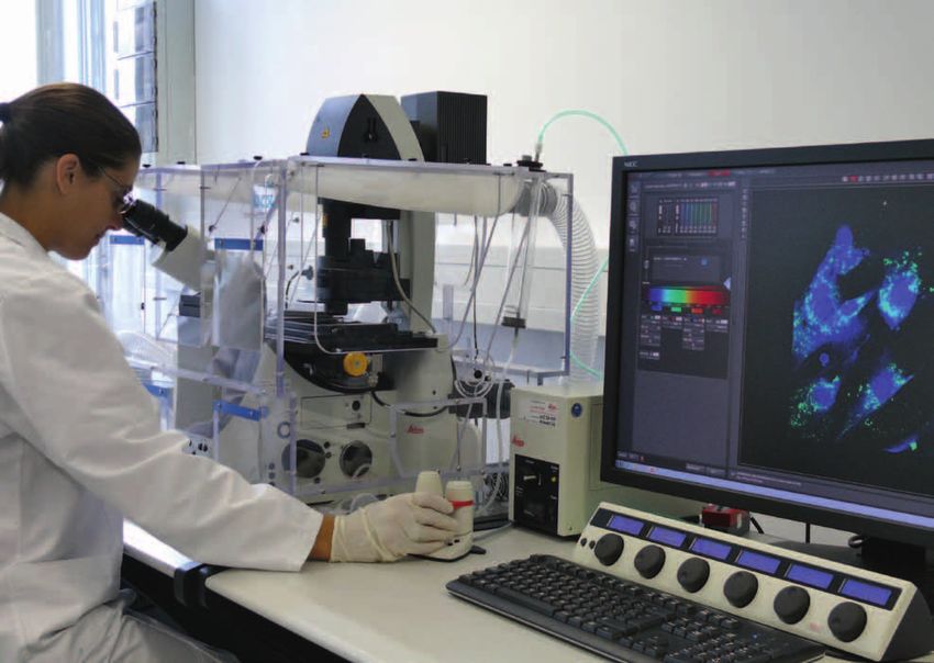
Sensitive Fluoreszenz-Mikrosko
pie und Live Cell Imaging mit geeignet für anspruchsvolle Sensitive fluorescence microscopy uous wave laser (405 nm) – suit
Hilfe des konfokalen Laserscan Proben und sehr hohe Aufnah and live cell imaging using the able for difficult samples and
ning-Mikroskops Leica TCS SP8 megeschwindigkeiten bei voller confocal laser scanning micro very high recording speeds at full
ausgestattet mit Weißlichtlaser konfokaler Auflösung scope Leica TCS SP8 equipped confocal resolution (Photo:
und Dauerstrichlaser (405 nm) – (Foto: Annette Maurer). with white light laser and contin Annette Maurer).
17Z e l l bi o l o g i e & A n g e w a n d t e V i r o l o g i e
Anfahrt St. Ingbert
How to find us in St. Ingbert
Mit dem Auto By car
Autobahn A 6/Ausfahrt St. Ingbert-West, links abbiegen in Autobahn A 6/exit St. Ingbert-West, turn left in the direction
Richtung Flughafen Saarbrücken-Ensheim, nach der Ampel of Saarbrücken-Ensheim Airport, after the traffic lights left in
links abbiegen in Richtung St. Ingbert-Süd (Ensheimer Straße), the direction of St. Ingbert-Süd (Ensheimer Strasse), straight
im Kreisverkehr geradeaus, nach ca. 1,5 km liegt das Institut through the roundabout, the Institute is on the left after
auf der linken Seite. around 1.5 km.
Autobahn A 1/bis Autobahnkreuz Saarbrücken, weiter Rich- Autobahn A 1/drive to Autobahn junction Saarbrücken, then
tung Karlsruhe/Mannheim auf der A 8 bis Autobahnkreuz continue in the direction of Karlsruhe/Mannheim on the A 8 to
Neunkirchen, weiter in Richtung Saarbrücken auf der A 6. Autobahn junction Neunkirchen, then in the direction of Saar-
Autobahn A 8/bis Autobahnkreuz Neunkirchen, weiter in Rich- brücken on the A 6.
tung Saarbrücken auf der A 6. Autobahn A 8/ drive to Autobahn junction Neunkirchen, then
Autobahn A 4/bis Autobahndreieck Saarbrücken, weiter in in the direction of Saarbrücken on the A 6.
Richtung Mannheim auf der A 6. Autobahn A 4/to Autobahn junction Saarbrücken, then in the
direction of Mannheim on the A 6.
Mit der Bahn By rail
Ab Saarbrücken Hauptbahnhof mit dem Taxi ca. 15 Minuten; From Saarbrücken-Hauptbahnhof (main station): either 15
mit dem Bahnbus oder mit dem Zug bis Bahnhof St. Ingbert, minutes by taxi, or first by bus or train to St. Ingbert station,
von dort mit dem Taxi ca. 1 Minute oder zu Fuß ca. 5 Minuten. then 1 minute by taxi or approx. 5 minutes on foot.
By air
Mit dem Flugzeug
5 to 10 minutes by taxi from Saarbrücken-Ensheim Airport.
Ab Flughafen Saarbrücken-Ensheim mit dem Taxi
5–10 Minuten.
18CELL B I OLOG Y & A P P L I ED V I ROLOG Y
Anfahrt Sulzbach
How to find us in Sulzbach
Mit dem Auto By car
Autobahn A 6 Richtung Saarbrücken, Ausfahrt St. Ingbert- Coming from the A 6 in the direction of Saarbrücken, exit
West, Hinweisschild: Richtung Sulzbach (ca. 6 km) folgen, vor St. Ingbert-West, follow the sign in the direction Sulzbach
Sulzbach Abfahrt »Industriegebiet Neuweiler« nehmen, dem (approximately 6 km); before Sulzbach take the exit "Indus-
Hinweisschild »Fraunhofer-Institut« folgend unter der Brücke triegebiet Neuweiler"; follow the sign "Fraunhofer-Institut",
durchfahren, erste Möglichkeit rechts, Hinweisschild »Fraun- drive under the bridge; take the first possible right, follow the
hofer-Institut«, nach 10 m rechts abbiegen, rechter Hand: sign "Fraunhofer-Institut"; turn right after 10 metres; on the
Einfahrt durch blaues Doppelflügeltor. right-hand side: entrance through the blue double gate.
19Fraunhofer-Institut für Biomedizinische Technik (IBMT) Fraunhofer Institute for Biomedical Engineering (IBMT) Ensheimer Straße 48 66386 St. Ingbert Germany Telefon: +49 (0) 6894/980-0 Fax: +49 (0) 6894/980-400 info@ibmt.fraunhofer.de www.ibmt.fraunhofer.de (deutsch/englisch) Leitung / Head of Institute Prof. Dr. Heiko Zimmermann heiko.zimmermann@ibmt.fraunhofer.de Prof. Dr. Günter R. Fuhr guenter.fuhr@ibmt.fraunhofer.de Presse- und Öffentlichkeitsarbeit / Redaktion Press and Public Relations / Editing Dipl.-Phys. Annette Eva Maurer Telefon: +49 (0) 6894/980-102 Fax: +49 (0) 6894/980-188 annette.maurer@ibmt.fraunhofer.de
Sie können auch lesen