30-year anniversary of ultrasound: Clinical use of 3D ultrasound in obstetrics and gynecology (1989-2019) 30-jähriges Ultraschalljubiläum: ...
←
→
Transkription von Seiteninhalten
Wenn Ihr Browser die Seite nicht korrekt rendert, bitte, lesen Sie den Inhalt der Seite unten
Editorial

      30-year anniversary of ultrasound: Clinical use of 3D ultrasound
      in obstetrics and gynecology (1989–2019)
      30-jähriges Ultraschalljubiläum: Klinischer Einsatz der 3D-Sonografie in Gynäkologie
      und Geburtshilfe (1989–2019)

                                                                        Correspondence
                                                                        Prof. Dr. Eberhard Merz
                                                                        Center for Ultrasound and Prenatal Medicine,
                                                                        Steinbacher Hohl 2 – 26, 60488 Frankfurt/Main, Germany

                                                                                                                                                                 This document was downloaded for personal use only. Unauthorized distribution is strictly prohibited.
                                                                        Tel.: ++ 49/69/76 01 35 79
                                                                        Fax: ++ 49/69/76 01 36 13
                                                                        ultraschallzentrum-frankfurt@web.de

                                                                        Prof. Dr. Rabih Chaoui
      Prof. Dr. Eberhard Merz                                           Center for Prenatal Diagnosis and Human Genetics,
                                          Prof. Dr. Rabih Chaoui
                                                                        Friedrichstrasse 147, 10117 Berlin, Germany
                                                                        chaoui@feindiagnostik.de

                                                                        Bibliography
                                                                        DOI https://doi.org/10.1055/a-0868-3760
                                                                        Published online: 2019
                                                                        Ultraschall in Med 2019; 40: 288–291
                                                                        © Georg Thieme Verlag KG, Stuttgart · New York
                                                                        ISSN 0172-4614

When the first commercial 3D ultrasound device (Combison 330,           [7, 11] not requiring the patient to be present, and loss-free
Kretztechnik, Zipf, Austria) became available in 1989 (▶ Fig. 1)        long-term storage of volumes [2, 11] represent such major advan-
[1], no one could have predicted just how important 3D ultra-           tages that experienced ultrasound examiners can’t imagine being
sound would become. While only the multiplanar mode with                without 3D ultrasound.
visualization of three orthogonal image planes was initially avail-        Of course, many diagnoses can also be made using 2D ultra-
able, various imaging modes have been developed in the last             sound. However, areas of interest can only be visualized on
three decades [2 – 11] allowing a wide range of imaging options         individual image planes. In contrast, 3D ultrasound offers not
in comparison to 2D ultrasound. Today, anatomical structures            only simultaneous visualization of multiple image planes as in CT
can be visualized with almost photographic quality (▶ Fig. 2a, b)       and MRI but also the demonstration of reconstructed image
[12 – 16]. The rapid advances in the field of computer technology       planes that cannot be visualized or can only be visualized less
and the design of special 3D probes have undoubtedly played an          effectively with conventional 2D ultrasound. Moreover, 3D ultra-
important role in this process [2, 7]. However, all of these devel-     sound allows the 3D display of structures that can be assessed
opments were only possible as a result of the vision of individual      more clearly in rotation mode and using movable light sources
ultrasound companies, the dedicated work of engineers, and the          [11]. 3D images are also easier for patients to understand than
ongoing collaboration between developers and users of 3D ultra-         individual sectional planes.
sound in the clinical routine.                                             3D ultrasound also has major advantages for ultrasound train-
    Nonetheless, the diagnostic potential of 3D/4D ultrasound has       ing [7, 11]. A volume containing a fetal abnormality or a tumor
still not yet been recognized in many countries and the method is       can be copied multiple times and loaded on numerous external
only used in prenatal imaging to produce nice images of fetal           workstations. As a result, multiple examiners can examine a
faces or as baby TV for parents-to-be [17, 18].                         volume with identical content at the same time.
    Of course, there is a learning curve involved in being able to         Finally, stored volumes are also suitable for data transfer via the
effectively utilize all that 3D ultrasound has to offer. Precise con-   Internet in order to obtain a second opinion from an expert.
trol of image planes, surface images of outer and inner surfaces,       Telemedicine is important particularly in countries without close
transparent images, 3D display of vessels in fetuses and tumors,        access to a specialized center.
the ability to perform virtual examination of stored volumes

288                                                                                 Merz E et al. 30-year anniversary of… Ultraschall in Med 2019; 40: 288–291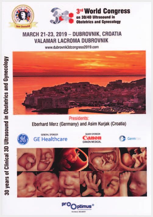
This document was downloaded for personal use only. Unauthorized distribution is strictly prohibited.
▶ Fig. 1 First commercially available 3D ultrasound system in 1989:
Combison 330 (Kretztechnik, Austria).
▶ Abb. 1 Erstes kommerziell erhältliches 3D-Ultraschallgerät, das
1989 auf den Markt kam: Combison 330 (Kretztechnik, Österreich).
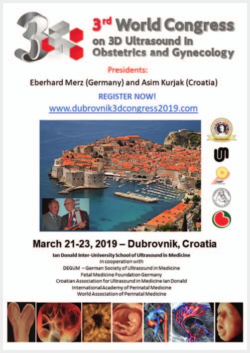
▶ Fig. 3 Announcement of the 3rd World Congress on 3D Ultra-
sound in Obstetrics and Gynecology.
▶ Abb. 3 Ankündigung des 3. Weltkongresses für 3D-Ultraschall in
Gynäkologie und Geburtshilfe.
to increase. 80 presentations by 42 speakers from around the world
provided the participants from more than 50 countries with a com-
prehensive overview of the current applications of 3D ultrasound in
prenatal and gynecological imaging as well as in breast imaging.
Cheaper ultrasound devices and faster matrix array probes as
▶ Fig. 2 Present quality of 3D images. a HDlive demonstration of a well as more intensive training are needed to allow increased use
fetal face at 35 weeks of gestation. b HDlive demonstration of a of 3D/4D ultrasound in the daily routine and to allow the tech-
cystic borderline tumor of the ovary with papillary projections
nique to be able to be utilized to its fullest potential. Moreover,
originating from the bottom of the inner wall of the cystic tumor.
standardization of image presentation [19] as well as of the no-
▶ Abb. 2 Heutige Qualität der 3D-Ultraschallbilder. a HDlive-Ober- menclature for the various imaging modes is needed [20]. Finally,
flächendemonstration eines fetalen Gesichts mit 35 Schwanger- the ability to exchange volumes between the different ultrasound
schaftswochen. b HDlive-Oberflächendemonstration eines companies would be highly advantageous.
zystischen Borderline-Tumors des Ovars mit papillären Wandstruk-
turen am Boden der Zystenwand.
30-jähriges Ultraschalljubiläum: Klinischer
All in all, 3D ultrasound currently offers so many advantages Einsatz der 3D-Sonografie in Gynäkologie
that, apart from the higher price of these ultrasound devices, it is
difficult to understand why it is often used only on a very basis. und Geburtshilfe (1989 – 2019)
The 3rd World Congress on 3D/4D Ultrasound in Gynecology and Als 1989 das erste kommerzielle 3D-Ultraschallgerät (Combi-
Obstetrics that was just held March 21 – 23, 2019 in Dubrovnik, Croa- son 330, Kretztechnik, Zipf, Österreich) auf den Markt kam
tia (▶ Fig. 3, 4) showed that interest in 3D/4D ultrasound continues (▶ Abb. 1) [1], konnte kaum jemand ahnen, dass sich die
Merz E et al. 30-year anniversary of… Ultraschall in Med 2019; 40: 288–291 289
Editorial

                                                                      nostik zur Herstellung netter Gesichtsbilder des Ungeborenen
                                                                      oder als Baby-TV für die werdenden Eltern verwendet [17, 18].
                                                                          Natürlich bedarf es einer Lernkurve, um das gesamte Potential
                                                                      der 3D-Sonografie ausnutzen zu können. Die exakte Kontrolle der
                                                                      Bildebenen, die Oberflächendarstellung von äußeren und inneren
                                                                      Oberflächen, die Transparenzdarstellungen, die räumliche Dar-
                                                                      stellung von Gefäßen beim Feten und bei Tumoren, wie auch die
                                                                      Möglichkeit der virtuellen Untersuchung von gespeicherten
                                                                      Volumina [7, 11], bei der die Patientin nicht anwesend sein muss,
                                                                      und die verlustfreie Langzeitspeicherung von Volumina [2, 11],
                                                                      stellen dabei so große Vorteile dar, dass der erfahrene Ultraschall-
                                                                      untersucher die 3D-Sonografie nicht mehr missen möchte.
                                                                          Natürlich können auch viele Diagnosen mit Hilfe der 2D-Sonogra-
                                                                      fie gestellt werden. Die Darstellung von Auffälligkeiten gelingt aller-

                                                                                                                                                               This document was downloaded for personal use only. Unauthorized distribution is strictly prohibited.
                                                                      dings immer nur in einzelnen Bildebenen. Die 3D-Sonografie bietet
                                                                      hingegen nicht nur die gleichzeitige Darstellung mehrerer Bildebe-
                                                                      nen wie beim CT und MRT, sondern auch die Demonstration von re-
                                                                      konstruierten Bildebenen, die sich mit der herkömmlichen 2D-Sono-
                                                                      grafie nicht oder weniger gut darstellen lassen. Hinzu kommt die
                                                                      räumliche Darstellung von Strukturen, die sich im Rotationsmodus
                                                                      wie auch in der Anwendung von beweglichen Lichtquellen [11] noch
                                                                      deutlicher beurteilen lassen. Räumliche Bilder sind auch für die Pa-
                                                                      tientinnen wesentlich besser verständlich als einzelne Schnittebenen.
                                                                          Für die sonografische Ausbildung bietet die 3D-Sonografie
                                                                      ebenfalls große Vorteile [7, 11]. Ein Volumen, in dem eine fetale
                                                                      Fehlbildung oder ein Tumor abgespeichert ist, kann mehrmals
                                                                      kopiert und an mehreren externen Workstations geladen werden.
                                                                      Damit können mehrere Untersucher ein Volumen mit identi-
                                                                      schem Inhalt gleichzeitig untersuchen.
                                                                          Letztlich bieten sich gespeicherte Volumina auch noch für den
   ▶ Fig. 4 Program cover of the 3rd World Congress on 3D/4D Ultra-
   sound in Obstetrics and Gynecology, March 21 – 23, 2019, Dubrov-   Datentransfer via Internet zu einem Zentrum an, um sich eine
   nik, Croatia.                                                      Zweitmeinung durch einen Experten einzuholen. Telemedizin ist
                                                                      vor allem in Ländern, in denen ein weiterführendes Zentrum in
   ▶ Abb. 4 Vorderseite des Programmheftes zum 3. Weltkongress        großer Entfernung liegt, von großer Bedeutung.
   für 3D/4D-Sonographie in Gynäkologie und Geburtshilfe, der vom         Alles in allem bietet die 3D-Sonografie heute so viele Vorteile,
   21. bis zum 23.3.2019 in Dubrovnik, Kroatien, stattfand.
                                                                      dass es – abgesehen vom höheren Preis dieser Ultraschallgeräte –
                                                                      kaum nachvollziehbar ist, dass diese Technik bei vielen nur in sehr
3D-Sonografie zu einem solchen Erfolgskonzept entwickeln              begrenztem Umfang zum Einsatz kommt.
würde. Während in der Anfangsphase nur der multiplanare Modus             Der 3. Weltkongress über 3D/4D-Ultraschall in Gynäkologie
mit der Abbildung der drei senkrecht aufeinander stehenden            und Geburtshilfe, der gerade vom 21. – 23.3.2019 in Dubrovnik,
Bildebenen zur Verfügung stand, konnten im Laufe der letzten          Kroatien, stattgefunden hat (▶ Abb. 3, 4), hat gezeigt, dass wei-
drei Jahrzehnte unterschiedliche Darstellungsmodi entwickelt          terhin ein wachsendes Interesse an der 3D/4D-Sonografie be-
werden [2 – 11], die im Vergleich zur 2D-Sonografie eine enorme       steht. Bei 80 Vorträgen von 42 Referenten aus der ganzen Welt
Abbildungsvielfalt ermöglichen und anatomische Strukturen             konnten sich die Teilnehmer aus mehr als 50 Ländern einen um-
heute in nahezu photografischer Abbildungsqualität zur Darstel-       fassenden Überblick über den aktuellen Einsatz der 3D-Sonografie
lung bringen (▶ Abb. 2a, b) [12 – 16]. Ohne Zweifel spielen dabei     in der pränatalen und gynäkologischen Diagnostik wie auch in der
auch die rasanten Fortschritte in der Computertechnologie und         Mammadiagnostik verschaffen.
die Konstruktion spezieller 3D-Schallköpfe eine wichtige Rolle [2,        Damit die 3D/4D-Sonografie in der täglichen Routine noch
7]. All diese Entwicklungen waren jedoch nur durch die Weitsicht      mehr angewandt wird und auch das breite Spektrum dieser Tech-
einzelner Ultraschallfirmen, dem engagierten Einsatz von Inge-        nik voll genutzt werden kann, bedarf es zukünftig nicht nur gün-
nieuren und der stetigen Interaktion zwischen Entwicklern und         stigerer Ultraschallgeräte und schneller Matrixsonden, sondern
3D-Anwendern im klinischen Alltag möglich.                            auch einer weiteren intensiven Ausbildung. Anzustreben ist
    Dennoch wird nach wie vor in vielen Ländern das diagnostische     außerdem neben der einheitlichen Standardisierung bei der Bild-
Potential, das in der 3D/4D-Sonografie steckt, bislang immer noch     darstellung [19] auch eine gemeinsame Nomenklatur für die
nicht erkannt und das Verfahren lediglich in der pränatalen Diag-     unterschiedlichen Darstellungsmodi [20]. Letztlich wäre auch
                                                                      die Austauschbarkeit von Volumina zwischen den verschiedenen
                                                                      Ultraschallfirmen von großem Nutzen.

290                                                                               Merz E et al. 30-year anniversary of… Ultraschall in Med 2019; 40: 288–291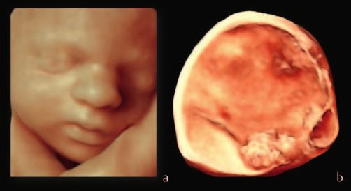
References                                                                      [11] Merz E, Kurjak A. 1989–2019: 30 Years of 3D Ultrasound in Obstetrics
                                                                                     and Gynecology. Donald School J Ultrasound Obstet Gynecol 2018; 12:
                                                                                     1–5
[1] Kirbach D, Whittingham TA. 3D ultrasound – the Kretztechnik Voluson®
                                                                                [12] Merz E, Pashaj S. Advantages of 3D ultrasound in the assessment of fetal
    approach. Eur J Ultrasound 1994; 1: 85 – 89
                                                                                     abnormalities. J Perinat Med 2017; 45: 643 – 650
[2] Merz E, Abramowicz JS. 3D/4D ultrasound in prenatal diagnosis: is it time
                                                                                [13] Pashaj S, Merz E. Detection of fetal corpus callosum abnormalities by
    for routine use? Clin Obstet Gynecol 2012; 55: 336 – 351
                                                                                     means of 3D ultrasound. Ultraschall in Med 2016; 37: 185 – 194
[3] Devore GR, Falkensammer P, Sklansky MS et al. Spatiotemporal image
                                                                                [14] Merz E. Physiological umbilical herniation—a distinctive sonographic
    correlation (STIC): new technology for evaluation of the fetal heart.
                                                                                     feature in the embryonic stage. Ultraschall in Med 2017; 38: 123 – 124
    Ultrasound Obstet Gynecol 2003; 22: 380 – 387
                                                                                [15] Merz E, Pashaj S. True or false umbilical cord knot? Differentiation via
[4] Chaoui R, Heling KS. New developments in fetal heart scanning: three-
                                                                                     3D/4D color Doppler ultrasound. Ultraschall in Med 2018; 39: 127 – 128
    and four-dimensional fetal echocardiography. Semin Fetal Neonatal Med
    2005; 10: 567 – 577                                                         [16] Salihagić-Kadić A, Kurjak A. Cognitive function of the fetus. Ultraschall in
                                                                                     Med 2018; 39: 181 – 189
[5] Benoit B, Chaoui R. Three-dimensional ultrasound with maximal mode
    rendering: a novel technique for the diagnosis of bilateral or unilateral   [17] EFSUMB Newsletter. Statement on the Use of Diagnostic Ultrasound for
                                                                                     Producing Souvenir Images or Recordings in Pregnancy. Ultraschall in

                                                                                                                                                                    This document was downloaded for personal use only. Unauthorized distribution is strictly prohibited.
    absence or hypoplasia of nasal bones in second-trimester screening for
    Down syndrome. Ultrasound Obstet Gynecol 2005; 25: 19 – 24                       Med 2007; 28: 100

[6] Kurjak A, Miskovic B, Stanojevic M et al. New scoring system for fetal      [18] Merz E. 3D/4D-Ultrasound in Obstetrics – Baby TV without Diagnostics?
    neurobehaviour assessed by three- and four-dimensional sonography.               Ultraschall in Med 2008; 29: 156 – 158
    J Perinat Med 2008; 36: 73 – 81                                             [19] Merz E, Benoit B, Blass HG et al. Standardization of three-dimensional
[7] Merz E. 25 years of 3D ultrasound in prenatal diagnosis (1989–2014).             images in obstetrics and gynecology: consensus statement. Ultrasound
    Ultraschall in Med 2015; 36: 2 – 8                                               Obstet Gynecol 2007; 29: 697 – 703

[8] Pooh RK. “See-through fashion” in prenatal diagnostic imaging. Donald       [20] Deng J. Nomenclature of three-dimensional and four-dimensional ultra-
    School J Ultrasound Obstet Gynecol 2015; 9: 111                                  sound in obstetrics, gynecology, and fetal echocardiography. In: Merz E,
                                                                                     Kurjak A, (eds) Donald School Textbook: Current status of clinical use of
[9] Hata T, AboEllail MA, Sajapala S et al. HDlive Silhouette mode with
                                                                                     3D/4D ultrasound in obstetrics and gynecology. New Delhi – London –
    spatiotemporal image correlation for assessment of the fetal heart.
                                                                                     Panama: Jaypee Brothers Medical Publishers; 2019: 61 – 74
    J Ultrasound Med 2016; 35: n1489 – 1495
[10] DeVore GR, Satou G, Sklansky M. 4D fetal echocardiography – an
     update. Echocardiography 2017; 34: 1788 – 1798

Merz E et al. 30-year anniversary of… Ultraschall in Med 2019; 40: 288–291                                                                                 291
Sie können auch lesen