Calbindin D28k-Immunoreactivity in Human Enteric Neurons
←
→
Transkription von Seiteninhalten
Wenn Ihr Browser die Seite nicht korrekt rendert, bitte, lesen Sie den Inhalt der Seite unten
Calbindin D28k-Immunoreactivity in Human
               Enteric Neurons
        Institut für Anatomie und Zellbiologie

            Der Medizinischen Fakultät
        der Friedrich-Alexander-Universität
                 Erlangen-Nürnberg
                         zur
       Erlangung des Doktorgrades Dr. med.
                   vorgelegt von
      Katharina Zetzmann aus Hildburghausen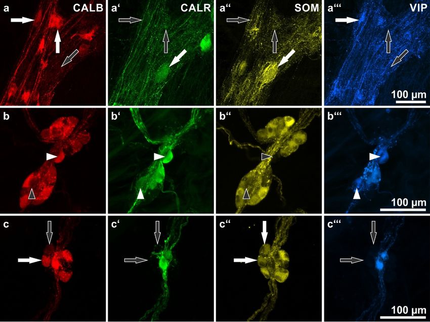
Als Dissertation genehmigt
von der Medizinischen Fakultät
der Friedrich-Alexander-Universität Erlangen-Nürnberg
Tag der mündlichen Prüfung: 29.06.2021

Vorsitzender des Promotionsorgans: Prof. Dr. med. Markus F.
Neurath

Gutachter/in:   Prof. Dr. Axel Brehmer
                Prof. Dr. Stefanie Kürten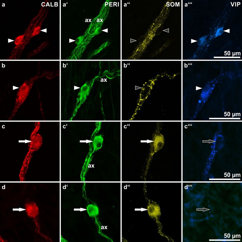
„Für meine Oma Elfi und meine Eltern“
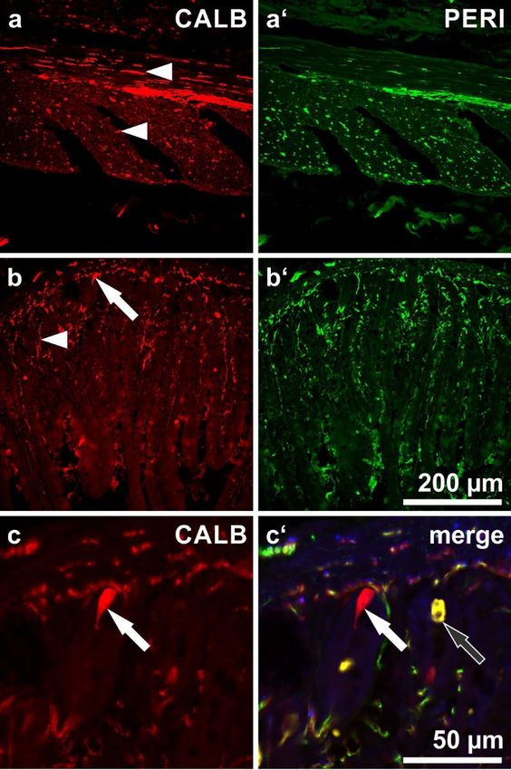
Table of content

1 Deutsche Zusammenfassung .......................................................................... 1

   1.1      Hintergrund und Ziele ............................................................................ 1

   1.2      Methoden .............................................................................................. 1

   1.3      Ergebnisse und Beobachtungen ........................................................... 2

   1.4      Schlussfolgerungen ............................................................................... 2

2 Einordnung in den fachwissenschaftlichen Kontext ......................................... 3

3 Original paper ................................................................................................ 13

   3.1      References .......................................................................................... 14

4 Attachement .................................................................................................. 17

   4.1 List of figures ........................................................................................... 17

   4.2 List of tables ............................................................................................ 18

5 List of publications ......................................................................................... 18

6 Acknowledgement ......................................................................................... 19

7 Curriculum vitae ............................................................................................ 19
1 Deutsche Zusammenfassung

1.1 Hintergrund und Ziele

Calbindin (CALB) ist ein calcium-bindendes Protein, das als
immunhistochemischer Marker für intrinsische primär-afferente Neuronen
(IPANs) im Meerschweinchendarm etabliert ist [5]. Studien im menschlichen
Enterischen Nervensystem zeigten, dass IPANs hier anders als im
Meerschweinchen chemisch kodiert sind [10], ein spezifisches Vorkommen in
anderen Neuronentypen konnte bislang nicht erbracht werden.

Ziel dieser Studie war eine quantitative Analyse calbindin-positiver Neuronen im
menschlichen Dünn- und Dickdarm sowie die Beantwortung der Frage, ob
Calbindin einer bestimmten Neuronenpopulation zugeordnet werden kann.
Dabei wurden mögliche Co-Lokalisationen mit weiteren Antikörpern gegen
Calretinin (CALR), Somatostatin (SOM) und Vasoaktives Intestinales Peptid
(VIP) berücksichtigt.

1.2 Methoden

Es wurden 26 Dick – und Dünndarmsegmente von Tumorpatient/innen
verwendet, die Zustimmung der Ethikkommission der Friedrich-Alexander-
Universität lag hierfür vor. Die Segmente wurden zu Präparaten des Plexus
myentericus und des Plexus submucosus verarbeitet („Häutchenpräparation“)
und vierfach immunhistochemisch gefärbt. Hierbei wurde ein „panneuronaler“
Marker antiHU C/D (HU) verwendet, um den Anteil von Teilpopulationen
bestimmen zu können. Markierte Ganglien in den Präparaten wurden mit einem
Konfokalen Fluoreszenzmikroskop digitalisiert, die quantitative Auswertung
erfolgte mithilfe der Software „Volocity“.

                                                                               1
1.3 Ergebnisse und Beobachtungen
CALB-positive Neuronen machten im Dünndarm 31% und im Dickdarm 25%
aller myenterischen Neuronen aus. In der Submukosa waren dies ca. 80% aller
Nervenzellen. Während hier CALB-positive Neuronen mehrheitlich (ca. 75%)
Co-Reaktivität mit VIP und CALR zeigten, war eine solche mit SOM nur
ausnahmsweise zu finden. Im Gegensatz dazu zeigten die myenterischen
CALB-positiven Neuronen keine Co-Reaktivität für die untersuchten Marker (HU
ausgenommen).
Co-Färbungen mit dem Strukturmarker Neurofilament (NF) zeigten, dass CALB-
Neuronen im Dünndarm oft eine Typ III-Morphologie zeigten: Neuronen dieses
Typs sind langdendritische uniaxonale Nervenzellen. Spezifisch ist dieser
Marker für Typ III-Zellen allerdings nicht, da das Protein auch in Nicht-Typ III-
Zellen gefunden wurde.
1.4 Schlussfolgerungen
CALB konnte immunhistochemisch in allen ganglionären Nervengeflechten des
menschlichen Dünn- und Dickdarms nachgewiesen werden. Ein besonders
hohes Vorkommen zeigte sich in den submukösen Plexus, was in Kontrast zu
früheren Studien steht [8].
Anders als im klassischen Modelltier des ENS, dem Meerschweinchen, ist
CALB im menschlichen Darm nicht als spezifischer Marker für „sensorische“
IPANs geeignet. Stattdessen kommt es in mehreren (sowohl myenterischen als
auch submukösen) Neuronenpopulationen vor, u.a. in den morphologisch
auffälligen Typ III-Neuronen des Dünndarms.
22 Einordnung in den fachwissenschaftlichen
Kontext
Zum Autonomen (Vegetativen, Viszeralen) Nervensystem des Menschen gehört
neben dem „Sympathikus“ und dem „Parasympathikus“ das Enterische
Nervensystem (ENS). Im Vergleich zum Somatischen Nervensystem
funktioniert das Vegetative Nervensystem weitgehend unwillkürlich (z.B. die
Innervation der glatten Muskulatur oder der endo – und exokrinen Drüsen) und
relativ autonom, jedoch nicht unabhängig vom Zentralen Nervensystem [45].
Das ENS stellt in diesem Zusammenhang eine Besonderheit dar. Schon der
Physiologe Langley beschrieb 1900 eine Aufteilung der autonomen Nerven in
sympathische, parasympathische und Eingeweidenerven [45]. Erst viel später,
manifestiert durch die erste (und vergleichsweise sehr späte) Monographie zum
ENS (Furness, Costa 1987), wurde diese Sicht bestätigt und das ENS als ein
System von Nervenzellen und Glia beschrieben, welches in den Wänden des
Gastrointestinaltraktes und der Gallenblase sowie in der Bauchspeicheldrüse
lokalisiert ist und in teils erheblichem Ausmaß autonom funktioniert [41].
Bemerkenswert sind z.B. die große Anzahl von enterischen Nervenzellen (ein
Mehrfaches der Neuronenzahl des Rückenmarks), deren funktionelle
Heterogenität sowie die Eigenschaften der enterischen Glia, die mehr der Glia
des Zentralen als der des restlichen Peripheren Nervensystem ähnelt [42].
Die Neuronen des ENS sind in intramuralen Nervengeflechten („Plexus“)
lokalisiert, dies sind mehretagige Nervennetze in den Wänden von Ösophagus-,
Magen- sowie Dünn- und Dickdarmwand. Ganglionäre Plexus enthalten die in
Gruppen („Ganglien“) beisammen liegenden Nervenzellkörper, welche durch
Bündel von langen Nervenzellfortsätzen („Axonen“) zu einem Geflecht
verbunden sind. Aganglionäre Plexus bestehen hauptsächlich aus Axonen ohne
regulär eingelagerte Nervenzellkörper, während enterische Gliazellen überall
das enterische Nervengewebe komplettieren. Beide Plexuskategorien
durchsetzen alle Schichten des Rumpfdarms (vom oberen Ösophagussphinkter
bis zum inneren Analsphinkter).
3Diese auch „ganglionäre Plexus“ genannten Strukturen sind die in der
Submukosa lokalisierten Plexus submucosus internus (Meissner) und externus
(Schabadasch) sowie der zwischen Längs- und Ringmuskulatur gelegene
Plexus myentericus (Auerbach).
Die erste Beschreibung des Plexus submucosus erfolgte durch Meissner
(1857). Er bezeichnete die Submukosa als „eine nervenreiche Schicht, die die
Muskelhaut des Darms an die Schleimhaut heftet“ [46]. Zuerst unterteilt wurde
der Plexus submucosus durch Schabadasch (1930), der beim Affen eine
Unterscheidung in zwei topografisch und strukturell unterschiedliche Geflechte
beschrieb [47]. Stach definierte den Plexus submucosus internus (Meissner)
und den Plexus submucosus externus (Schabadasch) [49] im
Schweinedünndarm. In dieser Spezies beherbergen beide submuköse
Geflechte qualitativ verschiedene Populationen von Nervenzellen, also
morphologisch, immunhistochemisch und auch elektrophysiologisch
unterscheidbare Nervenzellen. Obwohl auch in der menschlichen Submukosa
von Dünn- und Dickdarm eine topografische Trennung in ein internes
(Meissner-) und ein externes (Schabadasch-) Teilgeflecht bestätigt wurde, sind
hier (mit nur quantitativen Differenzen in der neuronalen Zusammensetzung)
wahrscheinlich beide für die Regulation von Schleimhautprozessen zuständig,
der äußere enthält womöglich zusätzlich auch Nervenzellen für die
Ringmuskulatur [12-15].
Der Plexus myentericus wurde erstmalig durch Auerbach (1862) beschrieben.
Dieser liegt zwischen Ring– und Längsmuskelschicht und ist v.a. für die
Peristaltik zuständig [32].
Aganglionäre Plexus sind regulär frei von Nervenzellkörpern und finden sich in
verschiedenen Gewebsschichten. Von luminal beginnend finden sich der
besonders nervenfaserreiche Plexus mucosus, weiter der Plexus muscularis
mucosae, der Plexus submucosus extremus (als äußerster submuköser
Plexus), die Plexus der Ring- und der Längsmuskulatur, die in der jeweiligen
Muskelfaserverlaufsrichtung angelegt sind, sowie der Plexus subserosus
unterhalb der Serosa [34].
4Die erheblich autonome Funktionsweise des Magen-Darm-Traktes beschrieben
bereits Bayliss und Starling (1899). Sie wiesen einen intrinsischen
Reflexmechanismus nach, der Darminhalt durch „Peristaltik“ von oral nach anal
transportieren kann: Darminhalt löst lokal eine Kontraktion der glatten
Muskulatur unmittelbar oral des Bolus aus sowie unmittelbar aboral (=anal)
davon Dilatation. Sympathikus und Parasympathikus können bremsend oder
(die Muskeltätigkeit) anregend einwirken, sind für den Grundreflex in Dünn- und
Dickdarm jedoch nicht zuständig [33]. Aufgrund seines komplexen
Reflexgeschehens, das auch die Steuerung muköser Funktionen einschließt,
wird das ENS auch als „Kleines Gehirn“ oder „Bauchgehirn“ bezeichnet [59].
Verschiedene Teilpopulationen enterischer Neuronen sind die
Hauptbestandteile dieses Reflexgeschehens.
Zur Identifizierung und Charakterisierung unterschiedlicher
Neuronenpopulationen steht ein vielfältiges Methodenspektrum zur Verfügung,
das sich auch gegenwärtig stetig erweitert. Entscheidend im hier behandelten
Zusammenhang sind dafür zwei morphologische Kriterien, die jeweils
unterschiedliche methodische Ansätze zu ihrer Analyse erfordern: die
dendritische Architektur der Neuronen als deren Hauptrezeptorregion für
eintreffende Signale [17;18;56] sowie die axonale Projektion, die man mittels
Tracing-Techniken zur Klärung von Zielzellen und -geweben (als Empfänger
der neuronalen Signale) bestimmenen kann [43;58]. Die chemische Kodierung
der Zellkörper (als Stoffwechselzentrum der Zelle auch zuständig für die
Produktion neuroaktiver Substanzen) ist in methodischer Kombination mit
jeweils einer der beiden vorgenannten Methoden und Kriterien das Bindeglied
(s.u.) für die Gesamtbeurteilung eines Neuronentyps und der Zuordnung zu
einer oder mehrerer Funktionen im Gesamtnetzwerk des ENS.
Die morphologische Beurteilung dendritischer Strukturen wurde durch den
russischen Histologen Alexander S. Dogiel (1899) begründet. Die schon von
ihm angewandte Häutchenpräparation ermöglicht durch Aufsicht auf die
gefärbten Ganglien (v.a. aus dem Meerschweinchendarm) die Beurteilung von
Länge und Form der Fortsätze unversehrter, nicht durch histologische Schnitte
zerteilter Nervenzellen [34].
5Dogiel‘s Typ I Neuronen haben kurze lamelläre Dendriten, im Gegensatz dazu
weisen Typ II Neuronen mehrere lange, jedoch keine kurzen Fortsätze auf. Da
Dogiel ein Anhänger der seinerzeit vielfach vertretenen Synzytiumtheorie des
Nervensystems war, führte seine Unterteilung in „kurze und lange“ Fortsätze
nicht zwangsläufig zur heutigen Vorstellung von „Dendriten und Axonen“ (siehe
hierzu im Gegensatz die Neuronentheorie des Nervensystems, faktisch von
Ramon y Cajal begründet [17]). Nur unklar wurden von Dogiel daher seine Typ
III Neuronen mit langen, dünnen Dendriten von Typ II Neuronen unterschieden
und deshalb für Jahrzehnte vergessen [17]. Heute scheint klar, dass Typ III-
Neuronen in verschiedenen Spezies verschiedene Funktionen ausfüllen (s.u.).
Die Erweiterung der morphologischen (Dogiel`schen) Klassifikation erfolgte
durch Stach in den 1980er Jahren durch Analyse von Neuronen in versilberten
Häutchen-(Plexus-)Präparaten aus dem Dünndarm des Schweins. Er
berücksichtigte dabei die Kombination zweier unabhängiger morphologischer
Parameter, die sich in den versilberten Häutchenpräparaten durch eindeutige
histologische Unterscheidung von Dendriten und Axonen der Nervenzellen
bestimmen ließen: der Architektur der kurzen Fortsätze („Dendriten“) und der
Projektionsrichtung der langen Fortsätze („Axone“) der Neuronen.
Typ I Neuronen im Schweinedarm sind multidendritisch und uniaxonal [51]. Die
Dendriten sind typisch lamellär endend, radiär angeordnet und kurz, die Axone
verlaufen größtenteils oralwärts („aszendierend“). Funktionell wurden Typ I
Neuronen dabei als Interneurone oder Motorneurone angesehen, sie sind
cholinerg und enthalten meist Enkephalin [44].
Typ II Neuronen sind adendritisch und multiaxonal. Die Axone verlaufen dabei
sowohl vertikal, also zwischen den Plexus myentericus und submucosus, als
auch zirkulär in der Ebene des Plexus myentericus [52]. Im Schweinedünndarm
enthalten sie Calcitonin Gene Related Peptide (CGRP) [38;48;57]. Aufgrund
von funktionellen Untersuchungen konnten multiaxonaler Charakter und
Projektionsrichtung im Meerschweinschendarm bestätigt werden, außerdem
werden sie (in allen bislang untersuchten Spezies) als IPANs angesehen [5].
Furness et al. (1990) konnten weiterhin im Meerschweinchendünndarm eine
Immunreaktivität der myenterischen Typ II Neuronen mit CALB nachweisen [5].
6Dieser Befund war ein grundlegender Ansatzpunkt zur Erstellung der
vorliegenden Arbeit. Für uns hat sich die Frage gestellt, ob CALB im
menschlichen Darm ebenfalls als Marker für einen bestimmten Neuronentyp
nutzbar ist (s.u.).
Typ III Neuronen zeigten sich im Schweinedünndarm uniaxonal und
multidendritisch [53]. Sie haben im Gegensatz zu Typ I-Neuronen lange,
schlanke, verzweigte, spitz endende Dendriten und unterschieden sich auch in
der Projektionsrichtung vom Typ I: im Gegensatz zu diesen projizierten Typ III
Neuronen nach anal. Im Schweinedünndarm wurden Typ III Neuronen in allen
Abschnitten in abnehmender Häufigkeit nach anal gefunden. Eine chemische
Kodierung ließ sich als nitrerg und serotoninerg (Serotonin, 5-HT)
charakterisieren [17].
Typ IV Neuronen des Schweinedünndarms zeigten einen exzentrisch
gelegenen Zellkern, kurze, spitz zulaufende Dendriten und – zur Abgrenzung
von den anderen Typen entscheidend - Axone, die ausnahmslos vertikal, über
die Submukosa zur Schleimhaut projizieren [35;50;54]. Sie sind cholinerg und
enthalten außerdem SOM [17].
Typ V Neurone zeigten sich ebenfalls multidendritisch und uniaxonal, sie treten
als Einzelzellen und in Aggregaten mit spezifischen Dendritenbündeln auf. Sie
sind immunhistochemisch durch die Co-Lokalisation von SOM und CGRP
gekennzeichnet (Brehmer 2002b: im Gegensatz zu Typ II: nur CGRP und im
Gegensatz zu Typ IV: nur SOM) und repräsentieren wahrscheinlich
deszendierende (nach anal projizierende) Interneuronen [39;55].
Als morphologisches Kennzeichen der Typ VI Neurone gelten feine, spitze sog.
„Axondendriten“, abgehend vom Axonursprung. Sie sind cholinerg, ihre
Projektionsrichtung ist größtenteils analwärts gerichtet. Sie sind, im Gegensatz
zu den anderen Typen, in hypertrophiertem Schweinedarm durch eine
ausgeprägte Soma- und Dendritenhypertrophie aufgefallen, was eine prä-
motorische (interneuronale) oder motorische Funktion nahelegt [34;36;37]. Sie
enthalten die periphere Variante der Cholin Acetyl-Transferase und gleichzeitig
der Stickstoffmonoxid-Synthase (NOS) [40].
7Stach et al. beschrieb 2000 einen weiteren speziellen Typ VII Neuronentyp, der
sich vorwiegend im Duodenum und im proximalen Jejunum auffinden ließ.
Diese Neuronentypen wurden im Hund- und Schweinedarm gefunden. Die
Neuronen projizierten vorwiegend nach anal und zeigten chemische
Kodierungen für VIP, CALB und CALR [19].
Die mit verschiedenen neurowissenschaftlichen Methoden gewonnenen
Kenntnisse über Strukturen und Funktionen des Enterischen Nervensystems in
verschiedenen Tiermodellen (v.a. Meerschweinchen, auch Schwein, Ratte
Maus) können aufgrund von teils signifikanten Speziesdifferenzen nicht
unmittelbar in der menschlichen Neurogastroenterologie genutzt werden. Für
die morphologische Identifizierung und Charakterisierung enterischer
Neuronenpopulationen im Menschen stehen zwei prinzipielle Methoden zur
Verfügung. Die ältere (seit Dogiel 1899) färbt mit unterschiedlichen
histologischen und neuerdings immunhistochemischen Techniken Zellkörper
und Fortsätze an. Hinzu kommen die seit den 1980/90er Jahren etablierten
Tracing-Techniken, die die axonalen Zielstrukturen identifizieren können. Da
beide jeweils mit der immunhistochemischen Markierung des Zellkörpers
kombiniert werden können, sind mit letzterer (der sog. chemischen Kodierung)
Aussagen über somanahe Fortsatzmorphologie und Ziel der axonalen
Projektion möglich.
Dabei zeigen sich grundsätzliche Übereinstimmungen mit den an Tiermodellen
erzielten Ergebnissen (z.B. die in allen Spezies übereinstimmende Morphologie
sog. intrinsischer primär-afferenter Neuronen – IPANs, die von Dogiel 1899
erstbeschrieben und deren multiaxonale Fortsatzstruktur von Stach 1981
erstcharakterisiert wurde) genauso wie Unterschiede (z.B. in der chemischen
Kodierung der o.g. IPANs).
Im menschlichen Plexus myentericus sind darüber hinaus zwei verschiedene,
vormals für (Dogiel-) Typ I-Neuronen gehaltene Populationen zu unterscheiden:
„Spiny“ Typ I Neuronen haben kurze, spitze Dendriten. Sie projizieren zumeist
nach anal und enthalten die neuronale-NOS und VIP [17] und sind
deszendierende Motor-, teils auch Interneuronen.
8„Stubby“ Typ I Neuronen haben kurze, aber plumpe Dendriten. Sie projizieren
(im Dünndarm) nach oral, sind cholinerg und enthalten teils zusätzlich
Enkephalin [31]. Sie sind wahrscheinlich aszendierende Inter- und/oder
Motoneuronen [17].
Die sog. „Dogiel-Typ-II-Neuronen“ sind insofern schon morphologisch auffällig,
als dass sie keine Dendriten, jedoch mehrere Axone besitzen (sog.
adendritische, multiaxonale Neuronen). Der Anteil dieser Typ II-Neuronen, die
beim Meerschweinchen als „sensorisch“ (IPANs) charakterisiert wurden und die
diese Funktion sehr wahrscheinlich auch beim Menschen ausfüllen, konnte im
menschlichen Dünndarm auf ca. 10% aller myenterischen Neuronen bestimmt
werden [10, 11].
Zu Typ III-Neuronen konnten in vorliegender Studie neue Befunde erhoben
werden (s.u.).
Menschliche Typ V Neuronen sind cholinerg und im Gegensatz zum
Schweinedarm, wo sie eher im Ileum gehäuft auftreten, eher im oberen
Dünndarm zu finden [17]. Über diese ebenfalls morphologisch auffälligen
Nervenzellen ist bislang wenig bekannt.
Von den oben beschriebenen myenterischen Neuronentypen sowohl
morphologisch als auch immunhistochemisch (und somit funktionell) zu
unterscheiden sind die bislang beschriebenen submukösen Neuronentypen, die
(nahezu) alle cholinerg sind. Submuköse adendritische Neuronen enthalten
zusätzlich Somatostatin, während die submukösen dendritischen Neuronen mit
Antikörpern gegen Vasoaktives Intestinales Peptid und (mehrheitlich) gegen
Calretinin färbbar sind. Axone mit diesen jeweiligen spezifischen Kodierungen
konnten ausschließlich in der Mukosa nachgewiesen werden, d.h., beide
Populationen sind muköse Effektorneuronen. Im Plexus submucosus externus
(Schabadasch) des Menschen konnte zusätzlich eine kleine, inkonstante
Population nitrerger Neuronen identifiziert werden, die möglicherweise in die
Ringmuskulatur projiziert [15]. Etwa 20% der menschlichen submukösen
Neuronen sind bislang weder morphologisch noch immunhistochemisch
charakterisiert.
9Wichtige Unterschiede zwischen oben beschriebenen Neuronentypen des
Menschen zeigten sich auch in pathologischer Hinsicht. Bei der v.a. in
ländlichen lateinamerikanischen Regionen endemische Chagas-Krankheit
bilden etwa 20% der chronisch Erkrankten ein „Chagas-Megakolon“ aus. Diese
durch den Verlust jeglicher Motorik bedingte chronische Erweiterung des
Kolons ist nach Untersuchungen der 1960er bis -70er Jahre durch
weitgehenden Verlust (myenterischer) Nervenzellen bedingt. Erneute Analysen
des Neuronenbesatzes betroffener Darmsegmente zeigten allerdings, dass (1)
im Plexus myentericus v.a. nitrerge Neuronen – v.a. in den Übergangszonen
zum chronisch dilatierten Darmsegment – überleben und, mehr noch, dass (2)
die VIP-positiven submukösen Neuronen gleichfalls überleben. Da für letztere
v.a. eine auf den Erhalt der mukösen Barriere der Kolon-Schleimhaut gerichtete
Funktion nachgewiesen werden konnte, ist erklärbar, warum Patienten mit
(abschnittsweisem) myenterischem Neuronenverlust und kompletter Lähmung
der Kolonmuskulatur Jahrzehnte überleben können – die (weitgehend)
erhaltene Barrierenfunktion der Kolonschleimhaut ermöglicht dies [3]. Es zeigte
sich im Rahmen dieser Studien zum einen, dass mit den pathologischen
Veränderungen des Enterischen Nervensystems (dem praktisch kompletten
Verlust nicht-nitrerger und nicht-VIPerger Neuronen) allein das Chagas-
Megakolon nicht erklärbar war, hierfür sind auch Schädigungen an der
Muskulatur selbst und deren Schrittmacherzellen (Interstitielle Cajal-Zellen) zu
berücksichtigen. Zum anderen ließ sich erklären, dass das „Chagas-
Megakolon“ einer völlig anderen Pathogenese unterliegt als das hierzulande
deutlich bekanntere „Hirschsprung-Megakolon“ [3].
Nach ihrer Stellung im neuronalen Netzwerk des ENS unterscheidet man drei
Gruppen von Neuronentypen: Die intrinsischen primär-afferenten Neuronen
(IPANs), mehrere Populationen von Interneuronen sowie die verschiedene
Zielzellen innervierenden Effektorneuronen.
IPANs nehmen Reize direkt oder indirekt, durch Zwischenschaltung anderer
Zelltypen, Veränderungen der Umgebung wahr (z.B. Dehnung der Darmwand
oder chemische Reize) und leiten Nervensignale an Inter- und direkt an
Effektorneuronen weiter. IPANS entsprechen dem morphologischen Typ II, sie
sind adendritisch und multiaxonal [5].
10Interneuronen können sowohl erregend als auch hemmend auf
Effektorneuronen wirken.
Letztere aktivieren oder hemmen wiederum nicht-nervale Zellen der glatten
Muskulatur, der Epithelien und Drüsen, der Blutgefäße und des Immunsystems.
Die Aktivierung bzw. Hemmung der effektorischen Neurone oder Motorneurone
erfolgt über Transmitter, wie beispielsweise Acetylcholin oder Substanz P (SP),
welche aktivierend auf muskarinerge Rezeptoren der glatten Muskelzellen
wirken. Als hemmende Transmitter sind Stickstoffmonoxid (NO) und VIP
beschrieben [34,59].
Das Ziel dieser Studie war zunächst die Darstellung der Verteilung der
Neuronen, die sich in ihrer chemischen Codierung für CALB in allen Dünn – und
Dickdarmabschnitten in Zusammenhang mit den drei Plexus des enterischen
Nervensystem positiv zeigten. Dafür wurde der Anteil der CALB-positiven
Neuronen in Relation zur gesamten Neuronenpopulation, welche durch den
Antikörper anti-HU C/D (HU) markiert wurden, bestimmt. Zusätzlich wurden
neuronale Co-Lokalisationen von CALB mit den Antikörpern für CALR, VIP und
SOM in allen Darmabschnitten untersucht. Weiterhin sollte die Frage
beantwortet werden, ob CALB als Marker für bestimmte Neuronentypen genutzt
werden kann.
Auch bei unserer Häutchenpräparation verblieb der Plexus myentericus auf der
Längsmuskulatur und der darunterliegenden Serosa haften. An diesen
(ungeschnittenen) Präparaten erfolgte die immunhistochemische 4-fach-
Inkubation mit den verschiedenen Antikörpern. Die Digitalisierung der gefärbten
Ganglien am konfokalen Fluoreszenzmikroskop ging der Quantifizierung der
Zellpopulationen (mithilfe des Programms „Volocity“) voraus.
Im Meerschweinchendünndarm wurde nachgewiesen, dass Typ II-Neuronen
den „sensorischen“ IPANs entsprechen sowie durch CALB- Antikörper
spezifisch anfärbbar sind. Im Schweinedünndarm konnten Typ II-Neuronen
(mutmaßliche IPANs) spezifisch durch CGRP markiert werden. Menschliche
myenterische Typ II-Neuronen dagegen zeigten sich immunreaktiv für CALR,
SOM und Substanz P. Daraus leitete sich die Frage ab, ob CALB im
11menschlichen ENS andere Neuronentypen anfärbt, wenn ja, ob womöglich
spezifisch (s.u.)?
Brehmer et al. (2004) konnten den Nachweis einer Co-Lokalisation von
Calretinin und Somatostatin im menschlichen, myenterischen Typ II Neuronen
finden [10]. Kustermann et al. (2011) fanden dagegen, dass beide Substanzen
in den submukösen Plexus morphologisch deutlich unterschiedliche
Neuronentypen markierten [13]. Beyer et al. (2013) zeigten, dass fast alle
submukösen Neuronen Acetycholintransferase-positiv waren (anders als im
Plexus myentericus) und, dass Somatostatin meist mit Substanz P kolokalisiert
war [14]. Beuscher et al. (2014) wiesen außerdem eine häufige Co-Färbung
CALR-positiver Neuronen mit vasoaktiven intestinalen Peptid (VIP) nach.
Aufgrund dieser Voruntersuchungen zur Charakterisierung morphologischer
Eigenschaften und der daraus resultierenden Auswahl von Antikörpern zur
immunhistochemischen Färbung der Neuronen galt unser Hauptaugenmerk
dem Verteilungsmuster des kalziumbindenden Proteins Calbindin D28k.
Neben der Beschreibung und Markierung der Typ II Neuronen im
Meerschweinchenmodell gibt es zahlreiche Befunde über das Vorkommen von
Calbindin D28k im zentralen Nervensystem des Menschen und in anderen
Spezies. Im Gegensatz dazu gab es bisher lediglich Hinweise zum Vorkommen
von Calbindin im menschlichen ENS, jedoch nicht zum spezifischen
Verteilungsmuster auf bestimmte Neuronenpopulationen. Somit untersuchten
wir auch in Hinsicht auf sein Potenzial als Marker spezifischer Neuronenytpen
seine Lokalisation in allen Unterregionen und Geflechten der menschlichen
Dünn- und Dickdarmsegmente.
123 Original paper
13International Journal of
Molecular Sciences
Article
Calbindin D28k-Immunoreactivity in Human
Enteric Neurons
Katharina Zetzmann 1 , Johanna Strehl 2 , Carol Geppert 2 , Stefanie Kuerten 1 ID
, Samir Jabari 1
and Axel Brehmer 1, *
1 Institute of Anatomy and Cell Biology, University of Erlangen-Nuremberg, Krankenhausstraße 9,
D-91054 Erlangen, Germany; katharina.zetzmann@gmx.de (K.Z.); stefanie.kuerten@fau.de (S.K.);
samir.jabari@fau.de (S.J.)
2 Institute of Pathology, University of Erlangen-Nuremberg, Krankenhausstraße 8-10, D-91054 Erlangen,
Germany; johanna.strehl@uk-erlangen.de (J.S.); carol.geppert@uk-erlangen.de (C.G.)
* Correspondence: axel.brehmer@fau.de; Tel.: +49-9131/852-2831
Received: 18 December 2017; Accepted: 4 January 2018; Published: 8 January 2018
Abstract: Calbindin (CALB) is well established as immunohistochemical marker for intrinsic primary
afferent neurons in the guinea pig gut. Its expression by numerous human enteric neurons has
been demonstrated but little is known about particular types of neurons immunoreactive for CALB.
Here we investigated small and large intestinal wholemount sets of 26 tumor patients in order to
evaluate (1) the proportion of CALB+ neurons in the total neuron population, (2) the colocalization
of CALB with calretinin (CALR), somatostatin (SOM) and vasoactive intestinal peptide (VIP) and
(3) the morphology of CALB+ neurons. CALB+ neurons represented a minority of myenteric neurons
(small intestine: 31%; large intestine: 25%) and the majority of submucosal neurons (between 72
and 95%). In the submucosa, most CALB+ neurons co-stained for CALR and VIP (between 69 and
80%) or for SOM (between 20 and 3%). In the myenteric plexus, 85% of CALB+ neurons did not
co-stain with the other markers investigated. An unequivocal correlation between CALB reactivity
and neuronal morphology was found for myenteric type III neurons in the small intestine: uniaxonal
neurons with long, slender and branched dendrites were generally positive for CALB. Since also
other neurons displayed occasional CALB reactivity, this protein is not suited as an exclusive marker
for type III neurons.
Keywords: calcium binding protein; calretinin; enteric nervous system; morphology; myenteric
plexus; submucosal plexus
1. Introduction
The combined application of various neuroscientific methods has enabled the identification
and characterization of different types of enteric neurons primarily in the guinea pig [1,2]. One of
these methods was the immunohistochemical distinction between several enteric neuron types by
different markers that deciphered their chemical codes. The value of these chemical codes consists
of the relatively simple possibility of representing neuron types in gut tissue samples, e.g., under
experimental or pathological conditions. This allows us to draw conclusions about possible selective
changes in neuron populations, i.e., a neurohistopathological diagnosis, by discriminating between
enteric neuron types [3].
In enteric neuroscience, calbindin (CALB) is a “famous” immunomarker since antibodies against
this calcium binding protein selectively label most morphological type II neurons, the intrinsic primary
afferent neurons in the guinea-pig small intestine (IPANs) [4–7].
Although CALB is also expressed by a substantial number of human enteric neurons (counted in
the duodenum [8]), immunohistochemical staining of CALB succeeded only in a few myenteric type II
Int. J. Mol. Sci. 2018, 19, 194; doi:10.3390/ijms19010194 www.mdpi.com/journal/ijmsInt. J. Mol. Sci. 2018, 19, 194 2 of 14
neurons [9]. We have shown that human myenteric type II neurons, the putative myenteric IPANs, are
immunohistochemically characterized by the colocalization of calretinin (CALR) with somatostatin
(SOM) [10,11].
The human enteric nervous system consists of three ganglionated (see below) and several
non-ganglionated plexus (e.g., in the muscle layers, the mucosa, etc. [12]). The ganglionated myenteric
plexus lies between the circular and longitudinal muscle layers. Human submucosal neurons are
located within two ganglionated subplexus [12]. The external (or outer) submucosal plexus (ESP) is
located under the inner border of the circular muscle layer and is mostly monolayered. The internal (or
inner) submucosal plexus (ISP) occupies the inner half of the submucosa and is frequently two- or even
three-layered [3,12]. As to the distribution patterns of neuron types within both plexus there are, as far
as we know, only quantitative differences between them. Two larger submucosal neuronal populations
are known to date [13]. One is non-dendritic, (pseudo-) uniaxonal and immunoreactive for SOM and,
partly, substance P [14]. The other one displays a multidendritic appearance and is immunoreactive
for vasoactive intestinal peptide (VIP) and, partly, CALR [15]. Both submucosal neuron types are
cholinergic and project into the mucosa. Additionally, there are some submucosal nitrergic neurons,
which are, however, occasionally absent [15].
Thus, immunohistochemistry for the calcium binding protein CALR has been proven to be
useful for the identification of particular human enteric neuron types: in the myenteric plexus (MP),
in colocalization with SOM, it labels morphological type II neurons, the putative IPANs; in the
submucosal plexus it is frequently colocalized with VIP and a marker for multidendritic neurons,
which are putative mucosal effector neurons (see above). Colocalization of CALR and VIP in these
neurons was almost complete in the colon but not in the small intestine [15].
The aim of this study was to address the question whether another calcium binding protein,
namely CALB, may also be a useful immunohistochemical marker for identifying a particular human
enteric neuron type. To this end, we quantified the proportion of CALB+ neurons in relation to the
putative total enteric neuronal population throughout all small intestinal and colonic subregions,
which was stained by the pan-neuronal marker HU C/D (HU) [16]. Furthermore, we estimated the
colocalization rates of CALB+ neurons with CALR, VIP and SOM: in the human MP colocalization of
CALR and SOM label the putative IPANs [10,11]; in the human submucosal plexus VIP and SOM label
the two larger different neuron populations known so far [14,15]. Finally, we analyzed the morphology
of CALB+ neurons by co-staining with neurofilament (NF) in the myenteric plexus [7,17] and by
peripherin (PERI) in the two submucosal plexus [12,13].
2. Results
2.1. Wholemounts Stained for HU C/D (HU) and Calbindin (CALB)
This staining combination allows for an estimation of the proportion of CALB+ neurons in relation
to the whole enteric neuron population (Figure 1, Table 1).
Overall, in both the small intestine and the colon, CALB+ neurons represented a minority in the
MP and the majority in the two submucosal plexus. The intensity of fluorescence labeling varied
considerably, some neuronal cell bodies were intensely fluorescent but others displayed weak labeling.
In the MP of the small intestine, the proportions varied between 25.1 and 36.7% (total 31.1%).
In the colon, the values ranged between 21.1 and 30.5% (total 25.2%).
In the ESP and ISP of the small intestine, the proportions ranged between 56.8 and 86.3% (total
ESP: 81%; ISP: 72.3%). In the colon, all values were higher than 90%, 93.1% in the ESP and 95% in
the ISP.Int. J. Mol. Sci. 2018, 19, 194 3 of 14
Int. J. Mol. Sci. 2018, 19, 194 3 of 14
Figure 1. Human enteric ganglia double immunostaining for HU C/D (HU; grey) and calbindin
Figure 1. Human enteric ganglia double immunostaining for HU C/D (HU; grey) and calbindin
(CALB; red): (a,a’) myenteric ganglion, (b,b’) external submucosal ganglion, (c,c’) internal
(CALB; red): (a,a’)
submucosal myenteric
ganglia. Pairs ganglion, (b,b’)point
of filled arrows external submucosalneurons
at representative ganglion, (c,c’) for
positive internal submucosal
both HU and
ganglia.
CALB; Pairs
pairsof
of filled arrows
arrowheads point
(filled at representative
or empty, respectively) neurons
point at HUpositive for− both
+ but CALB HU(Patients
neurons. and CALB;
pairsdata:
of arrowheads + but CALB− neurons. (Patients data:
(a) 70 years,(filled
ileum, or empty,
male; (b,c) respectively) point
70 years, sigmoid at HU
colon, female).
(a) 70 years, ileum, male; (b,c) 70 years, sigmoid colon, female).
Table 1. Mean values ± standard deviations of neuron numbers and proportions of means in 15
Tableganglia
1. Meanpervalues
wholemount (i.e., per
± standard subject) that
deviations were immunolabeled
of neuron numbers andfor the pan-neuronal
proportions marker
of means in 15HU
ganglia
alone as well as for both HU and CALB.
per wholemount (i.e., per subject) that were immunolabeled for the pan-neuronal marker HU alone as
Segment
well as for both HU and CALB. %
Plexus HU HU CALB
n = Patients CALB
Segment Duodenum MP 539.0 ± 23.9 165.0 ± 11.8 30.6 %
n = Patients
Plexus ESP 164.3HU
± 20.1 119.3 ±HU14.8CALB72.6 CALB
n=3
ISP 199.0 ± 35.4 164.3 ± 16.6 82.6
MP 539.0 ± 23.9 165.0 ± 11.8 30.6
Duodenum MP 367,7 ± 36.1 92.3 ± 11.3 25.1
Jejunum ESP 164.3 ± 20.1 119.3 ± 14.8 72.6
n=3 ESP 138.3 ±
± 22.8 119.3 ±164.3
16.8 ± 16.6
86.3
n=3 ISP 199.0 35.4 82.6
ISP 182.7 ± 31.2 151.7 ± 20.3 83
MP 367,7 ± 36.1 92.3 ± 11.3 25.1
Jejunum MP 435.3 ± 36.5 159.7 ± 15.6 36.7
Ileum ESP 138.3 ± 22.8 119.3 ± 16.8 86.3
n=3 ESP 128.0 ± 29.5 72.7 ± 15.0 56.8
n=3 ISP 182.7 ± 31.2 151.7 ± 20.3 83
ISP 185.7 ± 22.0 143.7 ± 18.7 77.4
Ileum Σ Small intestine
MP MP 447.3 ±
435.3 36.5
± 39.9 17.2 ± 15.6
139.0 ±159.7 31.1 36.7
ESP 128.0 ± 29.5 72.7 ± 15.0 56.8
n=3 ESP 143.5 ± 30.7 103.8 ± 18.1 72.3
n=9 ISP 185.7 ± 22.0 143.7 ± 18.7 77.4
ISP 189.1 ± 36.9 153.2 ± 22.8 81
Ascending MP
Σ Small intestine MP 413.4 ±
447.3 39.9
± 38.9 10.6 ± 17.2
101.4 ±139.0 24.5 31.1
ESP 143.5 ± 30.7 103.8 ± 18.1 72.3
n=9 colon ESP 120.2 ± 20.6 112.2 ± 16.1 93.3
ISP 189.1 ± 36.9 153.2 ± 22.8 81
n=5 ISP 220.0 ± 31.3 212.6 ± 19.7 96.6
Ascending Transverse MP MP 514.0 ±
413.4 38.9
± 36.6 14.0 ± 10.6
156.7 ±101.4 30.5 24.5
colon colon ESP ESP 202.5 ±
120.2 20.6
± 16.0 16.9 ± 16.1
188.5 ±112.2 93.1 93.3
n=5 ISP 220.0 ± 31.3 212.6 ± 19.7 96.6
n=4 ISP 203.7 ± 27.4 197.7 ± 19.7 97
Transverse Descending MP MP 430.3 ±
514.0 36.6
± 36.6 21.0 ± 14.0
91.0 ±156.7 21.1 30.5
colon colon ESP ESP 181.3 ±
202.5 16.0
± 29.9 20.1 ± 16.9
164.3 ±188.5 90.6 93.1
n=4 n=3 ISP ISP 217.7 ±
203.7 27.4
± 16.4 18.8 ± 19.7
201.3 ±197.7 92.5 97
Descending Sigmoid MP MP 418.8 ±
430.3 36.6
± 22.2 16.9 ± 21.023
96.4 ± 91.0 21.1
colon colon ESP ESP 173.4 ±
181.3 29.9
± 19.6 14.2 ± 20.1
163.8 ±164.3 94.5 90.6
n=3 n=5 ISP ISP 271.2 ±
217.7 16.4
± 24.4 18.0 ± 18.8
253.8 ±201.3 93.6 92.5
Sigmoid MP MP 441.6 ±
418.8 ± 40.0
22.2 24.2± 16.9
111.1 ±96.4 25.2 23
Σ Large intestine
colon ESP ESP 166.0 ±
173.4 19.6
± 33.5 22.9 ± 14.2
154.5 ±163.8 93.1 94.5
n = 17
n=5 ISP ISP 230.8 ±
271.2 24.4
± 30.7 20.6 ± 18.0
219.2 ±253.8 95 93.6
MP 441.6 ± 40.0 111.1 ± 24.2 25.2
Σ Large intestine
ESP 166.0 ± 33.5 154.5 ± 22.9 93.1
n = 17
ISP 230.8 ± 30.7 219.2 ± 20.6 95
MP = myenteric plexus; ESP = external submucosal plexus; ISP = internal submucosal plexus.Int. J. Mol. Sci. 2018, 19, 194 4 of 14
Int. J. Mol. Sci. 2018, 19, 194 4 of 14
MP = myenteric plexus; ESP = external submucosal plexus; ISP = internal submucosal plexus.
2.2. Wholemount Quadruple
2.2. Wholemount Quadruple Staining
Staining for
for Calbindin
Calbindin (CALB),
(CALB), Calretinin
Calretinin (CALR),
(CALR), Somatostatin
Somatostatin (SOM)
(SOM) and and
Vasoactive Intestinal Peptide (VIP)
Vasoactive Intestinal Peptide (VIP)
Here, colocalizations of CALB with CALR, VIP, and/or SOM immunoreactivities in both myenteric
Here, colocalizations of CALB with CALR, VIP, and/or SOM immunoreactivities in both
and submucosal neurons were studied (Figure 2, Table 2).
myenteric and submucosal neurons were studied (Figure 2, Table 2).
Most myenteric CALB-positive neurons did not co-stain for the other markers that were
Most myenteric CALB-positive neurons did not co-stain for the other markers that were
investigated. In the small intestine (total 86%), the proportions of these neurons varied between
investigated. In the small intestine (total 86%), the proportions of these neurons varied between 96.9%
96.9% (duodenum)
(duodenum) and 79.9% and (ileum).
79.9% (ileum). In the
In the large large intestine
intestine (total we
(total 71.8%), 71.8%),
foundwe found proportions
proportions between+
between 61% (descending colon) and 84.3% (sigmoid colon). Of the remaining
61% (descending colon) and 84.3% (sigmoid colon). Of the remaining minority of CALB neurons, minority +of CALB
neurons, most were co-reactive for VIP or CALR or both (total: 10.8% in the small intestine;
most were co-reactive for VIP or CALR or both (total: 10.8% in the small intestine; 27.5% in the colon). 27.5% in the
colon). Others co-stained for CALR and/or SOM (total: 2.6% in the small intestine;
Others co-stained for CALR and/or SOM (total: 2.6% in the small intestine; 0.5% in the colon). 0.5% in the colon).
Myenteric
Myenteric neurons
neurons co-reactive
co-reactive for
for all
all four
four markers
markers were
were only
only found
found exceptionally
exceptionally (altogether
(altogether
seven neurons: five in the small intestine; two in the ascending colon).
seven neurons: five in the small intestine; two in the ascending colon). Furthermore, Furthermore, only one myenteric
only one
neuron co-reactive for CALB, SOM and VIP was observed throughout
myenteric neuron co-reactive for CALB, SOM and VIP was observed throughout all specimens all specimens namely, in a
jejunal
namely,wholemount. These rare exceptions
in a jejunal wholemount. These rare were not listed
exceptions in Table
were 2. in Table 2.
not listed
Figure 2. Human enteric ganglia quadruple immunostaining for calbindin (CALB: a,b,c), calretinin
Figure 2. Human enteric ganglia quadruple immunostaining for calbindin (CALB: a,b,c), calretinin
(CALR: a’,b’,c’), somatostatin (SOM: a’’,b’’,c’’) and vasoactive intestinal peptide (VIP: a’’’,b’’’,c’’’). (a)
(CALR: a’,b’,c’), somatostatin (SOM: a”,b’’,c”) and vasoactive intestinal peptide (VIP: a”’,b”’,c”’).
Myenteric ganglion. Arrows point at three representative neurons displaying different combinations
(a) Myenteric ganglion. Arrows point at three representative neurons displaying different combinations
of immunolabeling. Horizontal arrows indicate a neuron positive for CALB and VIP but negative for
of immunolabeling. Horizontal arrows indicate a neuron positive for CALB and VIP but negative
CALR and SOM; vertical arrows show a neuron positive for CALB only; oblique arrows point at a
for CALR and SOM; vertical arrows show a neuron positive for CALB only; oblique arrows point
neuron positive for CALR and SOM but negative for CALB and VIP. (b) External submucosal
at a neuron positive for CALR and SOM but negative for CALB and VIP. (b) External submucosal
ganglion. Arrowheads point at two neurons positive for both VIP and CALR but negative for SOM,
ganglion. Arrowheads point at two neurons positive for both VIP and CALR but negative for SOM,
one neuron is positive for CALB (horizontal, filled arrowhead in (b), the other negative (vertical,
one neuron is positive for CALB (horizontal, filled arrowhead in (b), the other negative (vertical,
empty arrowhead in (b). (c) Internal submucosal ganglion. Arrows point at two neurons positive for
empty arrowhead in (b). (c) Internal submucosal ganglion. Arrows point at two neurons positive for
SOM but negative for VIP and CALR, one neuron is positive for CALB (horizontal, filled arrow in
SOM but negative for VIP and CALR, one neuron is positive for CALB (horizontal, filled arrow in
(c), the other one negative (vertical, empty arrow in (c). Patients data: (a) 69 years, ascending colon,
(c), the other one negative (vertical, empty arrow in (c). Patients data: (a) 69 years, ascending colon,
female; (b,c) 76 years, duodenum, male.
female; (b,c) 76 years, duodenum, male.Int. J. Mol. Sci. 2018, 19, 194 5 of 14
Table 2. Numbers (means ± standard deviations) of neurons stained for CALB in 15 ganglia per
wholemount (i.e., per subject) and proportions of CALB neurons without co-staining or displaying
colocalization with other markers, respectively.
CALB CALB CALB
Segment CALB CALB CALB CALB
Plexus Number CALR SOM
n = Patients Only VIP CALR SOM
Σ VIP CALR
MP 136.7 ± 19.0 96.90% 0.20% 1.20% 0.00% 0.20% 1.20%
Duodenum
ESP 68.0 ± 9.8 9.60% 3.80% 0.00% 53.80% 31.70% 0.90%
n=3
ISP 85.3 ± 14.6 0.60% 6.40% 12.20% 41.70% 39.10% 0.00%
MP 166.3 ± 22.0 83.00% 6.00% 4.80% 4.20% 0.60% 1.00%
Jejunum
ESP 79.3 ± 13.1 1.40% 1.40% 3.60% 65.90% 21.00% 2.90%
n=3
ISP 86.0 ± 12.0 0.00% 0.60% 5.10% 67.10% 17.70% 9.50%
MP 159.0 ± 18.8 79.90% 6.50% 7.30% 0.60% 3.60% 1.00%
Ileum
ESP 70.0 ± 12.4 1.40% 0.00% 7.10% 81.40% 9.50% 0.50%
n=3
ISP 95.3 ± 14.8 0.30% 0.70% 3.80% 85.00% 10.10% 0.00%
MP 154.0 ± 24.2 86.00% 4.50% 4.60% 1.70% 1.50% 1.10%
Σ Small intestine
ESP 72.4 ± 14.8 3.30% 1.30% 4.40% 70.30% 18.10% 1.30%
n=9
ISP 88.9 ± 16.0 0.30% 2.20% 6.30% 69.00% 19.70% 2.50%
Ascending MP 152.8 ± 18.0 65.40% 12.80% 13.70% 7.10% 0.60% 0.00%
colon ESP 91.0 ± 16.8 1.30% 11.00% 5.90% 77.80% 3.70% 0.00%
n=5 ISP 160.4 ± 22.2 2.00% 14.50% 2.20% 72.10% 8.70% 0.20%
MP 189.0 ± 19.7 72.90% 14.30% 7.90% 4.60% 0.00% 0.30%
Transverse colon
ESP 161.2 ± 17.9 1.80% 0.40% 8.90% 85.30% 3.20% 0.10%
n=4
ISP 184.0 ± 20.0 1.80% 0.30% 3.10% 87.50% 4.20% 2.40%
MP 65.0 ± 12.0 61.00% 31.30% 4.10% 3.10% 0.00% 0.50%
Descending colon
ESP 76.3 ± 14.9 1.70% 2.20% 5.20% 90.40% 0.40% 0.00%
n=3
ISP 84.7 ± 14.0 10.60% 4.70% 3.50% 79.10% 2.00% 0.00%
MP 99.4 ± 8.6 84.30% 2.40% 10.90% 1.60% 0.40% 0.40%
Sigmoid colon
ESP 101.8 ± 17.6 4.70% 15.70% 5.10% 72.30% 2.20% 0.00%
n=5
ISP 140.6 ± 18.1 0.80% 17.10% 2.30% 73.40% 6.10% 0.00%
MP 130.1 ± 22.0 71.80% 12.60% 10.30% 4.60% 0.30% 0.20%
Σ Large intestine
ESP 108.6 ± 19.3 2.50% 7.50% 6.70% 80.50% 2.70% 0.00%
n = 17
ISP 146.8 ± 24.3 2.50% 10.00% 2.70% 77.70% 6.00% 0.80%
Neurons co-labeled for all four markers or for CALB, SOM and VIP were only exceptionally observed and are
not included in the table but mentioned in the text. (MP = myenteric plexus; ESP = external submucosal plexus;
ISP = internal submucosal plexus).
In contrast to the MP, only a minority of submucosal CALB+ neurons did not co-stain for other
markers (total of ESP and ISP: 3.6% in the small intestine; 5% in the colon). The overwhelming majority
of submucosal CALB+ neurons co-stained for VIP and/or CALR (total: ESP 76% and ISP 77.5% in the
small intestine; ESP 94.7% and ISP 90.4% in the colon). A further, substantial population of CALB+
neurons co-stained for SOM (18.1% and 19.7% in the small intestine; 2.7% and 6% in the colon). As to
CALB+ neurons co-reactive for both SOM and CALR, only in the jejunum (ESP 2.9%; ISP: 9.5%; these
mainly occurred in a single submucosal wholemount) and in the ISP of the transverse colon (2.4%) a
noteworthy proportion was found.
Altogether 14 submucosal neurons co-reactive for all four markers were detected (four in jejunal
specimens; ten in colonic specimens) and one neuron (jejunum) was co-reactive for CALB, SOM and
VIP. These rare exceptions were not listed in Table 2.
2.3. Wholemount Quadruple Staining for Morphological Analysis
Next, we focused on a morphological analysis of myenteric neurons co-reactive for both CALB
and NF (Figure 3) and of submucosal neurons co-reactive for both CALB and PERI (Figure 4). Mainly
smaller myenteric CALB neurons displayed only weak or no reactivity for NF and could not be
evaluated morphologically.Int. J. Mol. Sci. 2018, 19, 194 6 of 14
Int. J. Mol. Sci. 2018, 19, 194 6 of 14
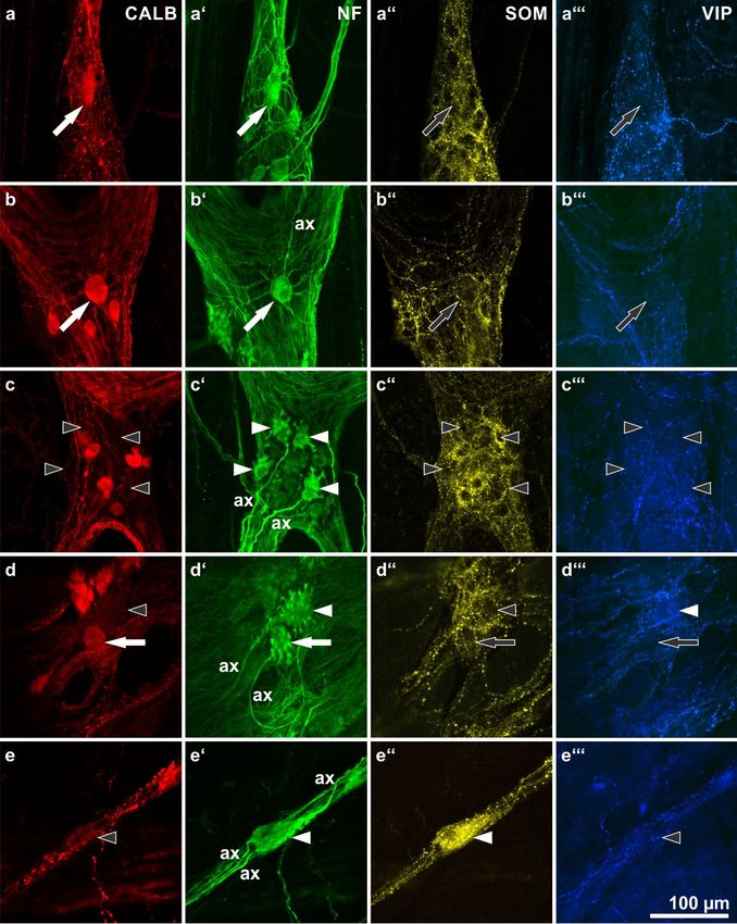
Figure 3. Calbindin (CALB: a–e) immunoreactivities of morphologically defined, neurofilament (NF:
Figure 3. Calbindin (CALB: a–e) immunoreactivities of morphologically defined, neurofilament (NF:
a’–e’)-labeled human myenteric neuron types and their co-reactivities for somatostatin (SOM: a’’–e’’)
a’–e’)-labeled human myenteric neuron types and their co-reactivities for somatostatin (SOM: a”–e”)
and vasoactive intestinal peptide (VIP: a’’’–e’’’). (ax = axons of marked neurons). (a,b) Two type III
and vasoactive intestinal peptide (VIP: a”’–e”’). (ax = axons of marked neurons). (a,b) Two type III
neurons with long, slender, branched dendrites (a’,b’: filled arrows) positive for CALB (a,b: filled
neurons with long, slender, branched dendrites (a’,b’: filled arrows) positive for CALB (a,b: filled
arrows) but negative for SOM and VIP (a’’,b’’: empty arrows). (c) Four stubby type I neurons (c’:
arrows) but negative for SOM and VIP (a”,b”: empty arrows). (c) Four stubby type I neurons (c’: filled
filled arrowheads) negative for all three other markers (c,c’’,c’’’: empty arrowheads). (d) A spiny (d’:
arrowheads) negative for all three other markers (c,c”,c”’: empty arrowheads). (d) A spiny (d’: filled
filled arrowhead) and a stubby (d’: filled arrow) type I neuron. The spiny one is negative for both
arrowhead) and a stubby (d’: filled arrow) type I neuron. The spiny one is negative for both CALB and
CALB and SOM (d,d’’: empty arrowheads) but positive for VIP (d’’’: filled arrowhead), the stubby
SOM (d,d”: empty arrowheads) but positive for VIP (d”’: filled arrowhead), the stubby one is positive
one is positive for CALB (d: filled arrow) but negative for SOM and VIP (d’’,d’’’: empty arrows). (e)
for CALB (d: filled arrow) but negative for SOM and VIP (d”,d”’: empty arrows). (e) A non-dendritic
A non-dendritic type II neuron displaying three axons (e’: filled arrowhead). It is co-reactive for SOM
type II neuron displaying three axons (e’: filled arrowhead). It is co-reactive for SOM (e”: filled
(e’’: filled arrowhead) but negative for both CALB and VIP (e,e’’’: empty arrowheads). (Patients data:
arrowhead) but negative for both CALB and VIP (e,e”’: empty arrowheads). (Patients data: (a–c)
(a–c) 59 years, ileum, male; (d) 57 years, ascending colon, male; (e) 42 years, sigmoid colon, female)
59 years, ileum, male; (d) 57 years, ascending colon, male; (e) 42 years, sigmoid colon, female)Int. J. Mol. Sci. 2018, 19, 194 7 of 14
Int. J. Mol. Sci. 2018, 19, 194 7 of 14
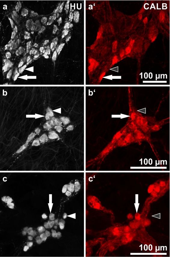
Figure 4. Calbindin (CALB)-immunoreactive human submucosal neurons: shapes as revealed by their
Figure 4. Calbindin (CALB)-immunoreactive human submucosal neurons: shapes as revealed by
peripherin (PERI)-immunoreactivities as well as co-immunolabeling for somatostatin (SOM) and
their peripherin (PERI)-immunoreactivities as well as co-immunolabeling for somatostatin (SOM) and
vasoactive intestinal peptide (VIP). (ax = axons of marked neurons). (a,b) Three neurons (arrowheads)
vasoactive intestinal peptide (VIP). (ax = axons of marked neurons). (a,b) Three neurons (arrowheads)
displaying a multidendritic/uniaxonal morphology (a’,b’: filled arrowheads) and co-reactivities for
displaying a multidendritic/uniaxonal morphology (a’,b’: filled arrowheads) and co-reactivities
VIP and CALB (a,a’’’,b,b’’’: filled arrowheads) but not for SOM (a’’,b’’: empty arrowheads). (c,d)
for VIP and CALB (a,a”’,b,b”’: filled arrowheads) but not for SOM (a”,b”: empty arrowheads).
Two neurons (arrows) displaying a non-dendritic/uniaxonal morphology (c’,d’: filled arrows) and
(c,d) Two neurons (arrows) displaying a non-dendritic/uniaxonal morphology (c’,d’: filled arrows)
co-reactivities for SOM and CALB (c,c’’,d,d’’: filled arrows) but not for VIP (c’’’,d’’’: empty arrows).
and co-reactivities for SOM and CALB (c,c”,d,d”: filled arrows) but not for VIP (c”’,d”’: empty arrows).
(Patients data: (a–c) 57 years, ascending colon, male; (d) 70 years, duodenum, female).
(Patients data: (a–c) 57 years, ascending colon, male; (d) 70 years, duodenum, female).
In the MP, a distinct correlation between CALB reactivity and morphological features of NF-
stainedIn neurons
the MP, could
a distinct correlation
exclusively betweenforCALB
be detected reactivity and
type III-neurons. morphological
These had one axonfeatures of
and long,
NF-stainedtapering
branched, neuronsdendrites
could exclusively
arrangedbe detected for typearound
circumferentially III-neurons. These(Figure
their soma had one axonThey
3a,b). and were
long,
branched, tapering dendrites arranged circumferentially around their soma (Figure
found only in the small intestinal MP and were generally positive for CALB. Other morphologically 3a,b). They were
found only
defined in thetypes
neuron smallwere
intestinal
mostly MP and were
negative forgenerally
CALB, withpositive
a fewforexceptions
CALB. Other morphologically
each. Stubby type I-
defined neuron types were mostly negative for CALB, with a few exceptions
neurons (Figure 3c), spiny type I neurons (Figure 3d) or type II neurons (Figure 3e) were each. Stubby type
frequently
I-neurons (Figure 3c), spiny type I neurons (Figure 3d) or type II neurons (Figure 3e) were
negative for CALB. However, in rare cases, neurons of these types were found to be positive for CALB frequently
negative
(e.g., for CALB.
a stubby type IHowever,
neuron ininFigure
rare cases,
3d). neurons of these types were found to be positive for CALB
(e.g., In
a stubby
the ESPtype
andI ISP
neuron in Figure
no distinct 3d).
correlation between CALB reactivity and a particular submucosal
neuron type could be found. Both multidendritic/VIP-reactive as well as nondendritic/uniaxonal/SOM-
reactive neurons frequently displayed coreactivity for CALB (Figure 4) but CALB− neurons of both
types were also commonly observed (Figure 2b,c).Int. J. Mol. Sci. 2018, 19, 194 8 of 14
In the ESP and ISP no distinct correlation between CALB reactivity and a particular
submucosal neuron type could be found. Both multidendritic/VIP-reactive as well as
nondendritic/uniaxonal/SOM-reactive neurons frequently displayed coreactivity for CALB (Figure 4)
butJ.CALB − neurons of both types were also commonly observed (Figure 2b,c).
Int. Mol. Sci. 2018, 19, 194 8 of 14
2.4. Sections
2.4. Sections Stained
Stained for
for Calbindin
Calbindin (CALB)
(CALB) and
and Peripherin
Peripherin (PERI)
(PERI)
In these + nerve fibers within the
In these specimens
specimens we we demonstrated
demonstrated thethe distribution
distribution pattern
pattern of
of CALB
CALB+ nerve fibers within the
gut wall +
gut wall (Figure
(Figure 5).
5). In
In addition
addition toto the
the ganglionated
ganglionated plexus,
plexus, CALB nerve fibers
CALB+ nerve fibers could
could be
be observed
observed
throughout all
throughout all gut
gut layers,
layers, including the longitudinal
including the longitudinal andand circular
circular sublayers
sublayers of of the
the muscular
muscular coat
coat
(Figure 5a) and the mucosa (Figure 5b). Casually, epithelial cells with a typical bottleneck
(Figure 5a) and the mucosa (Figure 5b). Casually, epithelial cells with a typical bottleneck shape shape
(enteroendocrine
(enteroendocrine cells)
cells)displayed
displayed CALB
CALBreactivity. Except
reactivity. from Figure
Except 5c’, we 5c’,
from Figure desisted
we from depicting
desisted from
the distribution patterns of SOM- and VIP-positive fibers (already published in [14,15]).
depicting the distribution patterns of SOM- and VIP-positive fibers (already published in [14,15]).
Figure 5. Section through the small intestinal wall, immunostained for calbindin (a,b,c: CALB; red),
Figure 5. Section through the small intestinal wall, immunostained for calbindin (a,b,c: CALB;
peripherin (a’,b’: PERI; green) and somatostatin (yellow;the latter is only depicted in (c’). (a) CALB-
red), peripherin (a’,b’: PERI; green) and somatostatin (yellow;the latter is only depicted in (c’).
immunoreactive nerve fibers in the longitudinal (upper arrowhead) and circular muscle layer (lower
(a) CALB-immunoreactive nerve fibers in the longitudinal (upper arrowhead) and circular muscle
arrowhead). (b) CALB-reactive mucosal nerve fibers (arrowhead). The arrow points at an
layer (lower arrowhead). (b) CALB-reactive mucosal nerve fibers (arrowhead). The arrow points
enteroendocrine cell lying in the base of a mucosal crypt and being positive for CALB. (c) The same
at an enteroendocrine cell lying in the base of a mucosal crypt and being positive for CALB.
enteroendocrine cell enlarged (filled arrow), not far from another endocrine cell reactive for
(c) The same enteroendocrine cell enlarged (filled arrow), not far from another endocrine cell reactive
somatostatin (empty
for somatostatin arrow).
(empty arrow).
3. Discussion
Beyond the bare registration of numbers and proportions of CALB+ neurons, this study aimed
to answer the question whether CALB may be a useful marker to label a particular human enteric
neuron population, as has been shown for the guinea pig intestine [2].
3.1. General Distribution of CALB in the Human Enteric Plexus
Our study has extended earlier findings obtained from the human duodenum [8] by showing
that CALB immunoreactivity is widely distributed throughout the myenteric and submucosal plexusSie können auch lesen