MECHANISTIC INSIGHTS INTO CARBON MONOXIDE AND COA - EDOC-SERVER
←
→
Transkription von Seiteninhalten
Wenn Ihr Browser die Seite nicht korrekt rendert, bitte, lesen Sie den Inhalt der Seite unten
“Mechanistic insights into carbon monoxide and CoA
binding at the Ni,Ni-[4Fe-4S] active site of the acetyl-CoA
 synthase from Carboxydothermus hydrogenoformans”

 Dissertation
 zur Erlangung des akademischen Grades

 docter rerum naturalium
 (Dr. rer. nat.)

 eingereicht an der
 Lebenswissenschaftlichen Fakultät
 der Humboldt-Universität zu Berlin

 von

 M.Sc. Julian Kreibich

 Präsident der Humboldt-Universität zu Berlin
 Prof. Dr.-Ing. Dr. Sabine Kunst

 Dekan der Lebenswissenschaftlichen Fakultät
 Prof. Dr. Dr. Christian Ulrichs

Gutachter/innen:

1. Prof. Dr. Holger Dobbek
2. Prof. Dr. Maria Andrea Mroginski
3. Prof. Dr. Christian Limberg

Tag der mündlichen Prüfung: 09.08.2021
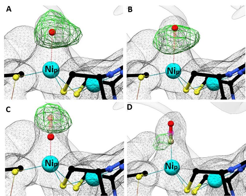
Table of Contents

Table of Contents
Zusammenfassung................................................................................................................................... iv

Abstract ................................................................................................................................................... vi

1 Introduction.......................................................................................................................................... 1

 1.1 Nickel containing enzymes ............................................................................................................ 1

 1.2 Autotrophic carbon fixation .......................................................................................................... 5

 1.2.1 Reductive acetyl-coenzyme A pathway (Wood-Ljungdahl-pathway) .................................... 6

 1.2.2 Acetyl-CoA synthase of C. hydrogenoformans (ACSCh) ........................................................... 8

 1.3 Research objective ...................................................................................................................... 12

2 Materials and Methods ...................................................................................................................... 14

 2.1 Anoxic work ................................................................................................................................. 14

 2.2 Metal free work ........................................................................................................................... 14

 2.3 Chemicals and materials.............................................................................................................. 14

 2.3.1 Media and antibiotics ........................................................................................................... 14

 2.3.2 Bacterial strains .................................................................................................................... 15

 2.4 Genetic methods ......................................................................................................................... 15

 2.4.1 Polymerase chain reaction (PCR) ......................................................................................... 15

 2.4.2 Molecular cloning of acsB .................................................................................................... 17

 2.4.3 Agarose gel electrophoresis ................................................................................................. 18

 2.4.4 Transformation ..................................................................................................................... 18

 2.4.5 Isolation of plasmid DNA: ..................................................................................................... 19

 2.5 Biochemical methods .................................................................................................................. 19

 2.5.1 Expression of the gene encoding for ACSCh .......................................................................... 19

 2.5.2 Purification of ACSCh ............................................................................................................. 20

 2.5.3 Sodium dodecyl sulfate polyacrylamide gel electrophoresis (SDS-PAGE) ........................... 20

 2.5.4 UV/vis spectroscopy ............................................................................................................. 21

 2.5.5 Synthesis of methylcobinamide ........................................................................................... 21

 2.5.6 Determination of the iron content of ACSCh ......................................................................... 21

 i
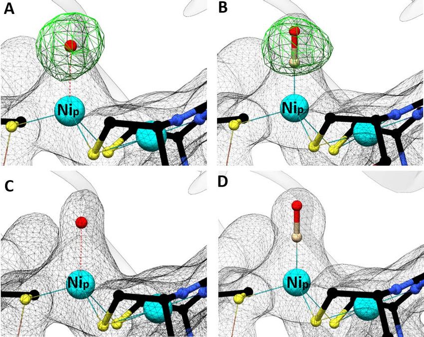
Table of Contents

 2.5.7 Determination of protein concentration.............................................................................. 22

 2.5.8 Measurement of ACSCh activity (acetyl-CoA formation) ...................................................... 22

 2.5.9 Isothermal titration calorimetry (ITC) .................................................................................. 22

 2.6 Protein crystallization .................................................................................................................. 23

 2.7 Data collection and processing.................................................................................................... 24

 2.8 Structure determination and refinements .................................................................................. 24

 2.9 Graphic representation ............................................................................................................... 24

3 Results ................................................................................................................................................ 25

 3.1 Expression and purification of ACSCh ........................................................................................... 25

 3.2 UV/vis spectroscopy of ACSCh ...................................................................................................... 27

 3.3 Activity of ACSCh ........................................................................................................................... 28

 3.4 Structural and biochemical analysis of ACSCh-closed....................................................................... 29

 3.4.1 Crystallization of ACSCh-closed and structure determination ................................................... 29

 3.4.2 ACSCh-closed structure and metal identification of cluster A ................................................... 30

 3.4.3 Molecular tunnel and observation of CO ligand binding ..................................................... 33

 3.4.4 Hydrophobic CO-binding patch ............................................................................................ 35

 3.4.5 CO-binding on ACSCh in solution ........................................................................................... 36

 3.5 Structural and biochemical analysis of ACSCh-open ........................................................................ 37

 3.5.1 Crystallization of ACSCh-open with CoA co-crystallized ............................................................ 37

 3.5.2 CoA ligand modeling............................................................................................................. 38

 3.5.3 CoA-binding to ACSCh in solution .......................................................................................... 42

 3.6 Comparison of cluster A coordination ........................................................................................ 43

4 Discussion ........................................................................................................................................... 46

 4.1 ACSCh-closed and ACSCh-open conformational states.......................................................................... 46

 4.2 CO- and CoA-binding on ACSCh .................................................................................................... 48

 4.3 Role of Nip in the mechanism of ACS........................................................................................... 50

5 Outlook ............................................................................................................................................... 53

Appendix................................................................................................................................................ 54

 ii
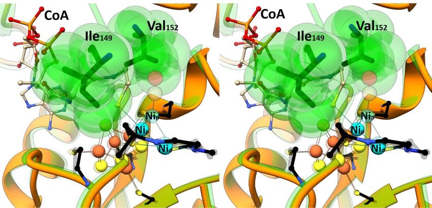
Table of Contents

Literature ............................................................................................................................................... 63

List of Abbreviations .............................................................................................................................. 71

List of Figures......................................................................................................................................... 74

List of Tables .......................................................................................................................................... 76

Acknowledgements ............................................................................................................................... 77

Declaration ............................................................................................................................................ 78

 iii
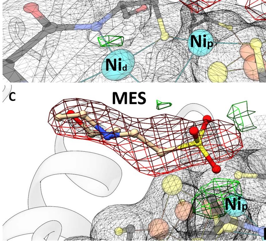
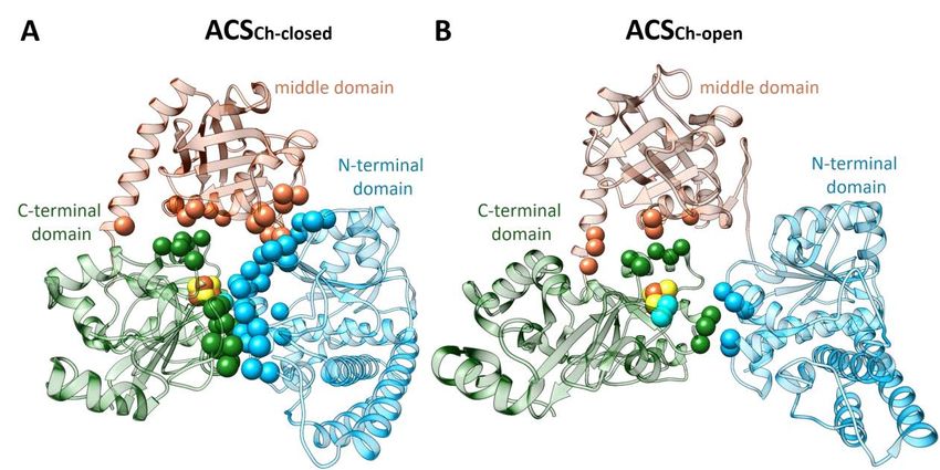
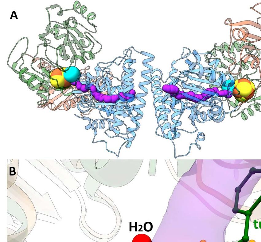
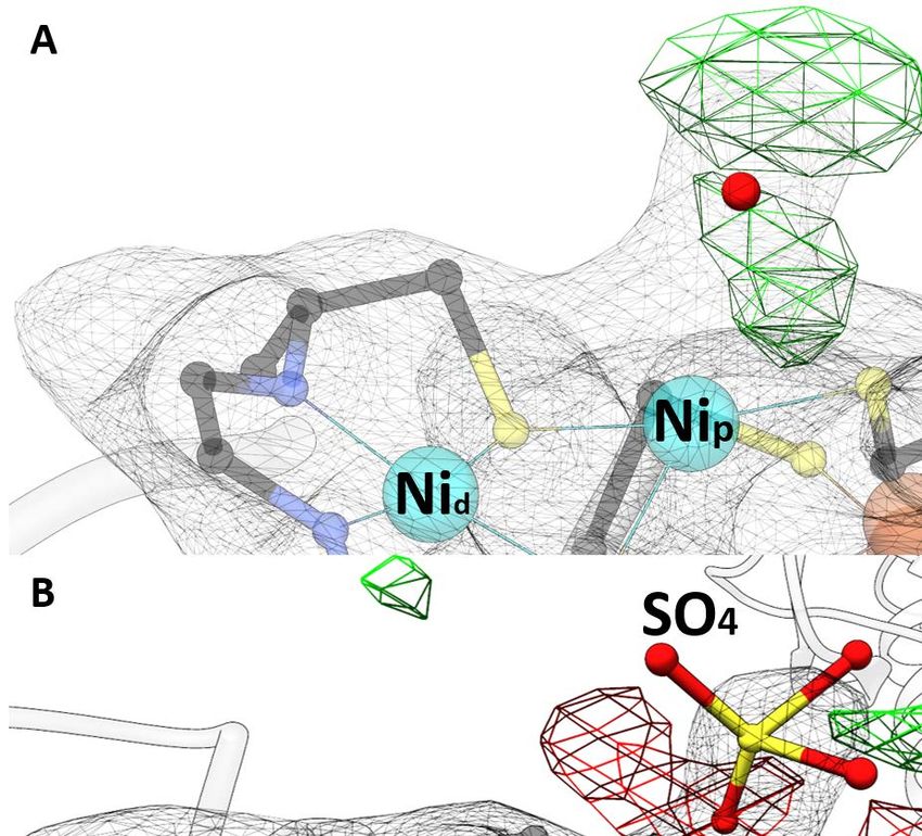
Zusammenfassung

Zusammenfassung

Die Acetyl-CoA Synthase (ACS) beinhaltet ein einzigartiges [4Fe-4S]-(μ2-SCys)-[Ni((μ2-SCys)2Gly)Ni]
Metallcluster (Cluster A) in seinem aktiven Zentrum, welches wichtig für ein autotrophes Wachstum
von Bakterien und Archaeen ist, die den reduktiven Acetyl-CoA-Weg zur Energiekonservierung oder
Kohlenstofffixierung nutzen. Der letzte Schritt dieses Syntheseweges wird von der ACS am proximal
zum [4Fe-4S] Cluster liegenden Ni ion katalysiert (Nip), in dessen Kondensationsreaktion aus CO, CoA
und einem Methylkation Acetyl-CoA synthetisiert wird.

In meiner Arbeit wurde die monomere ACS von Carboxydothermus hydrogenoformans (ACSCh)
heterolog in Escherichia coli exprimiert und aufgereinigt. Diese zeigte ein vollständig besetztes
[4Fe-4S] Cluster, jedoch keine vollständige Besetzung der beiden Ni-Positionen, im Cluster A. Eine
aktive ACSCh mit einem vollständig besetzten und intakten Cluster A konnte mithilfe chemischer
Rekonstitution generiert werden, seine Ligandenbindung untersucht und in verschiedenen
Konformationen kristallisiert werden.

Um die Interaktion der ACSCh mit seinen Substraten besser zu verstehen, wurde ACSCh in drei
verschiedenen Stadien kristallisiert. Zuerst wurde die aufgereinigte ACSCh in einer neuen Konformation
kristallisiert und deren Struktur mit einer Auflösung von 2.1 Å mit der Raumgruppe P212121 bestimmt.
Diese Konformation (ACSCh-closed) wies einen geringeren Kontakt zur Solventumgebung auf als die
vorher bekannte Struktur der ACSCh (ACSCh-open, PDB-ID: 1RU3). Das aktive Zentrum des Clusters A der
ACSCh-closed unterscheidet sich von der ACSCh-open in der Koordination des Nip, welches verzerrt
tetraedrisch koordiniert in ACSCh-closed vorliegt und quadratisch planar in ACSCh-open, wobei die Liganden
in beiden Konformationen identisch sind. Eine Analyse des Modells wies einen molekularen Tunnel
auf, der nur in ACSCh-closed vorhanden ist, welcher als CO-Kanal zur Substratversorgung dienen könnte
und besitzt einen zusätzlichen Schließmechanismus, welcher durch die Bewegung von Phe515 zwischen
ACSCh-open und ACSCh-closed gesteuert wird.

Zweitens wurde die Kristallstruktur einer CO gebunden ACSCh-closed mit einer Auflösung von 2.0 Å gelöst.
Darin wurde eine Elektronendichte am Nip identifiziert, die zu einem zweiatomigen Liganden passt. Die
Modellierung dieser Elektronendichte mit CO führte zu einer überzeugenden Beschreibung der
Elektronendichte. CO bindet am ebenfalls verzerrt tetraedrisch koordinierten Nip anstelle eines
Wassermoleküls. Im Vergleich zu ACSCh-closed unterscheidet sich die CO gebundene Struktur nur
geringfügig sowohl vom Cluster A als auch von der Gesamtstruktur des Proteins. Dies weist darauf hin,
dass die Konformation der ACSCh-closed sich in einem Stadium befindet, welches bereit für die CO

 iv
Zusammenfassung

Bindung ist. Die Konformationsänderung von ACSCh-open zu ACSCh-closed ist dabei der entscheidende
Schritt, um CO zur katalytischen Bindungsstelle zu leiten.

Weiterhin wurde Ni rekonstituierte ACSCh mit CoA kokristallisiert. Die daraus folgende CoA gebundene
ACSch Struktur mit der Raumgruppe H32 wurde mit einer Auflösung von 2.3 Å gelöst und zeigt ACSCh in
der offenen Konformation mit einer zusätzlichen positiven Elektronendichte über dem Nip des
Cluster A. Die Gesamtstruktur der CoA gebundenen ACSCh ist vergleichbar mit der ACSCh-open Struktur.
CoA bindet am Nip über seine Thiolgruppe und erstreckt sich zwischen zwei Domänen, wobei Nip
quadratisch planar koordiniert wird. Die Gegenwart von CoA wurde mit Berechnungen verschiedener
Elektronendichte-Karten für CoA: feature-enhanced, omit und polder maps zusätzlich validiert und gibt
damit erste Einsichten in die Interaktion und Koordinierung von CoA in ACSCh.

Die Bindung von CO und CoA an ACSCh wurde zudem mittels isothermaler Titrationskalorimetrie weiter
charakterisiert. Dabei bindet CoA enthalpisch getrieben mit einem KD von 3.1 µM und CO entropisch
getrieben mit einem KD von 9.4 µM an ACSCh. Der Konformationswechsel zwischen
ACSCh-open und ACSCh-closed für die Bindung unterschiedlicher Substrate ist dabei wichtig für die
Substratspezifität, welche mit einer Änderung der Koordination des Nip einhergehen. ACSCh-open zeigt
eine quadratisch planare Koordinierung des Nip und bietet genug Raum für eine CoA-Bindung. Im
Vergleich dazu ist in ACSCh-closed das Nip verzerrt tetraedrisch koordiniert und das Cluster A ist tiefer in
der ACSCh verborgen. Dies resultiert in einem geringeren Kontakt mit der Solventumgebung und der
Ausbildung einer hydrophoben Bindungstasche für die CO-Bindung, welche aus dem molekularen
Tunnel in ACSCh-closed zum aktiven Zentrum transportiert wird, wodurch die Bindung größerer Substrate
wie CoA am Nip blockiert wird.

Die Ergebnisse dieser Arbeit verschaffen damit erste Einblicke in den Mechanismus der
Substratbindung in ACSCh und unterstreicht die wichtige Funktion des Koordinierungswechsels des Nip
für die Substratbindung, welche mit einer Domänenumlagerung der ACSCh einhergeht.

 v
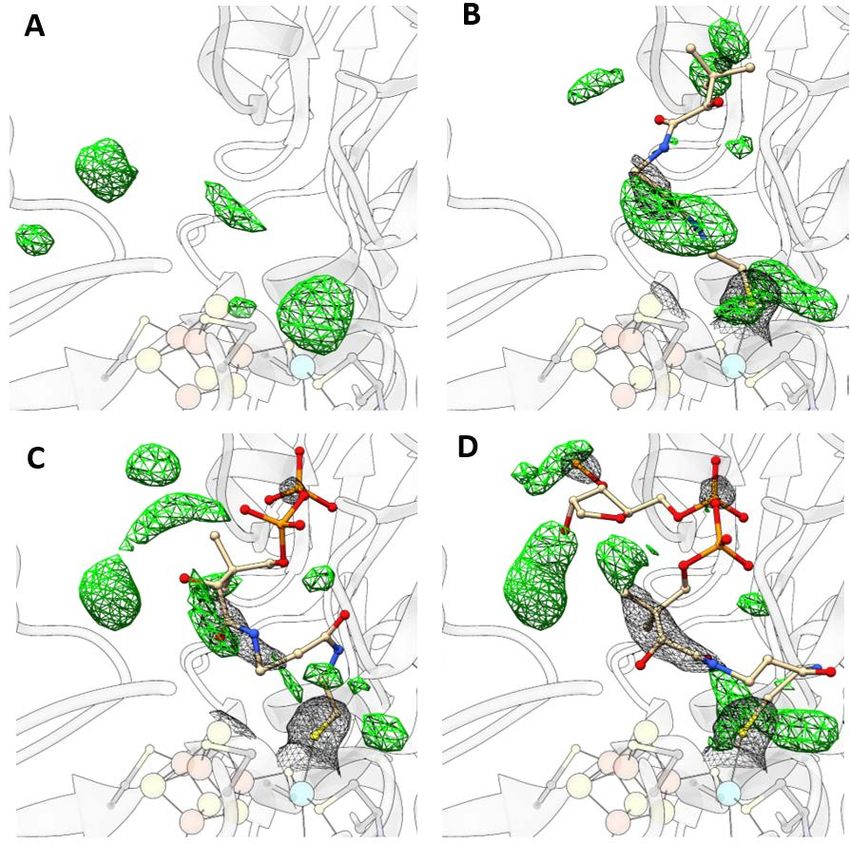
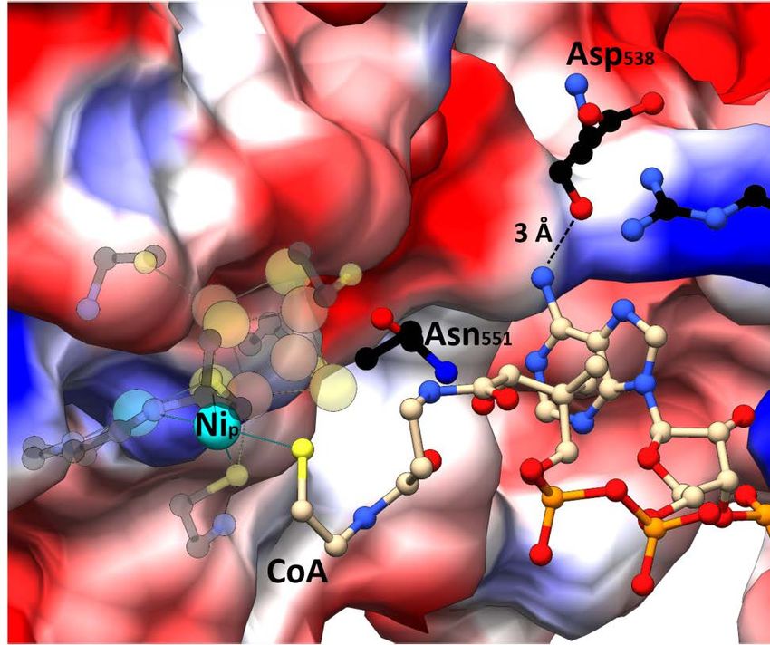
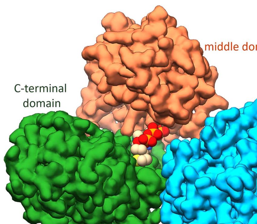
Abstract

Abstract

The acetyl-CoA synthase (ACS) harbors a unique [4Fe-4S]-(μ2-SCys)-[Ni((μ2-SCys)2Gly)Ni] cluster (cluster
A) in its active site, which is important for bacteria and archaea to survive in autotrophic growth using
the reductive acetyl-CoA pathway to conserve energy or fix carbon. The last step of this pathway is
catalyzed by ACS in a final condensation reaction of CO, CoA and a methyl cation at the Nip, the Ni ion
proximal to the [4Fe-4S] cluster.

In my study, the monomeric ACS of Carboxydothermus hydrogenoformans (ACSCh) was heterologously
expressed in Escherichia coli and purified with [4Fe-4S] cluster, but lacking binuclear nickel. An intact
and active cluster A containing binuclear Ni was generated by chemical reconstitution of ACSCh, which
was used to study ligand binding and crystallized in different forms.

Crystal structures of three different states of ACSCh were resolved to understand how ACS accomplishes
the interaction with those substrates. At first, as-isolated ACSCh structure was solved in a new
conformation at dmin of 2.1 Å with a space group of P212121 and less solvent exposed (called ACSCh-closed)
than the known structure of ACSCh (called ACSCh-open, PDB-ID:1RU3). The active site cluster A of
ACSCh-closed differs to that of ACSCh-open in the coordination of the proximal Ni, which is distorted
tetrahedrally coordinated in ACSCh-closed and square planar coordinated in ACSCh-open without changing
the coordinating ligands. Analysis of the model revealed a molecular tunnel that is only present in
ACSCh-closed, which might act as CO channel for substrate delivery at the Nip site with an additional gating
mechanism executed by the movement of the Phe515 between ACSCh-open and ACSCh-closed conformation.

Secondly, a CO-bound ACSCh-closed crystal was obtained and solved at a resolution of 2.0 Å. An electron
density fitting with a diatomic ligand at the Nip site was clearly identified and modeled as a CO
molecule, which clearly satisfied the observed electron density. CO binds at the Nip site completing the
tetrahedral coordination geometry of Nip by replacing a water molecule. Compared to ACSCh-closed, the
CO-bound structure differs only slightly at cluster A and also in overall structure, indicating that the
conformation observed in ACSCh-closed is a ready state to bind CO and that the conformational switch
between open and closed is responsible for CO migration and binding to the catalytic site.

Fully Ni-reconstituted ACSCh was co-crystallized with CoA. The CoA-bound ACSch structure was solved
at 2.3 Å resolution in a space group of H32, depicting ACSCh in the open conformation with an additional
positive electron density above Nip of cluster A. Overall structure of the CoA-bound ACSCh is similar to
the ACSCh-open state. CoA binds at the Nip site through its thiol group and flexibly extends to a gap
between two domains, where the Nip shows square planar geometry. This was further supported by

 vi
Abstract

calculating different electron density maps for CoA, feature-enhanced, omit and polder maps. This
structure gives first insights into interaction and coordination of CoA inside ACSCh.

The binding of CO and CoA to ACSCh has been further characterized by isothermal calorimetry
experiments. While CoA binding is enthalpically driven with a KD of 3.1 µM, CO binds to ACSCh by
entropic contribution with a KD of 9.4 µM. The observed conformational switch between ACSCh-open and
ACSCh-closed for the binding with different substrates is of importance in substrate specificity,
accompanied by a different coordination of Nip. ACSCh-open has a square planar coordination of Nip and
enough space for CoA binding. In comparison, ACSCh-closed depicts Nip in a distorted tetrahedral
coordination and cluster A is more buried into the ACSCh interior, generating a hydrophobic binding
pocket for CO that is delivered through the molecular tunnel in ACSCh-closed, consequently blocking
access of large substrates like CoA.

The results of this work provide initial insights into the mechanism of substrate binding in ACSCh and
point out the important role of the coordination switch of Nip coupled to domain rearrangement for
substrate engagement.

 vii
Introduction

1 Introduction

1.1 Nickel containing enzymes

Nature evolved biocatalysts which allows cells to perform a diverse range of chemical reactions to
realize complex metabolism in cells. One way to obtain this variety is the complexation of transition
metals in proteins. The chemical reactivity of the metal depends on the coordination geometry and
oxidation state and proteins are used to coordinate metals in a framework to control these
parameters. Additionally, metals play a crucial role in physiology and pathology of biological systems
[1]. The composition of all life forms on earth consist of 25 essential elements including many metals
e.g. Fe, Zn, Cu, Co, Se [2]. Bioinformatic studies of 1371 different enzymes with known structures show
that 47% of these enzymes contain a metal and 40% of them do so at their catalytic site [3]. Metals are
not only essential as catalysts for reactions, but they are also associated with proteins in storage and
transport systems (e.g. methallothionine, Hpn and ferritin [4, 5]) as well as in signal transduction,
regulation of transcription and chemotaxis [6]. Metallochaperones are a distinct class of chaperons
that protect and facilitate the maturation of metalloproteins (e.g. HypA, Atx1, UreE [7-9]) and other
metal binding proteins that are involved in the intracellular control of metal ions (e.g. Fur, NikR [10,
11]).

Although the metabolic importance of metals has been known since the beginning of the last century
[12, 13], nickel was only discovered to be an important element in biological systems in 1965 [14].
Since then, nickel-associated proteins have been found in sensing, transport systems, acetogenesis
and nickel insertion [15-18]. Despite the advantages of nickel as a transition metal, organisms have
also to deal with the toxicity of nickel [19].

Thus far, eleven enzymes are known carrying nickel as an active site component: glyoxalase I from
Escherichia coli [20], acireductone dioxygenase [21], urease [22], mononuclear nickel superoxide
dismutase [23], [NiFe]-hydrogenase [24], carbon monoxide dehydrogenase (CODH) [25], acetyl-
coenzyme A (CoA) synthase (ACS) [26], methyl-coenzyme M (CoM) reductase [27], lactate racemase
[28], some quercetinase e.g. in Streptomyces sp. FLA [29] and glycerol-1-phosphate dehydrogenase
from Bacillus subtilis [30]. The structural and catalytic complexity of these enzymes at the metal sites
varies by the coordination and redox property, which ranges from mononuclear redox (in)active metals
to heteronuclear metal cluster or a part of an organometallic cofactor (Fig. 1 and Tab. 1).

As depicted in Figure 1A-C, the nickel ion is in an octahedral geometry coordinated by His, Glu and
water in the acireductone dioxygenase, quercetinase and glyoxalase I [31-33]. In urease, two nickel

 1
Introduction

ions are bridged by a carbamylated Lys and an additional water molecule. Each nickel ion is coordinated
by two His residues, one of which is ligated by an additionally Asp residue. A water molecule completes
coordination for each nickel as a square pyramidal and an octahedral (Fig. 1D) [34]. In the Ni-containing
superoxide dismutase, the nickel ion is bound by an amide from a Cys residue, a primary amine from
the N-terminus, two thiolate ligands from Cys residues and an additional His residue, forming a square
pyramidal geometry (Fig. 1E) [35]. Upon reduction the Ni ion forms a square planar coordination by
displacement of the His residue [36]. The lactate racemase from Lactobacillus plantarum uses the
organic cofactor pyridinium-3-thioamide-5-thiocarboxylic acid mononucleotide, which is ligated by a
lysine (Fig. 1F) to bind the nickel using two sulfur and the pyridinium C4 carbon atom and completes
the nickel coordination by a histidine residue [37]. The nickel in the methyl-CoM reductase is mainly
coordinated by the tetrapyrrole nitrogens in the porphyrin and only one axial coordination from a Gln
residue, where the sixth axial coordination is lighted by CoM (Fig. 1G) [38]. In [NiFe]-hydrogenase the
nickel ion is coordinated by four Cys thiolates and an oxygen, where two of the thiolates and the
oxygen are bridged to the iron (Fig. 1H). The iron is further coordinated by two cyanide and one carbon
monoxide [39]. Another hetero nuclear cluster with nickel in a protein is depicted in CODH and ACS
(Fig. 1I and J), in which the nickel ion is part of a cluster consisting of other common metal clusters
[40]. The [Ni4Fe4SOHx] cluster (cluster C) of CODH can be described as a [3Fe-4S] cluster that is bridged
to a Ni-Fe center and the nickel ion is coordinated by additional thiolates from Cys residues [41]. In
ACS the [4Fe-4S]-(μ2-SCys)-[Ni((μ2-SCys)2Gly)Ni] cluster (cluster A) consists of a [4Fe-4S] cluster bridged
by a Cys residue to a binuclear Ni site. Both Ni ions are in a square planar coordination sphere. The Ni
proximal to the [4Fe-4S] cluster is named proximal Ni (Nip) and coordinated by three Cys residues and
a water ligand. In contrast, the distal Ni (Nid) is coordinated by two Cys residues and two nitrogen
atoms from the protein backbone [40].

 2
Introduction

Figure 1: Schematic overview of nickel coordination in structural known nickel enzymes. The nickel
ion is octahedral coordinated in the enzymes acireductone dioxygenase (A), glyoxalase I (B),
quercetinase (C). A square pyramidal coordination is known for the enzymes urease (D) and Ni
superoxide dismutase (E). An organic cofactor is bound to lactate racemase (F) and methyl-CoM-
reductase (G). In [NiFe]-hydrogenase (H), CODH (I) and ACS (J) the nickel ion is part of a unique metal
cluster. See text for detailed information. Atoms are color-coded distinctively: Fe in orange, Ni in cyan,
C in black, N in blue, S in yellow, O in red, P in orange, C atoms of pyridinium-3,5-bisthiocarboxylic acid
mononucleotide (P2TMN) and F430 are light brown.
 3
Introduction

Table 1: Nickel containing enzymes and their biochemical properties.

 Enzyme Biological Reaction Structural
 importance representative
 and redox activity
 Glyoxalase I Detoxification Hemithioacetal ⇋ mononuclear
 S-D-lactoylglutathione non-redox active
 Acireductone Methionine 1,2-dihydroxy-3-keto-5- mononuclear
 dioxygenase metabolism methylthiopentene +O2 ⇋ HCOO- non-redox active
 +CO + 3-methylthiopropionate
 Urease Nitrogen (NH2)2CO + H2O ⇋ 2 NH4+ + HCO3- mononuclear
 metabolism non-redox active
 Nickel superoxide Oxidative 2O2-+2H+ ⇋ H2O2 + O2 mononuclear
 dismutase stress redox active
 [NiFe]-hydrogenase Energy H2 ⇋ 2 H+ + 2e- hetero nuclear
 metabolism cluster
 redox active
 ACS Carbon CH3-Co(III)FeSP + CO + CoA-SH ⇋ hetero nuclear
 metabolism Acetyl-CoA + Co(I)FeSP cluster
 redox active
 CODH Carbon CO + H2O ⇋ CO2 + 2e- + 2H+ hetero nuclear
 metabolism cluster
 redox active
 Methyl-CoM Methane CH3-S-CoM + CoB-SH ⇋ CH4 + Organic cofactor
 reductase formation CoM-S-S-CoB heterodisulfide redox active
 Lactate racemase Pyruvate L-lactic acid ⇋ D-lactic acid Organic cofactor
 metabolism
 Quercetinase Flavonoid Quercetin + O2 ⇋ CO + H+ + 2- mononuclear
 degradation protocatechuoylphloroglucinol non-redox active
 carboxylic acid
 Glycerol-1-phosphate Phospholipid sn-glycerol 1-phosphate + NADH ⇋ not known
 dehydrogenase biosynthesis glycerone phosphate + NAD+

As shown in Table 1, nickel containing enzymes vary not only structurally, but they perform different
enzymatic reactions (redox reaction, condensations and racemization) and the redox state of the Ni
ion can be changed between Ni(0,+1,+2 and +3) states with a range of the potential of 1.5 V [42]. They
play a crucial role in detoxification (glyoxalase I, nickel superoxide dismutase), carbon (ACS, CODH) and
nitrogen (urease) metabolism and in the synthesis of important precursors (acireductone dioxygenase,
lactate racemase, glycerol-1-phosphate dehydrogenase). Nevertheless, these proteins are often found
to rely on additional helping proteins for nickel delivery, metallocentre assembly or for the synthesis
of organic cofactors [43-45]. Genomic analysis of nickel metalloenzymes revealed that these enzymes
are widely distributed among bacteria and archaea and the most common are urease and the
[NiFe]-hydrogenase. Eucaryotes show nickel enzymes mostly in fungi and plants [46]. In animals, no
physiologically relevant nickel dependent enzyme is known.

 4
Introduction

1.2 Autotrophic carbon fixation

Carbon is the most important element for all cells to build new biomass. Therefore, assimilation of
carbon is one of the fundamental processes for all life forms on earth. There are two different types of
carbon fixation: autotrophic and heterotrophic [47]. Organisms that can grow autotrophically are able
to produce complex organic compounds from inorganic nutrients such as CO2. They can be further
divided into photoautotrophs and chemolithoautotrophs depending on their energy source.
Photoautotrophs use light [48] and chemoautotrophic organisms can oxidize reduced inorganic
compounds (chemolithotroph) or organic compounds like formate (chemoorganotroph) to provide the
energy for CO2 fixation [49]. The autotrophic carbon fixation is distributed among many prokaryotes
and plants, which are able to assimilate carbon from inorganic CO2 to build biomass. Through this
assimilation, 2∙1011 t carbon is turned into organic compounds per year, which corresponds to 10 % of
the CO2 of the atmosphere. This biomass is an essential carbon source for heterotrophic organisms,
which utilize organic carbon compounds for carbon fixation [50].

Table 2: Mechanisms for autotrophic carbon fixation. Table was modified from [51]

 Metabolic pathway ATP/ Reductant CO2-Fixing enzymes Active Oxygen
 pyruvate CO2 sensitivity
 species
 Reductive pentose 7 NADP(H) RubisCO CO2 aerobic
 phosphate cycle
 2- Oxoglutarate CO2
 synthase
 Reductive citric acid 2 NADP(H), Isocitrate CO2 anaerobic,
 cycle Ferredoxin dehydrogenase microaerobic
 Pyruvate synthase CO2
 PEP carboxylase HCO3-
 3-hydroxypropionate 7 NAD(P)H Acetyl-CoA and HCO3-
 bicycle propionyl-CoA aerobic
 carboxylase
 3-hydroxypropionate- 9 NAD(P)H Acetyl-CoA and HCO3- microaerobic,
 4-hydroxybutyrate propionyl-CoA anaerobic
 cycle carboxylase
 Dicarboxylate-4- 5 NADP(H), Pyruvate synthase CO2 anaerobic,
 hydroxybutyrate ferredoxin PEP carboxylase HCO3- microaerobic
 cycle
 Reductive acetyl-CoA max. 1 NAD(P)H, Acetyl-CoA synthase- CO2
 pathway ferredoxin, CO dehydrogenase
 F420H2 Formylmethanofuran CO2 strict
 dehydrogenase anaerobic
 Pyruvate synthase HCO3-

 5
Introduction

There are six known autotrophic CO2 fixation mechanisms: reductive pentose phosphate cycle,
reductive citric acid cycle, 3-hydroxypropionate bicycle, 3-hydroxypropionate-4-hydroxybutyrate
cycle, dicarboxylate-4-hydroxybutyrate cycle and the reductive acetyl-CoA pathway [52]. They all
require energy and reducing agents to reduce CO2 into a formaldehyde (Equation 1) [50].

 2 + 4[ ] + ↔ ( 2 ) + 2 + + ℎ ℎ (1)

As shown in Table 2, the reductive acetyl-CoA pathway (also called Wood-Ljungdahl-pathway), is the
most energy efficient autotrophic carbon fixation pathway, in which only one ATP is hydrolyzed to
generate one molecule of pyruvate. Nevertheless, this pathway is used only by evolutionarily primitive
organisms, including archaea and eubacteria [53].

1.2.1 Reductive acetyl-coenzyme A pathway (Wood-Ljungdahl-pathway)

The reductive acetyl-CoA pathway is proposed to be one of the oldest metabolic pathways used to
conserve energy and to fix carbon autotrophically [54]. Because this pathway was conserved in
carboxydotrophic and acetogenic bacteria, methanogenic archaea, sulfate-reducing bacteria and
archaea for 3.5 billion years before the great oxygenation event [55], most of the enzymes are oxygen
sensitive. Therefore, all of these organisms live in the absence of dioxygen.

For instance, Carboxydothermus hydrogenoformans is the most extensively studied representative of
the hydrogenogenic gram-negative bacteria that utilizes CO as carbon and energy source under
anaerobic chemolithoautotrophic conditions [56]. It was first isolated from a hot swamp of Kunashir
Island in 1991, and is able to grow under a 100% CO atmosphere in the gas phase. The optimal growth
temperature is between 70-72 °C. It oxidizes CO to CO2 under H2 formation in equimolar quantities and
uses H2O as electron acceptor ( + 2 → 2 + 2) [57].

C. hydrogenoformans uses the reductive acetyl-CoA pathway, which consists of two branches, the
methyl (Eastern) and carbonyl (Western) branch [58] (Fig. 2). In the methyl branch, one CO2 molecule
is reduced to formate by formate dehydrogenase and transferred to tetrahydrofolate (THF) as methyl
carrier. While bacteria utilize THF as acceptor for the methyl group, archaea use
tetrahydromethanopterin (H4MPT) as a methyl acceptor. Stepwise multiple reductions of THF results
in formation of (6S)-5-methyltetrahydrofolate that is used to transfer the methyl group through the
methyl transferase (MeTr) to the corrinoid cofactor of the corrinoid iron-sulfur protein (CoFeSP) [59].

In the carbonyl branch, one CO2 molecule is reduced by the carbon monoxide dehydrogenase (CODH)
with two electrons to a CO molecule. Finally, the methyl group of CoFeSP and the CO molecule from
 6
Introduction

CODH are transferred to the active site of the acetyl-CoA synthase (ACS) and condensed with CoA to
acetyl-CoA. This pathway is used in some bacteria and archaea in the reverse direction to degrade
acetate to CO2 and THF/H4MPT bound methyl for example in Archaeoglobus fulgidus [60].

Figure 2: The reductive acetyl-CoA pathway in C. hydrogenoformans. The methyl group is carried by
tetrahydrofolate (THF) to (6S)-5-methyltetrahydrofolate, trough to the methyltransferase (MeTr).
Afterwards, the methyl group is further delivered to the corrinoid iron-sulfur protein (CoFeSP) and
finally transferred to the active site of acetyl-CoA synthase (ACS). One CO2 is reduced by the carbon
monoxide dehydrogenase (CODH) to CO and delivered to ACS for the final conversion of the methyl
group, CO and CoA to acetyl-CoA.

In the genome of C. hydrogenoformans, genes encoding for proteins for the final steps of the reductive
acetyl-CoA pathway are located in one operon (Fig. 3). Although they are arranged in an operon in
bacteria and archaea as well [55], the functional interaction of these enzymes is arranged in different
compositions between the archaea and bacteria phyla. For instance, in methanogens and other
archaea the key enzymes are arranged in one multienzyme complex of 2 MDa [61]. In contrast, in
bacteria a bifunctional ACS/CODH complex of 310 kDa is arranged in a heterotetrametric complex e.g.
in Moorella thermoacetica [62].

Figure 3: Gene cluster in C. hydrogenoformans encoding proteins for the reductive acetyl-CoA
pathway. Genes encoding for proteins of the methyl branch are further downstream and not shown.
The gene cooC1 encodes a maturation protein for CODH and acsF encodes a maturase for ACS, in which
both are responsible for nickel insertion [63-65]. Orf7 is translated into the reductive activator of
CoFeSP (RACo), which is responsible to reduce Co(II) of CoFeSP to Co(I) [66]. This figure is adapted and
modified from Figure 1 of Jeoung et al. [67].
 7
Introduction 1.2.2 Acetyl-CoA synthase of C. hydrogenoformans (ACSCh) ACS from C. hydrogenoformans (ACSCh) has a calculated molecular weight of 82.2 kDa (732 amino acids) and is the central enzyme of the reductive acetyl-CoA pathway. It connects the carbonyl and methyl branch for the final condensation of CO, a methyl cation and CoA to acetyl-CoA according to Equation 2. It consists of three globular domains [62, 68], the N-terminal domain (residues 1 to 315), the middle domain (residues 316 to 500) and the C-terminal domain (residues 501 to 732) (Fig. 4A). The N-terminal domain exhibits a Rossmann-fold, while the other two domains contain and folds without any known structural motif. Only residues of the C-terminal domain coordinate the unique active site (cluster A) of ACSCh, which consists of a cubane-type [4Fe-4S] cluster, bridged to two nickel ions forming a [4Fe-4S]-(μ2-SCys)-[Ni((μ2-SCys)2Gly)Ni] cluster (Fig. 4B) [40]. ( ) – 3 + + → ( ) + – (2) The distal nickel ion (Nid) is square planar coordinated by two sulfur atoms from cysteine residues and two nitrogen atoms from the protein backbone in a Cys-Gly-Cys motif. The second nickel ion is named proximal Ni (Nip) because of the proximity from the [4Fe-4S] cluster and has square planar coordination by three cysteine residues and a water ligand. Figure 4: Structure of ACS and cluster A. A) Monomeric ACSCh from (PDB-ID: 1RU3 [40]) in the open conformation. The N-terminal domain is depicted in blue, the middle domain in orange and the C-terminal domain in green. The cluster A is depicted as spheres (Fe in orange, S in yellow and Ni in cyan). B) Cluster A of ACSCh (C in black, N in blue and O in red, other atoms has the same color code as in A). C) Superposition of the open and closed form of ACS of M. thermoacetica (ACSMt). The open conformation (ACSMt-open) is orange and the closed conformation (ACSMt-closed) dark green. D) Bifunctional ACS/CODH complex from M. thermoacetica (PDB-ID: 1OAO [62]). Subunits of CODH are depicted in grey and rose. ACS has the same color code as in C. Zn is shown as violet sphere. The cluster A of ACSMt and cluster B, C and D of CODH are depicted as spheres. 8
Introduction In C. hydrogenoformans, ACS exists as a monofunctional enzyme during growth under CO conditions [40], but it can also be isolated as a heterotetramer in complex with CODH [41]. In contrast to M. thermoacetica, a homoacetogenic bacteria, where ACS is always associated with CODH as the ACS/CODH bifunctional complex (Fig. 4D) [62]. In this complex, each CODH subunit is flanked by one ACS subunit and the active sites of each CODH subunit are connected via a tunnel to cluster A of ACS to guide CO to the active site [69]. Several crystal structures are available for the bifunctional complex (PDB-IDs: 1MJG [68], 1OAO [62], 2Z8Y [69], 3I04 [70], 6X5K [71], 6YTT [72]). The first crystal structure of the ACS/CODH (PDB-ID: 1MJG) was obtained from M. thermoacetica, in which the proximal position of cluster A (Mp) is occupied with a tetrahedral coordinated Cu [68]. However, another crystal structure from M. thermoacetica depicts the same complex with two different conformations of the ACS (ACSMt) subunits (Fig. 4C and D) [62]. One exhibits a conformation with both metal positions in the cluster A occupied by Ni ions, referred to the open conformation (ACSopen), because ACS is more solvent exposed compared to the previously known conformation of the Cu containing ACS. The other ACS subunit differs from the first ACS/CODH structure (PDB-ID: 1MJG) only in the composition of cluster A, where the proximal position is occupied with a tetrahedral coordinated Zn ion instead of a Cu ion. Because of the reduced solvent exposure is this conformation called the closed conformation (ACSclosed). The different solvent exposure is accomplished by a rearrangement of the domains, resulting through a relative repositioning of the N-terminal domain by 39.2 Å (Fig. 4C). ACS is part of a 2 MDa ACDS complex in archaea whose structure is unknown. The ACDS complex is composed of five different subunits (22)48()8 [61]. The −subunit is an ACS analogue of 53 kDa in size and lacks the N-terminal domain of the mono- and bifunctional ACS [73-75]. The CODH analogue is represented by the and CoFeSP by the subunits. In addition to its different oligomeric state, the ACDS complex contains an additional archaea specific subunit () which is proposed to bind FAD as electron donor for CO oxidation [76]. However, archaea lack a gene encoding for the methyl transferase (acsE) that is bacteria specific [55]. There was a long scientific discussion about the correct metal composition of cluster A. Some proposed a tetrahedral coordinated Cu ion as active state of ACS [68, 77], but later it was shown that Cu acts as an inactivator of the catalysis [40, 78-80]. Instead of the Cu-Ni, ACS requires a Ni-Ni centre to be fully active [40]. The inactive Zn and Cu forms of ACS are a result of metal impurities, which are caused by the labile property of the Nip and can be replaced by other divalent metals like Zn and Cu or removed by 1,10-phenanthroline [78, 81, 82]. Only Ni is capable to restore the activity of ACS [78]. Although different structures of ACS are available, there is no generally accepted mechanism for ACS catalysis at the cluster A. The only general agreements are that CoA binds as the last substrate in the catalytic reaction and that the oxidation state of the [4Fe-4S] cluster and the Nid remains in 9
Introduction

the +2-oxidation state. The isolation of a methylated [83, 84] and acetylated [85] species of ACS which
are electron paramagnetic resonance (EPR) silent are arguments for a Nip (II) state. Pulse-chase studies
of ACS propose a random-sequential mechanism as shown in Figure 5 [86]. The redox state of the Nip
during catalysis is discussed in the scientific community and the basis of two proposed main
hypotheses of the ACS mechanism: The diamagnetic and paramagnetic mechanism [87, 88], where the
Nip undergoes redox change during the catalysis.

Figure 5: Proposed random sequential mechanism of ACS. The binding order of the methyl group or
carbon monoxide is variable, only the CoA substrate binds in an ordered way as last.

Both mechanisms have in common that the oxidation state of the cluster A in the inactive state is as
follows: [4Fe-4S]2+-(Nip2+)-(Nid2+) [89]. Despite being a random sequential description, both will be
described in the order that CO binds first to the active site to simplify the mechanism. In the
paramagnetic mechanism the binding of CO to Nip occurs with a simultaneously reduction of the Nip2+
to the Nip+ state [90]. This state can be tracked by infrared spectroscopy and EPR. The EPR signal for
the Nip+ is called the NiFeC signal, which is the evidence that the Nip acts as CO binding site [73, 78, 91,
92]. The transmethylation occurs in a nucleophilic SN2-type reaction [93-95] and results in a formation
of CH3-Nip3+-CO state, which is expected to be highly oxidizing and unstable state and undergoes a rapid
reduction to Nip2+ that is EPR inactive by an internal electron transfer whose mechanism is not known.
Because of the rapid reduction, the Ni3+ state may have never been observed. The methylation is
followed by the C-C bond formation to generate the acetyl species. A CoA-dependent thiolysis results
in production of the final product and release of acetyl-CoA, which regenerates the active species
(Fig. 6) [90].

 10
Introduction

Figure 6: Paramagnetic mechanism. Although the mechanism is a random sequential mechanism, it is
simplified as an ordered reaction, where CO binds first. See text for details. Figure adopted and
modified from [90].

In contrast to the paramagnetic mechanism, every intermediate in the diamagnetic mechanism is EPR
silent. The NiFeC species is described in this theory as an reversible CO inhibited state [96] and Nip
cycles between the Nip0 and Nip2+ state during catalysis. The reaction starts with a two-electron
reduction of the Nip2+ to the Nip0 state. Afterwards, one CO molecule binds to Nip and generates a
Nip0-CO species that is followed by a methyl transfer to generate a CH3-(CO)-Nip2+ intermediate that
undergoes the C-C bond formation. A subsequent attack of CoA results in the release of acetyl-CoA
and regeneration of the Nip0 state, which is ready for another catalytic cycle (Fig. 7).

 11
Introduction

Figure 7: Diamagnetic mechanism. Although the mechanism is proposed as random sequential
mechanism, it is simplified as ordered reaction, where CO binds first. See text for details. Figure
adopted and modified from [97].

The ACS reaction is the biological equivalent of the industrially important Monsanto and Cativa
processes. In these processes, expensive rhodium and iridium catalysts are used to synthesize acetic
acid from methanol and carbon monoxide [98, 99]. In comparison, the metals used in the biological
system are cheaper catalysts and more convenient for industrial processes, which is one reason why
ACS is an important research subject in the biotechnological industry. However, the unclarified
mechanism and its oxygen sensitivity are continuing to limit the ACS usage for industrial applications.
Another challenge is the labile property of the Nip. Even though AcsF is known as the cluster A
maturase, there are still open questions regarding the mechanism of the Ni insertion [63].

1.3 Research objective

ACS is the central enzyme of the reductive acetyl-CoA pathway and is responsible for the final reaction:
the synthesis of acetyl-CoA. This reaction takes place at a unique metal cluster in ACS, catalyzing a
process that is similar to the industrial Monsanto and Cativa processes, and ACS matches the
requirement to be an attractive alternative catalyst for this reaction. As such, molecularly
understanding of ACS mechanism is of high interest. Decades of research of ACS were carried out and
provided the first insights into the protein structure and the metal cluster. Nevertheless, information
regarding the reaction cycle itself is contradictory and much discussed in the scientific community.

 12
Introduction

There are large numbers of spectroscopic and biochemical data, but lacking structural data, especially,
on enzyme-substrate complex, which could help us to understand the catalytic mechanism of ACS at
the molecular level.

Therefore, the main objective of this thesis is to obtain new crystals structures of ACS, especially, with
bound-substrate states in high-resolution to unambiguously identify a small ligand like CO. Such ligand-
enzyme interaction is further confirmed by biochemical and biophysical methods, leading to a deeper
understanding of the nature of the catalysis by ACS.

 13
Materials and Methods

2 Materials and Methods

2.1 Anoxic work

The protein purification and crystallization were done under anoxic conditions in a glove box (model
B, COY Laboratory) under a N2/H2 (95%/5%) atmosphere. The oxygen content inside the glove box was
always below 10 ppm and monitored with the anaerobic monitor CAM 12 (Coy Laboratory). To remove
oxygen from solutions, they were all degassed under vacuum and gas-flushed with nitrogen in a gas
train with at least seven cycles and 18 min incubation for each step.

2.2 Metal free work

To avoid contamination by other divalent metals, especially Zn, all buffers and crystallization solutions
were treated with 5% w/v Chelex 100 resin (Biorad) for one hour and afterwards removed by filtration.
Additionally, all glassware was washed with 38% v/v HCl and subsequently rinsed with deionized water
(Milli-Q H2O).

2.3 Chemicals and materials

Unless otherwise noted, all chemicals were purchased from Fluka (Neu-Ulm), AppliChem (Darmstadt),
Carl Roth (Karlsruhe), Merck (Darmstadt), Biorad (München) and Sigma-Aldrich (Deisenhofen) and had
at least an analytical purity grade. The gases N2 (99,99%) an N2/H2 (95%/5%) were ordered from Air
Liquide (Berlin) and CO (99.9%) from Riessner Gase (Lichtenfels). All column materials for protein
purification were from GE Healthcare.

2.3.1 Media and antibiotics

Media for cultivation of E. coli are shown in Table 3. Super optimal broth medium (SOB) media was
purchased from Roth. All media were autoclaved for 20 min at 120 °C.

The antibiotics that were used for this work had the following concentration:
Carbenicillin (Cb), 50 µg/ml
Kanamycin (Km), 50 µg/ml
Tetracyclin (Tet), 100 µg/ml

 14
Materials and Methods

Table 3: Composition of cultivation media.

 Medium Composition for 1 l
 Lysogeny broth medium (LB) 10 g tryptone
 5 g yeast extract
 10 g NaCl

 Terrific broth medium (TB) 12 g tryptone
 24 g yeast extract
 10 ml glycerol
 10% phosphate buffer (0.17 M KH2PO4, 0.72 M K2HPO4)

 Super optimal broth medium (SOB) 20 g tryptone
 5 g yeast extract
 0.96 g MgCl2
 0.5 g NaCl
 0.186 g KCl

2.3.2 Bacterial strains

The bacterial strain E. Coli M15 DZ291 [100] with the genotype (F- Φ80ΔlacM15 thi lac- mtl- recA+) was
purchased from Qiagen and used for the cloning and expression of the gene acsB. The pREP4 plasmid
in E-coli M15 carries a Km resistance and constantly expresses the lac repressor protein encoded by
the lac I.

2.4 Genetic methods

2.4.1 Polymerase chain reaction (PCR)

PCRs were used to amplify DNA fragments from plasmids. The reaction mix and the thermo cycler
setting are summarized in the following Table 4.

Table 4: PCR for cloning purposes. a) reaction mix. b) PCR thermo cycler program.

a)

 Reagent Volume [µl]
 Forward primer (10 µM) 1
 Reversed primer (10 µM) 1
 Phusion Mastermix (2-fold) 25
 Template (100-300 ng/µl) 1
 milliQ-H2O ad 50

 15
Materials and Methods

b)

 Step Temperature [°C] Time [s] Cycle number
 Initial Denaturation 98 30 -
 Denaturation 98 10
 Amplification 61 30 30
 Elongation 72 30/kb
 Final Elongation 72 300 -
 storage 4 - -

Colony PCR

A successful transformation of E. coli was verified by colony PCR. Single colonies were picked from an
LB-agar plate and resuspended in 20 µl MilliQ-H2O. This suspension was used for PCR (Table 5).

In case of a positive clone, the leftover of the suspension was used to inoculate a 5 ml LB culture,
containing the respective antibiotics.

Table 5: Colony PCR. a) reaction mix. b) PCR thermo cycler program.

a)

 Reagent Volume [µl]
 Forward primer (10 µM) 0.5
 Reversed primer (10 µM) 0.5
 Taq Polymerase 0.5
 Template (resuspended colony) 2
 dNTPs (2 mM) 2.5
 Reaction Buffer (10-fold) with Mg2+ 2.5
 milliQ-H2O 16.5

b)

 Step Temperature [°C] Time [s] Cycle number
 Initial Denaturation 98 120 -
 Denaturation 98 10
 Amplification 51 10 30
 Elongation 72 60/kb
 Final Elongation 72 60/kb -
 storage 4 - -

Site directed mutagenesis

Point mutations were introduced by using the QuickChange method [101]. The PCR product was
cleaned with the GeneJET PCR Purification Kit (Thermo Scientific) and incubated with 10 u of DpnI for
1 h at 37 °C with the respective buffer. After the digestion of the template DNA, 5 µl of the reaction

 16
Materials and Methods

mix was used for transformation of E. coli M15 pREP4. The PCR reaction mix and the thermo cycler
settings are summarized in Table 6.

Table 6: Site directed mutagenesis (QuickChange method). a) reaction mix b) PCR thermo cycler
program

 Reagent Volume [µl]
 Forward primer (10 µM) 1
 Reversed primer (10 µM) 1
 Pfu DNA Polymerase 1
 Plasmid (10-30 µg/µl) 1
 dNTPs (2 mM) 5
 10-fold Pfu-buffer with Mg2+ 5
 milliQ-H2O 36

b)

 Step Temperature [°C] Time [s] Cycle number
 Initial Denaturation 95 30 -
 Denaturation 95 30
 Amplification 55 45 18
 Elongation 72 780
 Storage 4 - -

2.4.2 Molecular cloning of acsB

Because the gene acsB encoding the ACSCh protein was already cloned into a pET28a vector
(pET28a-twinst-ACSCh) [63] from the genomic DNA of C. hydrogenoformans [102], the gene was
sub-cloned into a modified pQE30 vector (generated by B. Sc. Lena Swiatek, AG Dobbek, Humboldt
Universität zu Berlin) without His-tag. Amplification of the gene was done by PCR using the Phusion
DNA Polymerase. The pET28a-twinst-ACSCh was used as a template for the PCR reaction. Because the
pet28a vector that contained the ACSCh gene was cloned with an N-terminal twinstrep-tag sequence
including a tobacco etch virus (TEV) protease cleavage site for the purification, this sequence was
amplified together with the ACSCh gene. The plasmid was kindly provided by Dr. Jae-Hun Jeoung (AG
Dobbek, Humboldt Universität zu Berlin).

The PCR product was cleaned with a Nucleo Spin Gel and PCR Clean Up kit (Machery-Nagel).
Furthermore, the cleaned PCR product and the cloning vector were both cleaved with the restriction
Fast Digest enzymes BamHI and PstI for 30 min at 37 °C. An additional dephosphorylation step was
taken with the vector by adding the Fast thermosensitive alkaline phosphatase (Fast AP, Thermo
Scientific) enzyme to the reaction mix. After incubation, both samples were loaded on an agarose gel.
The DNA fragments were isolated with the Gene JET Gel Extraction Kit (Thermo Scientific). The ligation
step of the gene of ACSCh into the cloning vector was done with the T4-DNA-Ligase at 4 °C for 4 h in a
 17
Materials and Methods

ratio of 3:1 (twinstrep-tag-ACSCh fragment to vector). All kits and enzymes were used according to the
manufacturer’s manual. All DNA samples were eluted from spin columns with MilliQ-H2O.

Forward Primer of acsB (restriction site underlined):

5´-CTC ATC GGA TCC ATG GGC AGC AGC CAT C-3´

Reverse Primer of acsB (restriction site underlined):

5´-TAG TCC TGC AGT TAG AGT AGT GGC TCC ATG G-3´

A successful cleavage of the twinstrep-tag by using the TEV protease would form an N-terminal
Gly-His brace, which could act as a possible metal binding site, such as Cu and Ni [103, 104]. To
minimize the risk of an additional binding site, the histidine was mutated to an asparagine by using the
QuickChange method (as described above in 2.4.1).

Forward primer acsB H45N:

5´-GAA AAC TTG TAT TTC CAG GGC AAT ATG AGC GAA G-3´

Reverse primer acsB H45N:

5´-CTT CGC TCA TAT TGC CCT GGA AAT ACA AGT TTT C-3´

2.4.3 Agarose gel electrophoresis

50x-fold Tris-acetate-ethylenediaminetetraacetic acid (EDTA) Buffer (TAE-Buffer): 242 g Tris,
57 ml glacial acetic acid, 100 ml 0.5 M EDTA, ad 1,000 ml H2O

6-fold loading buffer: 0.25% (w/v) bromophenol blue / 0.25% (w/v) xylene cyanol / 30% (v/v) glycerol

The electrophoresis was performed using 0.8% (w/v) agarose gel in 1x TAE-buffer. To follow the
migration of the DNA bands, 4 µl of Midori Green (Nippon Genetics Europe GmbH) was added to 60 ml
of a gel. Each sample was mixed with 6x-fold loading dye. To estimate the size of the bands, one well
was loaded with 3-5 µl DNA marker (Gene Ruler 1 kb DNA ladder, Fermentas). The electrophoresis was
performed in an OwlTm D4 chamber at 100 V for 30 min.

2.4.4 Transformation

100 µl of chemical competent cells were thawed on ice and 1 µl plasmid (for plasmid
maintenance/expression pre culture) or 10 µl of the ligation reaction mix were added. After 30 min
 18
Materials and Methods incubation on ice, the suspension was heat shocked for 45 s at 42 °C and again incubated on ice for 2 min. 1000 µl of prewarmed SOB medium was added and the cells were incubated at 37 °C and 140 rpm for 1 h. For a transformation of cells that were further used for expression, 50 µl of the cell suspension and in case of a transformation with a ligation product 90% and 10% of the cell suspension was transferred to different LB-Agar plates with the appropriate antibiotics and incubated over night at 37 °C. 2.4.5 Isolation of plasmid DNA: Plasmid DNA was isolated from 5 ml bacterial culture grown in LB media with appropriate antibiotics using the Nucleo Spin Plasmid Easy Pure kit (Machery-Nagel). The culture was inoculated one day prior with a single colony from a LB-agar plate. The elution step was performed with 40 µl MilliQ-H2O. 2.5 Biochemical methods 2.5.1 Expression of the gene encoding for ACSCh For the expression of the gene encoding for ACSCh, competent E. coli M15 pREP4 cells were transformed with the plasmid construct containing acsB and the pISC plasmid [105]. One colony was used to inoculate into TB media containing the appropriate antibiotics. This culture was grown over night at 37 °C by 140 rpm. The culture flask was sealed with a butyl rubber septum to induce a transition from aerobic to anaerobic growth over time. The next day, fresh TB medium was supplemented with 40 mM sodium fumarate, 0.5 mM cysteine, 0.1 mM FeSO4, 1% glucose and the appropriate antibiotics. Afterwards, the flask was attached to a gas train and bubbled with nitrogen to generate an oxygen-free medium. After over pressurizing the overnight culture with nitrogen, both flasks were connected by a needle with two-way out through the septa and the pre-culture was released into the main culture by pressure difference to prevent oxygen contamination. The culture grew at 37 °C in a water bath under constant nitrogen flow. After the culture reached an optical density of 0.9-1.2 at 600 nm (OD600), the culture was induced with 0.5 mM isopropyl -D-1-thiogalactopyranoside (IPTG) and the temperature adjusted to 30 °C. Cells were harvested after 16 to 20 h by centrifugation at 6000 rpm for 20 min and the cell pellets frozen in liquid N2 and stored at -80°C until use. 19
Materials and Methods 2.5.2 Purification of ACSCh Frozen cells were resuspended in 5 ml buffer A (50 mM Tris/HCl pH 8.0, 100 mM NaCl, 2 mM TCEP) per gram of cells, supplemented with a small amount of DNase A, lysozyme and avidin and stirred for 1 h. The suspended cells were lysed in a rosette cell on ice by sonification (Bandelin Sonoplus HD 2070) with a KE 76 sonotrode three times for 5 min (amplitude 50%, 5 cycles). The cell lysate was centrifuged at 30,000 rpm for 45 min at 12 °C. The supernatant was loaded on a strep-tactin column, washed with buffer A containing 1 mM sodium dithionite (DT) and equilibrated with buffer A. After loading the supernatant, the column was washed with 10 column volume (CV) of buffer A. Elution was performed with 2.5 mM desthiobiotin in the same buffer. To remove the twinstrep-tag of ACSCh, the eluted protein solution was incubated with strep-tagged TEV protease in a ratio of 1:100 for 16 h and supplemented with −mercaptoethanol to a final concentration of 10 mM. Subsequently, the protein solution was applied to a Q-Sepharose anionic exchanger column. The column had been equilibrated before with buffer B (50 mM Tris/HCl pH 8.0, 10 mM NaCl). After the protein solution was loaded on the Q-Sepharose column, the column was washed with a mixture of 70% buffer B and 30% buffer C (50 mM Tris/HCl pH8, 500 mM NaCl) for at least 10 CV. The elution was performed with 100% buffer C. To remove uncut ACSCh from the elution fraction, a strep-tactin column was tentatively attached after the Q-Sepharose column during elution. The eluted protein fraction was reconstituted with NiCl2. Reconstitution was performed by adding a 6-fold excess of NiCl2 to the protein solution and incubation for 60-72 h at 45 °C. The solution was also supplemented with -mercaptoethanol to a final concentration of 10 mM. Reconstituted ACSCh was subsequently loaded on a sephadex S200 pre-grade column (16 mm x 600 mm) for size exclusion chromatography (SEC) using buffer A without TCEP. Fractions that correspond to monomeric ACSCh were collected, concentrated, frozen and stored in liquid N2. 2.5.3 Sodium dodecyl sulfate polyacrylamide gel electrophoresis (SDS-PAGE) Separating-gel: 12% (w/v) acrylamide/bis-acrylamide (29:1) 375 mM Tris-HCl pH 8.8 0.1% (w/v) Sodiumdodecylsulfate (SDS) 0.08% (v/v) N,N,N´,N´-tetramethylethylendiamin (TEMED) 0.05% (w/v) Ammonium persulfate (APS) Stacking-gel: 6% (w/v) acrylamide/bis-acrylamide (29:1) 125 mM Tris/HCl (pH 6.8) 0.1 (w/v) SDS 0.08% (v/v) TEMED 0.05% (w/v) APS 20
Sie können auch lesen