A 73-year-old woman presented to the dermatology clinic with an 11-month history of an evolving pruritic, erythematous rash on her thighs and ...
←
→
Transkription von Seiteninhalten
Wenn Ihr Browser die Seite nicht korrekt rendert, bitte, lesen Sie den Inhalt der Seite unten
A 73-year-old woman presented to the
dermatology clinic with an 11-month history of
an evolving pruritic, erythematous rash on her
thighs and buttocks. On physical examination
she was noted to have polycyclic erythematous
plaques. What is the most likely diagnosis?

Dermatomyositis

Subacute lupus erythematosus

Erythema gyratum repens

Tinea versicolor

Necrolytic migratory erythema

    The patient received a clinical diagnosis of erythema gyratum repens, which is a rare
    paraneoplastic rash associated with breast, lung, or esophageal cancer. She underwent
    imaging and colonoscopy before being diagnosed with anal squamous cell carcinoma. The
    patient was treated with topical glucocorticoids and gabapentin for pruritus. The rash
    improved after initiation of chemotherapy and radiation.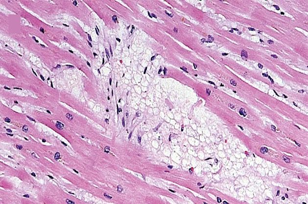
Die Dermatomyositis (DM, auch Lilakrankheit, Wagner-
Unverricht-Syndrom) ist eine idiopathische Myopathie
(=Muskelerkrankung) bzw. Myositis (=Muskelentzündung) mit
Hautbeteiligung und gehört zu der Gruppe der Kollagenosen. Ist
nur die Muskulatur betroffen, so spricht man in der Regel von
einer Polymyositis (PM). Aktuelle Studien zur Pathogenese der
DM und PM widersprechen sich. Da noch nicht gesichert ist, ob
DM und PM die gleiche Pathogenese haben, wird im Folgenden
die Dermatomyositis getrennt besprochen und auf die
Polymyositis sei nur verwiesen.
Die Erkrankung ist selten und kann in jedem Lebensalter
auftreten. Das Häufigkeitsmaximum ist das 50. Lebensjahr,
Frauen sind öfter betroffen als Männer (Gynäkotropie 2:1). Bei
Kindern liegt das Altersmaximum bei vier bis zwölf Jahren. Bei
Haushunden tritt die Dermatomyositis nahezu ausschließlich bei
Welpen und Junghunden auf.
Sie wurde von Ernst Leberecht Wagner (1821–1888) 1863
beschrieben und weiter von Heinrich Unverricht untersucht.
Ungefähr 50 % der Dermatomyositiden sind mit Tumoren
assoziiert. Besonders hervorzuheben sind dabei
Ovarialkarzinome. Es ist deswegen nötig, bei neu aufgetretener
Dermatomyositis nach Tumoren zu suchen. Allerdings ist der
zeitliche Bezug zum Auftreten der Dermatomyositis zu dem des
Tumors extrem variabel – die Haut- und Muskelerkrankung kann
dem Tumor sowohl vorausgehen, als auch nachfolgen oder sich
zeitgleich manifestieren. In manchen Fällen kann das
Wiederauftreten einer Dermatomyositis nach zwischenzeitlicher
Heilung von einem Tumor dessen Rezidiv, d. h. Wiederkehr,
anzeigen. Die im Rahmen einer Neoplasie auftretende
Dermatomyositis ist nach Entfernung der Neoplasie vollständig
reversibel.
Der Lupus erythematodes (lateinisch lupus, deutsch
‚Wolf‘, und altgriechisch ἐρυθηματώδης erythēmatṓdēs
aus ἐρύθημα erýthēma, deutsch ‚Röte‘, und Suffix -
ώδης -ṓdēs, deutsch ‚ähnlich wie‘), auch
Schmetterlingsflechte, ist eine seltene
Autoimmunerkrankung. Beim Lupus erythematodes ist
das körpereigene Immunsystem fehlreguliert: Es richtet
sich hierbei gegen gesunde körpereigene Zellen.
Dadurch werden Organe und Organsysteme, z. B. die
Haut, geschädigt.
Es gibt unterschiedliche Formen des Lupus
erythematodes: Die verschiedenen Formen des
kutanen Lupus erythematodes (CLE) befallen
üblicherweise nur die Haut. Der systemische Lupus
erythematodes (SLE) kann alle Organe befallen. Er
gehört zur Gruppe der Kollagenosen. Als Kollagenose
gehört der systemische Lupus erythematodes zu den
Erkrankungen des rheumatischen Formenkreises. Der
neonatale Lupus erythematodes bei Neugeborenen ist
eine Folge der mütterlichen Lupus-erythematodes-
Erkrankung.
Der Name Lupus wurde von dem lombardischen
Chirurgen Roger Frugardi (um 1140–1195) eingeführt,
ist aber auch schon im 10. Jahrhundert belegt. Der
Begriff Lupus leitet sich vom lateinischen Namen für
den Wolf ab. Früher verglich man die Narben, die nach
dem Abheilen der Hautschäden verbleiben, mit Narben
von Wolfsbissen.
Das Erythema gyratum repens gehört zu den figurierten
Erythemen. Es tritt paraneoplastisch auf, vor allem bei
Bronchial- und Mammakarzinomen sowie bei malignen
Tumoren des weiblichen Genitales, des Ösophagus
oder des Magens. Die Hautveränderungen können dem
Tumorleiden 4-8 Monate vorausgehen.
Das Eyrthema gyratum repens tritt vor allem bei
Erwachsenen zwischen dem 40. und 60. Lebensjahr
auf.
Das Erythema gyratum repens präsentiert sich als
urtikariell eleviertes, rasch wanderndes (in Stunden), 1-
2 cm breites, streifiges Erythem. Die Effloreszenzen
sind anulär, girlandenartig oder spiralig ineinander
geschwungen und erinnern an Holzmaserungen. Im
Randbereich weisen sie eine halskrausenartige
Schuppung auf. Das Erythema gyratum repens tritt
bevorzugt im Bereich des Körperstamms und an den
proximalen Extremitätenabschnitten auf.
Die Ursache für das Erythema gyratum repens ist
unklar. Vermutet wird eine immunologische Reaktion
auf Antigene wie z.B. Verbindungen, die von Tumoren
produziert werden. Die Hautveränderung kann schon
mehrere Monate auftreten, bevor andere Symptome
einer Tumorerkrankung auffallen. Der Primärtumor ist
meist ein Karzinom (Mamma-, Magen-, Lungen-,
Prostata-, Ösophagus-, Genitalkarzinome), seltener
werden andere Neoplasien wie ein Melanom oder ein
Plasmozytom gefunden.
Pityriasis versicolor (Kleienpilzflechte, auch:
Kleieflechte) ist eine häufig vorkommende Pilzinfektion
der obersten Hautschicht (Epidermis). Der Erreger
dieser Hautmykose ist Malassezia furfur (früher u. a.:
Pityrosporum orbiculare bzw. Pityrosporum ovale). Der
Pilz bleibt als Hefe im einzelligen Stadium, bildet also
keinen Fruchtkörper und kein Mycel aus. Es gibt von
Land zu Land unterschiedliche Prävalenzen für die
versch. Malassezia-Spezies. Die Erkrankung ist
harmlos und nicht ansteckend.
Malassezia-Hefen gehören bei annähernd 100 % der
Bevölkerung zur normalen Hautflora. Die Gründe,
warum sie bei manchen Menschen pathogen
(krankhaft) werden, sind nicht ganz geklärt. Es wird
jedoch beobachtet, dass die Hautmykose verstärkt in
den Sommermonaten und bei Menschen mit Neigung
zu starkem Schwitzen auftritt. Auch eine Verbindung mit
hoher Schilddrüsenfunktion wird angenommen. Es
bildet sich ein Pilzrasen, der zum einen physikalisch
Licht blockiert, zum anderen toxisch die
Melaninproduktion hemmt. Bei Sonnenkontakt bräunt
die befallene Haut weit weniger als die umgebenden
Partien, wodurch weiße Flecken (Maculae) entstehen.
Diese können linsengroß sein oder sich zu einer
landkartenartigen Marmorierung der Haut ausweiten.
Die Diagnose wird vom Dermatologen meist als
Blickdiagnose gestellt und kann durch das Abkratzen
von Hautschuppen und die Begutachtung unter dem
Mikroskop bestätigt werden. Die Pilzzellen sind als
traubenförmige Kugelhäufchen zu erkennen.
Erythema necrolyticum migrans

Bei Glukagon-produzierenden Tumoren (Glukagonom)
auftretendes paraneoplastisches Syndrom der Haut mit
erythematösen bogenförmigen Arealen mit Krusten-
bildung und langsamer Ausbreitungstendenz. Weitere
mögliche Erscheinungen sind Cheilitis, Nageldystrophi-
en, Stomatitis, Diarrhö, Thromboseneigung, Diabetes
mellitus, Hyperglukagonämie und B-Symptomatik.
Diagnostiziert wird klinisch, laborchemisch und his-
tologisch. Das Glukagonom wird gesucht und saniert.
Bizarr geformte zirzinäre Erytheme mit zentrifugaler
Ausbreitung und Pustelbildung.
Erythema necroticans migrans; Fünfte obligate kutane
Paraneoplasie; Impetigo circinata; Necrolytic migratory
erythema; Nekrolytisches migratorisches Erythem;
Paraneoplasie fünfte obligate kutane; Staphylodermia
superficialis circinata.

Postuliert wird durch das erhöhte Glucagon eine
katabole Stoffwechsellage mit einem Mangel an Zink
und verschiedenen Aminosäuren. Für diese These
spricht das rasche Ansprechen der Hautverändeurngen
auf Aminosäure-Substitutionen. Bemerkenswert ist,
dass sich das klinische und histologische Bild nicht von
Hautverändeurngen bei Zinkmangelsyndrom oder
Biotin-Mangel unterscheidet.
Die Sichelzellkrankheit oder Sichelzellenanämie (medizinisch Drepanozytose), auch Sichelzellanämie ist
eine erbliche Erkrankung der roten Blutkörperchen (Erythrozyten). Sie gehört zur Gruppe
der Hämoglobinopathien (Störungen des Hämoglobins) und führt zu einer korpuskulärenhämolytischen Anämie.
Bei den Betroffenen liegt eine Mutation der β-Kette des Hämoglobins vor. Es können entweder alle β-Ketten
betroffen sein (schwere, homozygote Form) oder nur ein Teil (mildere, heterozygote Form). Die Krankheit tritt vor
allem bei dunkelhäutigen Personen aus Subsahara-Afrika und deren Nachfahren, aber auch in Teilen des
Mittelmeerraums und des Nahen Ostens bis Indien auf und wurde durch Migration global verbreitet. Sie ist nach
wie vor in den Entwicklungsländern mit einer hohen Mortalität verbunden. Die Krankheit wurde 1910 von James
Herrick und Ernest Lyons bei einem Patienten aus der Karibik beschrieben und die Bezeichnung
Sichelzellenanämie wurde zuerst von Vernon Mason 1922 benutzt. Die Betroffenen bilden ein abnormes
Hämoglobin (Sichelzell-Hämoglobin, HbS), das bei Sauerstoffmangel zur Bildung von Fibrillen neigt. Dabei
verformen sich die roten Blutzellen durch die enthaltenen Fasern zu sichelförmigen Gebilden, verklumpen
miteinander und verstopfen kleine Blutgefäße, wodurch eine Entzündungentsteht. Durch die Verklumpung und
Gefäßverstopfung kann es bei der homozygoten Form zu anfallsartigen schmerzhaften, z. T. lebensbedrohlichen
Durchblutungsstörungen (Sichelzellkrisen) kommen, die unter anderem zu venösen Thrombosen führen können.
Aufgrund einer Punktmutation auf Chromosom 11 ist bei der Sichelzellenanämie an der Position sechs der β-
Globin-Protein-Untereinheit des Hämoglobins die Aminosäure Glutaminsäure durch Valin ersetzt. Die
Bezeichnung dieser Variante in offizieller genetischer Nomenklatur lautet HBB-p.E6V. Die betroffenen
Erythrozyten verformen sich bei abnehmendem Sauerstoffpartialdruck sichelförmig, verfangen sich leicht in
den Kapillaren und lysieren überdies sehr schnell. Durch die Hämolyse werden Hämoglobin, Arginase und
freie Sauerstoffradikale freigesetzt. Freies Hämoglobin bindet Stickstoffmonoxid etwa 1000-mal stärker als
intrazelluläres und Arginase verwandelt Stickstoffmonoxid zu Nitrit und Nitrat. Stickstoffmonoxid ist der
wichtigste Vasodilatator, und die Konzentrationsabnahme führt zur Gefäßverengung und somit zu
Durchblutungsstörungen.
Hydroxycarbamid (INN),
auch Hydroxyharnstoff oder Hydroxyurea, ist
ein Zytostatikum, das zur Behandlung insbesondere
von malignen Bluterkrankungen
(Leukämien, Myeloproliferative Neoplasien) eingesetzt
wird. Es ist auch für die Behandlung
der Sichelzellanämie zugelassen. Im Rahmen von
experimentellen Studien wurde es für die antiretrovirale
Behandlung bei HIV-Infektion getestet. Die Wirkung der
Substanz beruht auf der Hemmung des
Enzyms Ribonukleotidreduktase, welche die Ribose zur
Desoxyribose reduziert. Diese verläuft über einen
radikalischen Mechanismus, der die Bildung eines
Tyrosinradikals im aktiven Zentrum des Enzyms
erfordert. Das stabile Tyrosinradikal entsteht durch ein
nahegelegenes Eisenzentrum, welches aus zwei
Fe3+ besteht. Hydroxyharnstoff komplexiert das Eisen
und bewirkt die Reduktion des Eisens zum Fe2+,
wodurch die DNA-Synthesekapazität der jeweiligen
Zelle deutlich eingeschränkt wird. Durch die
europäische Arzneimittelbehörde EMA wurde die
Substanz auch zur Behandlung der Sichelzellanämie
zugelassen. Hydroxycarbamid erhöht die fetale
Hämoglobin-Synthese (Hb F) im Blut. Ein prozentual
erhöhter Anteil von Hb F im Blut wirkt protektiv
gegenüber der Polymerisation von Sichelzellen. In
mehreren klinischen Studien konnte die Wirksamkeit im
Rahmen vaso-okklusiver Krisen gezeigt werden.
Modification of the Pathophysiology of Sickle
Cell Disease by Hydroxyurea.
All the manifestations of sickle cell disease
depend, directly or indirectly, on sickling, which in
turn is produced, as red cells deliver oxygen to
tissues, by the polymerization of
deoxyhemoglobin S. Sickled red cells may
become stuck in capillaries and, by jamming small
vessels, may cause vaso-occlusion and
consequent pain crises. Sickled red cells may
undergo lysis within the bloodstream
(intravascular hemolysis), whereby hemoglobin
released in the plasma will bind nitric oxide (NO),
resulting in vasoconstriction that will further favor
vaso-occlusion. Sickled red cells will also be
phagocytosed by macrophages (extravascular
hemolysis); together with intravascular hemolysis,
this causes anemia, often severe. Sickled red
cells will also adhere to the endothelium, causing
a chronic inflammatory state that is associated
with neutrophil leukocytosis. The main action of
hydroxyurea is to cause an increase in the
intracellular concentration of fetal hemoglobin
(HbF), which interferes with the deoxyhemoglobin
S polymer formation, reducing the rate of sickling.
Through this mechanism, hydroxyurea affects the
very source of all the pathologic features of sickle
cell disease. In addition, hydroxyurea lowers the
neutrophil count, thus reducing the chronic
inflammatory state.
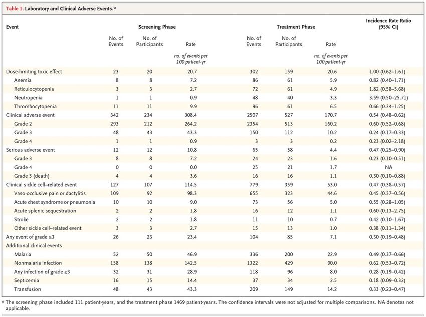
Hydroxyurea for Children with Sickle Cell Anemia in Sub-Saharan Africa
Hydroxyurea is an effective treatment for sickle cell anemia, but few studies have been conducted in sub-
Saharan Africa, where the burden is greatest. Coexisting conditions such as malnutrition and malaria may
affect the feasibility, safety, and benefits of hydroxyurea in low-resource settings. We enrolled children 1 to
10 years of age with sickle cell anemia in four sub-Saharan countries. Children received hydroxyurea at a
dose of 15 to 20 mg per kilogram of body weight per day for 6 months, followed by dose escalation. The
end points assessed feasibility (enrollment, retention, and adherence), safety (dose levels, toxic effects, and
malaria), and benefits (laboratory variables, sickle cell–related events, transfusions, and survival).
Retention of Participants in the Trial. The shaded area represents the 95% confidence interval for death or
withdrawal from the trial. The inset shows the same data on an enlarged y axis.
During hydroxyurea treatment, the white-cell count, absolute neutrophil count, and absolute reticulocyte
count significantly decreased, reflecting the intended mild bone marrow suppression, and these effects were
sustained over time. The overall rate of sickle cell–related events was significantly reduced (114.5 vs. 53.0
events per 100 patient-years; incidence rate ratio, 0.47; 95% CI, 0.38 to 0.57), and the rates of vaso-
occlusive pain and the acute chest syndrome were both reduced. The rates of infection also declined,
including rates of nonmalaria infection (142.5 vs. 90.0 events per 100 patient-years; incidence rate ratio,
0.62; 95% CI, 0.53 to 0.72) and severe infection of grade 3 or higher (28.9 vs. 8.0 events per 100 patient-
years; incidence rate ratio, 0.28; 95% CI, 0.19 to 0.42). Analyses of additional key clinical events revealed
significant reductions during hydroxyurea treatment in the rate of malaria infections (46.9 vs. 22.9 events per
100 patient-years; incidence rate ratio, 0.49; 95% CI, 0.37 to 0.66), blood transfusion (43.3 vs. 14.2 events
per 100 patient-years; incidence rate ratio, 0.33; 95% CI, 0.23 to 0.47), and death (3.6 vs. 1.1 events per 100
patient-years.
Adverse Events before and during
Hydroxyurea Treatment. Error bars indicate
68% confidence intervals, which correspond
to approximately 1 standard error.

In this trial involving children with sickle cell
anemia living in sub-Saharan Africa, we
found that hydroxyurea treatment was
feasible, reasonably safe, and had both
laboratory and clinical benefits. Specifically,
as compared with pretreatment rates, the
rates of clinical events, including vaso-
occlusive pain, infection, malaria,
transfusion, and death, declined after 1 year
of hydroxyurea treatment.

In conclusion, our results show that daily
hydroxyurea treatment was feasible and
safe for children with sickle cell anemia in
sub-Saharan Africa. Moreover, hydroxyurea
treatment reduced the rates of painful
events, infection, malaria, transfusion, and
death. Despite the recognition that 50 to
90% of affected children in Africa die before
the age of 5 years, sickle cell anemia
remains a neglected disease for which safe
and effective treatment options are needed.
The choice of the graft conduit for coronary artery
bypass grafting (CABG) has significant implications
both in the short- and long-term. The patency of a
coronary conduit is closely associated with an
uneventful postoperative course, better long-term
patient survival and superior freedom from re-
intervention. The internal mammary artery is regarded
as the primary conduit for CABG patients, given its
association with long-term patency and survival.
However, long saphenous vein (LSV) continues to be
utilized universally as patients presenting for CABG
often have multiple coronary territories requiring
revascularization. Traditionally, the LSV has been
harvested by creating incisions from the ankle up to the
groin termed open vein harvesting (OVH).
These concerns regarding wound morbidity and patient
satisfaction led to the emergence of endoscopic vein
harvesting (EVH). Published experience comparing
OVH with EVH suggests decreased wound related
complications, improved patient satisfaction, shorter
hospital stay, and reduced postoperative pain at the
harvest site following EVH. Despite these reported
advantages concerns regarding risk of injury at the time
of harvest with its potential detrimental effect on vein
graft patency and clinical outcomes have prevented
universal adoption of EVH.
Randomized Trial of Endoscopic or Open Vein-Graft Harvesting for Coronary-
Artery Bypass
The saphenous-vein graft is the
most common conduit for coronary-
artery bypass grafting (CABG). The
influence of the vein-graft harvesting
technique on long-term clinical
outcomes has not been well
characterized. We randomly
assigned patients undergoing CABG
at 16 Veterans Affairs cardiac
surgery centers to either open or
endoscopic vein-graft harvesting.
The primary outcome was a
composite of major adverse cardiac
events, including death from any
cause, nonfatal myocardial
infarction, and repeat
revascularization. Leg-wound
complications were also evaluated.
Leg-wound infections occurred in 18
patients (3.1%) in the open-harvest group
and in 8 patients (1.4%) in the
endoscopic-harvest group (absolute
difference, 1.7 percentage points; relative
risk, 2.26; 95% CI, 0.99 to 5.15).
Incisional leg pain had little or no effect on
functioning at 6 weeks after surgery in
62.2% of the patients in the open-harvest
group, as compared with 79.1% of those
in the endoscopic-harvest group (relative
risk, 0.79; 95% CI, 0.73 to 0.85).
Antibiotics were administered at follow-up
to 14.4% of the patients in the open-
harvest group and in 4.6% of the patients
in the endoscopic-harvest group (relative
risk, 3.15; 95% CI, 2.06 to 4.82).
In this trial, in which vein-graft harvesting
for CABG was performed by operators with
documented experience, we did not find
any significant difference between open
and endoscopic vein-graft harvesting in the
rate of major adverse cardiac events over
a median follow-up of 2.78 years. We
found a trend toward lower rates of major
adverse cardiac events in association with
the endoscopic technique when recurrent
events were compared between the two
treatment groups, although longer-term
follow-up will be necessary to determine
whether this finding is persistent.
Endoscopic harvesting resulted in better
harvest-site healing than did the open
approach, a finding consistent with
previous observations.
In conclusion, our trial did not show a
significant difference between endoscopic
and open vein-graft harvesting in the rate
of major adverse cardiac events among
patients undergoing CABG surgery during
a follow-up period with a median duration
of 2.78 years. The rate of wound
complications was lower in the endoscopic-
harvest group than in the open-harvest
group. Further studies are needed to
establish standards for harvester expertise
to ensure the safety of patients and
effectiveness of the procedure.
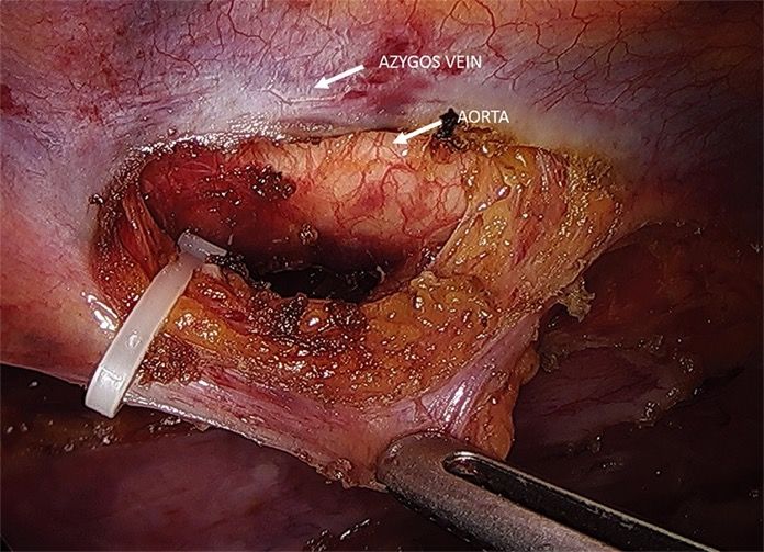
Hybrid and total minimally invasive esophagectomy: how I do it
Esophagectomy is a major surgical procedure
associated with a significant risk of morbidity and
mortality. Minimally invasive esophagectomy is
becoming the preferred approach because of the
potential to limit surgical trauma, reduce
respiratory complications, and promote earlier
functional recovery. Various hybrid and total
minimally invasive surgical techniques have been
introduced in clinical practice over the past 20
years, and minimally invasive esophagectomy has
been shown equivalent to open surgery
concerning the short-term outcomes.
Implementation of a minimally invasive
esophagectomy program is technically demanding
and requires a significant learning curve and the
infrastructure of a dedicated multidisciplinary
center where optimal staging, individualized
therapy, and perioperative care can be provided to
the patient. Both hybrid and total minimally
invasive techniques of esophagectomy have
proven safe and effective in expert centers. The
choice of the surgical approach should be driven
by preoperative staging, tumor site and histology,
comorbidity, patient’s anatomy and physiological
status, and surgeon’s experience.
Preoperative staging and tumor characteristics influence the choice of the surgical strategy,
i.e., a 2-stage or a 3-stage procedure. In some circumstances, starting with laparoscopy or
thoracoscopy may be useful to provide the ultimate staging. Initial laparoscopic approach for
gastric conduit preparation, as part of a hybrid or total minimally invasive Ivor Lewis operation,
is feasible in the majority of patients with esophageal adenocarcinoma.
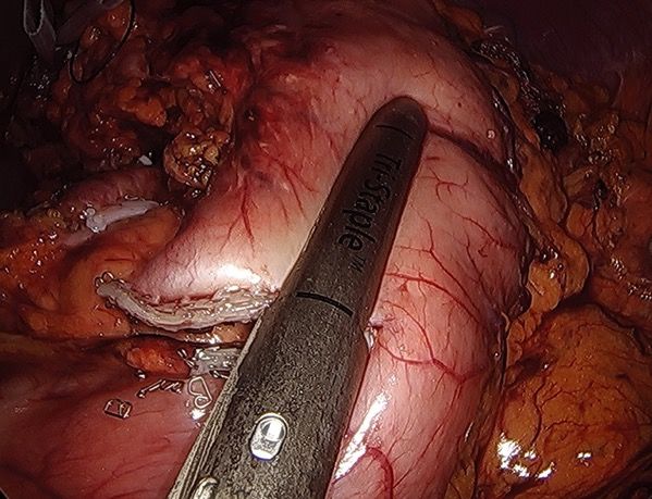
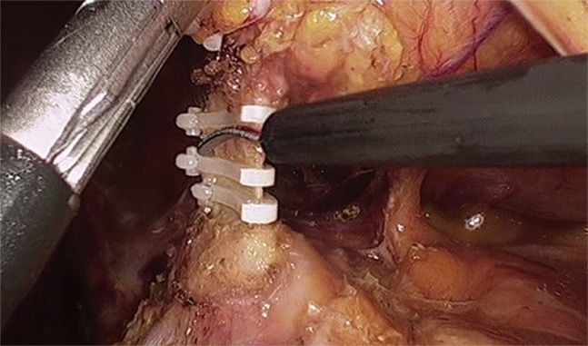
Laparoscopic division of the left gastric artery between
Hemolock clips.
Laparoscopic gastric tubulisation.
Trans-thoracic esophago-gastric anastomosis. (A)
Circular stapler introduced into the gastric tube through
a gastrotomy at the apex of the lesser curve; (B) the
anastomosis as viewed through the gastrotomy site.
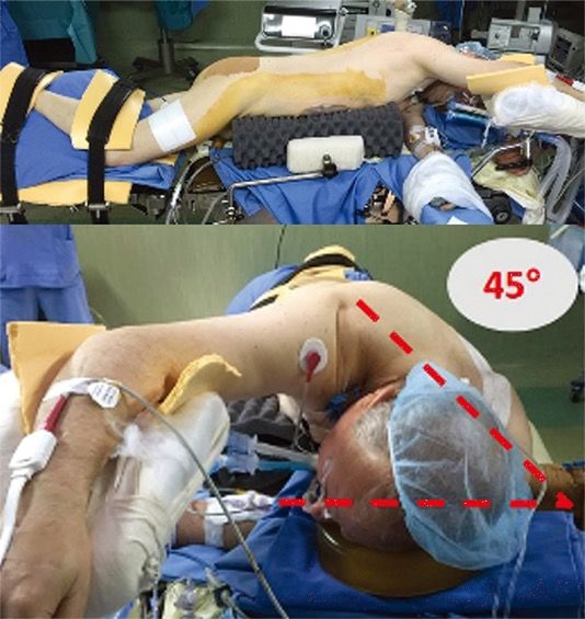
Semi-prone patient positioning with a typical 45°
angle.
The thoracic duct is secured with Hemolock clip at the
level of the diaphragm.
Hybrid Minimally Invasive Esophagectomy for Esophageal Cancer
Postoperative complications, especially pulmonary
complications, affect more than half the patients who
undergo open esophagectomy for esophageal cancer.
Whether hybrid minimally invasive esophagectomy
results in lower morbidity than open esophagectomy is
unclear. We performed a multicenter, open-label,
randomized, controlled trial involving patients 18 to 75
years of age with resectable cancer of the middle or
lower third of the esophagus. Patients were randomly
assigned to undergo transthoracic open
esophagectomy (open procedure) or hybrid minimally
invasive esophagectomy (hybrid procedure). Surgical
quality assurance was implemented by the
credentialing of surgeons, standardization of technique,
and monitoring of performance. Hybrid surgery
comprised a two-field abdominal–thoracic operation
(also called an Ivor–Lewis procedure) with laparoscopic
gastric mobilization and open right thoracotomy. The
primary end point was intraoperative or postoperative
complication of grade II or higher according to the
Clavien–Dindo classification (indicating major
complication leading to intervention) within 30 days.
Analyses were done according to the intention-to-treat
principle.
The percentage of patients receiving
neoadjuvant therapy was similarly high in
the two groups (75% in the hybrid-procedure
group and 72% in the open-procedure
group). A total of 3 patients (3%) who had
been assigned to the hybrid-procedure
group underwent intraoperative conversion
to the open procedure: 1 underwent
laparotomy without resection because of
advanced disease, 1 underwent
intraoperative conversion to the open
procedure because of subcutaneous
emphysema, and 1 underwent intraoperative
conversion to the open procedure, as
decided by the surgeon on the basis of
intraoperative physiological stress of the
patient. According to the intention-to-treat
principle, these patients were included in the
hybrid-procedure group.
After adjustment for age, sex, American
Society of Anesthesiologists risk score,
neoadjuvant therapy use, tumor location,
histologic subtype, resection-margin status,
pathological tumor and node stages, and trial
center, we found that minimally invasive
surgery was associated with a 77% lower risk
of major intraoperative and postoperative
complications within 30 days than open
surgery (adjusted odds ratio, 0.23; 95% CI,
0.12 to 0.44; P
In this multicenter, randomized, controlled
trial, we found that hybrid minimally
invasive esophagectomy was associated
with a 77% lower risk of major
intraoperative and postoperative
complications than open esophagectomy.
Furthermore, minimally invasive surgery
was associated with a 50% lower risk of
major pulmonary complications than open
surgery. Overall survival and disease-free
survival were at least as good with
minimally invasive surgery as with the
open procedure. In parallel to previous
findings regarding colorectal resection and
gastrectomy, we found that a minimally
invasive approach to the abdominal phase
of an Ivor–Lewis two-field abdominal–
thoracic esophagectomy was associated
with substantially lower major morbidity,
specifically pulmonary morbidity. In
conclusion, this multicenter, randomized,
controlled trial showed that hybrid
minimally invasive esophagectomy
resulted in a lower incidence of major
complications (specifically, pulmonary
complications) during or after
esophagectomy for cancer than did open
surgery. The hybrid procedure also
resulted in overall survival and disease-
free survival that were similar to those
observed with open esophagectomy.
Das WHIM-Syndrom (kurz für Warzen-Hypogammaglobulinämie-
Immundefizienz-Myelokathexis-Syndrom) ist eine vererbbare,
seltene Immunschwächekrankheit. Charakteristisch für das
WHIM-Syndrom ist eine Immunschwäche, die sich in
wiederkehrenden bakteriellen und viralen Infektionen äußert.
Davon sind insbesondere die Atemwege
mit Nasennebenhöhlenentzündungen, Mandelentzündungen und
Lungenentzündungen betroffen. Die Patienten sind anfällig für
Infektionen mit humanen Papillomaviren, die sich in zahlreichen
Warzen, insbesondere im Hand- und Fußbereich, äußern. WHIM-
Syndrom-Patienten haben darüber hinaus ein erhöhtes Risiko, an
viral-bedingten Krebsarten, wie beispielsweise
demCervixkarzinom, zu erkranken. Im Blutserum der Patienten
können erniedrigte IgG-Konzentrationen gemessen werden
(Hypogammaglobulinämie). Histologisch erscheint
das Knochenmark der WHIM-Patienten voller T-Vorläuferzellen.
Dem gegenüber kann eine Neutropenie beobachtet werden, die
auf eine gestörte Auswanderung und somit
Zurückhaltung neutrophiler Granulozyten aus dem Knochenmark
zurückgeführt werden kann (Myelokathexis). Das WHIM-Syndrom
ist eine autosomal-dominant vererbte Krankheit. Als häufigste
Ursache, die bei 92 % der betroffenen Patienten gefunden wurde,
werden Mutationen eines Gens auf dem Genlocus 2q21, das
den Chemokinrezeptor CXCR4 codiert, angesehen. Diese
Mutationen im intrazellulären Teil des membranständigen
Rezeptors für das Zytokin CXCL12 (SDF-1) führen zu einem
verkürzten Rezeptorprotein, dem die Fähigkeit der Internalisierung
nach Aktivierung fehlt. Somit sind Mechanismen der negativen
Selbstregulation unterbrochen und der Rezeptor kann
dauerstimuliert werden.
Plerixafor for the Treatment of WHIM Syndrome
WHIM syndrome (warts, hypogammaglobulinemia, infections, and myelokathexis), a primary immunodeficiency
disorder involving panleukopenia, is caused by autosomal dominant gain-of-function mutations in CXC
chemokine receptor 4 (CXCR4). Myelokathexis is neutropenia caused by neutrophil retention in bone marrow.
Patients with WHIM syndrome are often treated with granulocyte colony-stimulating factor (G-CSF), which can
increase neutrophil counts but does not affect cytopenias other than neutropenia. In this investigator-initiated,
open-label study, three severely affected patients with WHIM syndrome who could not receive G-CSF were
treated with low-dose plerixafor, a CXCR4 antagonist, for 19 to 52 months. Myelofibrosis, panleukopenia,
anemia, and thrombocytopenia were ameliorated, the wart burden and frequency of infection declined, human
papillomavirus–associated oropharyngeal squamous-cell carcinoma stabilized, and quality of life improved
markedly. Adverse events were mainly infections attributable to the underlying immunodeficiency. One patient
died from complications of elective reconstructive surgery.
The periods of treatment with granulocyte colony-stimulating factor
(G-CSF) and plerixafor are demarcated by the red and blue bars at
the top of each corresponding column of graphs. The dotted
portion of the red line for Patient 1 denotes the time when high-
dose G-CSF treatment (300 μg subcutaneously every day) was
interrupted owing to severe thrombocytopenia and the drug was
given at a reduced dose (150 μg subcutaneously every other day)
only during episodes of cellulitis. Horizontal dashed black lines in
each graph designate the upper and lower limits of the normal
range for each variable, as determined by the National Institutes of
Health (NIH) Clinical Center Clinical Hematology Laboratory.
Baseline values were assessed when neither drug was being taken
(solid circles). All values for Patients 1 and 2 at baseline and during
plerixafor treatment were determined at the NIH. Peak refers to
values obtained approximately 3 hours after plerixafor
administration, the time of the peak white-cell count, as defined
previously. Trough refers to values obtained approximately 12
hours after a dose of plerixafor was administered. Most white-cell
values for Patient 3 were determined at trough by the local provider
in Germany owing to travel limitations; exceptions are designated
by the peak and trough symbols in the figure key, and these values
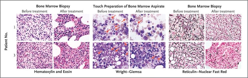
were determined at the NIH.Amelioration of Myelokathexis and Myelofibrosis during Long-Term, Low-Dose Plerixafor Treatment. Core bone marrow–biopsy samples were obtained from Patients 1 and 3 approximately 3 days before starting plerixafor (before treatment) and 24 and 52 months after starting plerixafor (after treatment) for Patients 1 and 3, respectively. Pretreatment hematoxylin and eosin–stained biopsy samples from both patients show markedly hypercellular marrow with granulocytic hyperplasia, right-shifted myelopoiesis, an elevated myeloid-to-erythroid ratio of approximately 5:1, and abundant neutrophils consistent with myelokathexis. This pattern was found in approximately 90% of the pretreatment marrow but in only 40 to 50% of the post-treatment marrow. The post- treatment images depict areas of normocellular marrow with normal myelopoiesis and a normal myeloid-to- erythroid ratio of 2:1. The pretreatment touch preparations of bone marrow aspirate (Wright–Giemsa stain) show frequent atypical neutrophils with pyknotic nuclear segments connected by thin, wispy strands of chromatin that are characteristic of myelokathexis (red arrows). These neutrophils can still be seen in the post- treatment samples but are less frequent. Both patients had severe myelofibrosis as defined by pretreatment dense reticulin staining of bone marrow; myelofibrosis was ameliorated after plerixafor treatment. All images are at 1000× magnification.
Amelioration of Skin Pathologic Conditions during Long-Term, Low-Dose Plerixafor Treatment.

Panel A shows the left medial lower leg, left foot, and right medial lower leg of Patient 1 both before starting
plerixafor and after 36, 36, and 28 months of plerixafor therapy for the left, middle, and right post-treatment
images, respectively. The fingers of Patient 2 are shown before plerixafor was started and after 9 months of
treatment, and the hand of Patient 3 is shown before plerixafor was started and after 32 months of treatment.
Patient 1 had chronic eczematoid dermatitis associated with recurrent cellulitis for 6 years. The left and middle
post-treatment images of this patient show resolution of inflammation, with residual areas of
hyperpigmentation probably representing uncleared hemosiderin; recurrent cellulitis ceased. The right
pretreatment image of this patient shows a chronic inflammatory mass centered at a site of recurrent cellulitis
and saphenous-vein insufficiency that was removed surgically 6 months after plerixafor was started. The
surgical wound (16×10×2 cm) healed completely (right post-treatment image). Patients 2 and 3 had a
reduced cutaneous wart burden after plerixafor therapy. Both patients also received topical imiquimod, and
Patient 2 received human papillomavirus (HPV) vaccination during treatment. Panel B shows clearance of 17
HPVs and Trichodysplasia spinulosa polyomavirus in Patient 1 after 18 months of plerixafor therapy. The
relative abundance of each of the viruses detected is conveyed by the arc length on the donut plot. The
number in the center is the number of different HPV types plus polyomavirus species detected.
We describe the CXCR4 antagonist plerixafor as a mechanism-based therapy for three
patients with WHIM syndrome who could not receive G-CSF. There was a reduction in the
frequency of infection in all three patients; resolution of chronic, progressive, multifocal
eczematoid and follicular lesions in Patient 1, associated with clearance of TSPyV and 17 HPV
types; a reduction in the wart burden in Patients 2 and 3; and a partial response of head and
neck squamous-cell carcinoma in Patient 2. All three patients reported improved quality of life.
Further controlled assessment of the safety and efficacy of plerixafor in patients with WHIM
syndrome is a challenge because the disease is extremely rare. Nevertheless, our phase 3
trial of G-CSF versus plerixafor in WHIM syndrome is designed to permit this assessment.
Acute Infection and Myocardial Infarction
Until the early 20th century, the human life expectancy was less than 50 years, and infections were often
fatal. Only in the past century have humans, on average, lived long enough for cardiovascular disease to
develop regularly and have antimicrobial therapies made survival from infection the norm. Furthermore,
sophisticated techniques for assessing myocardial damage have evolved during the past 50 years. It is
therefore not surprising that an association between acute infections and myocardial infarction has been
appreciated only in the past few decades. We will review the evidence that acute bacterial and viral
infections are associated with an increased risk of myocardial infarction in the short, intermediate, and long
term, and we will then discuss mechanisms that might explain this association.
Temporal Pattern of Cardiovascular
Risk after the Onset of Acute Infection.
The risk of a cardiovascular event is
several times higher after the onset of
respiratory infection than in the
absence of infection. The risk of a
cardiovascular event is proportional to
the severity of the infection. The risk
returns to baseline over a period of
weeks after an upper respiratory tract
infection. However, the time required
for the risk to return to baseline is
prolonged after a severe infection,
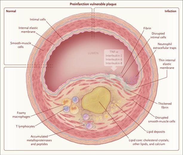
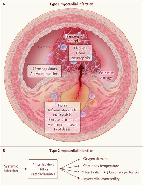
such as pneumonia.Features Present at the Time of Cardiac Involvement in Acute Infection. Shown is a vulnerable plaque (thin-cap fibroatheroma) during early infection before the development of myocardial infarction. Lipids have already accumulated in the wall of the coronary artery, with thinning of the internal elastic membrane, disruption of intimal and smooth- muscle cells, and fibrin deposition. Also present are foamy macrophages, T lymphocytes, metalloproteinases, peptidases, and neutrophil extracellular traps. In the lumen are inflammatory cytokines, including tumor necrosis factor α (TNF-α) and interleukins 1, 6, and 8, that result from sepsis elsewhere in the body.
Mechanisms of Cardiac Involvement in Acute
Infection.

Panel A shows rupture of an atheromatous
plaque, the mechanism of type 1 myocardial
infarction. As a result of the inflammation that
develops with infection, the thin-cap atheroma
ruptures, releasing inflammatory cells and fibrin
into the lumen. In the presence of circulating
procoagulants and activated platelets, this
release causes immediate accumulation of
platelets, fibrin, and neutrophils and trapping of
red cells, all of which cause acute obstruction of
the coronary arteries. Panel B shows the
process of demand ischemia, the mechanism of
type 2 myocardial infarction. Acute infection
causes the release of interleukin-1, TNF-α, and
catecholamines, which increase the core body
temperature, oxygen demand, and heart rate.
Coronary perfusion declines because of
decreased filling time. Cytokines also act to
suppress cardiac output. These factors, taken
together, cause a mismatch of oxygen needs
and oxygen supply, resulting in demand
ischemia.
Features Present after Cardiac Involvement in Acute Infection. Shown is an example of direct
myocardial involvement in pneumococcal pneumonia. In the heart of a patient who was treated with
antibiotic agents but still died from pneumococcal pneumonia, there are disrupted myocytes and
there is a relative absence of neutrophil infiltration. In addition, in experimentally induced infection
and without treatment, microcolonies of Streptococcus pneumoniae were present.
Vaccination

A meta-analysis of five randomized trials showed a 36% lower risk of a composite of cardiovascular events
among adults who had received influenza vaccine than among those who had not. The benefit was even greater
when the analysis was limited to persons with known coronary artery disease. In contrast, there are limited data
from randomized trials regarding the effect of pneumococcal vaccination on cardiovascular risk. A meta-analysis
of eight observational studies, all of which were published after 2000, showed a 17% lower risk of myocardial
infarction among patients 65 years of age or older who had received pneumococcal polysaccharide vaccine than
among those who had not.40 The lack of a more prominent effect may reflect the decline in the prevalence of
pneumococcal pneumonia in recent decades.

Summary

Practitioners may be able to influence the risk of postinfection myocardial infarction if they remain mindful of the
increased risk of myocardial infarction during and after acute infections and if they do not dismiss elevated
troponin levels as “troponin leak.” Among patients with acute infection who have clinical indications for statins
and aspirin, these medications should be continued (if the patient is already receiving them) or may be initiated if
no contraindications are present.

Finally, because the risk of other cardiovascular events — such as heart failure, arrhythmias, and strokes — also
increases after acute infection, the mechanisms that account for these associations need to be characterized.
This is especially important in the case of heart failure, because after pneumonia the risk of worsening heart
failure is even higher than the risk of myocardial infarction. An integrated understanding of the interplay between
acute infections and the cardiovascular system should facilitate efforts to reduce the risk of myocardial infarction
and other cardiovascular events after acute infections.
Ophthalmia Neonatorum
              A 2-week-old baby girl with a 3-day history of
              purulent discharge from both eyes was brought by
              her parents to the ophthalmology clinic. The baby
              had been born at full term by means of
              spontaneous vaginal delivery. She had not
              received ocular prophylaxis after delivery, and the
              mother had not undergone prenatal testing for
              chlamydia or gonorrhea infection. An eye-
              discharge sample obtained from the baby and an
              endocervical swab obtained from the mother
              tested positive for Chlamydia trachomatis DNA and
              negative for Neisseria gonorrhoeae DNA by
              polymerase chain reaction. Perinatal transmission
              of C. trachomatis or N. gonorrhoeae can result in
              neonatal conjunctivitis, known as ophthalmia
              neonatorum. The ongoing incidence of ophthalmia
              neonatorum caused by C. trachomatis or N.
              gonorrhoeae can be addressed by routine
              maternal prenatal screening for and treatment of
              sexually transmitted infections and by postpartum
              neonatal ocular prophylaxis against N.
              gonorrhoeae. In addition to treatment of the baby,
              which included a 2-week course of oral
              erythromycin, a single dose of oral azithromycin
              was given to each parent. The baby’s symptoms
              resolved within 5 days after the initiation of
              treatment, and she remained healthy at follow-up 2
              weeks later.
“Trachealization” of the Esophagus A 32-year-old man presented to the emergency department with difficulty swallowing oral secretions and the feeling that food was stuck in his throat after he ate a pizza roll. The patient reported that similar episodes had occurred previously, but in each instance the feeling resolved spontaneously, and he did not seek medical care. At the time of presentation, the patient was drooling. Upper endoscopy revealed impacted food material (Panel A) and prominent mucosal rings extending 20 cm from the incisors to the level of the gastroesophageal junction, with two discrete areas of narrowing and associated linear furrows (Panel B). Biopsy specimens were obtained, and esophagitis was observed, with more than 40 eosinophils per high-power field. An endoscopic finding of fixed esophageal rings, or “trachealization,” is suggestive of eosinophilic esophagitis, although a definitive diagnosis is made on the basis of clinical presentation, histologic findings, and the exclusion of other causes of esophageal eosinophilia, such as proton-pump inhibitor–responsive esophageal eosinophilia. The patient was treated with an 8-week course of omeprazole, but there was no symptom resolution or histologic improvement on repeat endoscopic biopsies, which confirmed the diagnosis of eosinophilic esophagitis. He was started on an 8-week course of both swallowed fluticasone and a six-food elimination diet (elimination of the six most commonly identified types of allergenic food — wheat, milk, soy, nuts, eggs, and seafood). No additional endoscopies were performed after completion of treatment with fluticasone and the elimination diet. At a 1-year follow-up visit, the patient reported no further symptoms of food impaction.
Das United States Army Special Forces Command
(Airborne) (USASFC; deutsch Luftlande-
Sondereinsatzkommando des Heeres der Vereinigten
Staaten; kurz Special Forces oder USSF) ist die
dienstälteste Spezialeinheit der US Army. Ihre etwa
10.000 Soldaten werden aufgrund ihres
grünen Baretts auch Green Berets genannt. Es handelt
sich dabei ausschließlich um Kampftruppen, die für ihre
Aufträge von anderen Einheiten des United States
Army Special Operations Command (USASOC)
„Sondereinsatzkräfte des Heeres der Vereinigten
Staaten“ unterstützt werden. Bis zum 27. November
1990 hieß die Einheit United States Army 1st Special
Operations Command. In der Diktion der US-
Streitkräfte steht der Begriff Special Forces traditionell
ausschließlich für das USASFC (Green Berets). Die
anderen Sondereinsatzkräfte des Heeres, zum Beispiel
das 75th Ranger Regiment oder das 160th SOAR,
sowie die Spezialeinheiten der
anderen Teilstreitkräfte werden unter dem
Begriff Special Operations Forces subsumiert. Seit ihrer
Aufstellung waren die Special Forces an allen
militärischen Konflikten und Kriegen der Vereinigten
Staaten mit Ausnahme des Einsatzes in Mogadischu
1993[A 1] beteiligt und leisteten weltweit in über 70
Nationen Militärberatung, infrastrukturelle und
humanitäre Hilfe.
A 34-Year-Old Veteran with Multiple Somatic Symptoms
A 34-year-old man was evaluated at this hospital because of headaches, cognitive changes, mood symptoms,
flashbacks, chest pain, arm tingling, and gastrointestinal symptoms. The patient had served as a special
operations combat medic in the U.S. Army Rangers for 8 years. He was wounded several times.
He served in Operation Iraqi Freedom, completing three tours of duty. The patient had had multiple traumatic
injuries and experiences during training and deployment. Nine years before the current evaluation, during a
parachute-jump training, he had a syncopal episode. Afterward, he could recall only that he had awoken on the
ground while a colleague was packing his parachute. He had 3 weeks of headaches, stiffness of the cervical
and thoracic spine, and difficulty sleeping. Eight years before the current evaluation, the patient was hit by an
explosive blast wave. Afterward, he reported “cloudy” mentation. While he served as the company medic, he
was a first responder in two cases in which a soldier had committed suicide. Three years later, during his third
deployment, the patient was involved in a motor vehicle accident as a helmeted back-seat passenger. The
armored fighting vehicle rolled approximately 9 m into a canal, and the patient was pinned under several men.
He had blunt trauma to the head and reportedly lost consciousness for 20 minutes; he had a concussion and
traumatic injuries of the head and face, including a hard-palate fracture. He subsequently had headaches and
difficulty eating and breathing because of lip and nose swelling.
After the patient’s third deployment ended, his wife noticed that he placed kitchen items in the wrong location,
became lost while grocery shopping, and was unable to recall the birth of his first daughter. The patient
expressed difficulty adjusting to a postdeployment routine, which included child care. He mentioned that he
missed his level of entrusted responsibility in the Army Rangers platoon. He reported having “deep, lingering
pain at deaths of friends and exposures to children in dire circumstances” and “thinking philosophically about
death” but did not report suicidal or homicidal ideation.
Between 3 and 5 years before the current evaluation, while the patient was still in the military, he sought
medical and psychiatric care on three occasions. During the first evaluation, duloxetine was prescribed, but it
resulted in a rash and peeling of the skin. Three years before the current evaluation, he received inpatient
treatment at a military hospital, which included psychotherapy sessions and muscle relaxation and breathing
exercises. Two years before the current evaluation, various medications — including sumatriptan, ibuprofen,
prednisone, topiramate, and amitriptyline — were tried, with varying degrees of success.
Twenty-two months before the current
evaluation, the patient was honorably
discharged from the military, and he
moved to New England. Thirteen
months before the current evaluation,
his condition was assessed by a social
worker, psychologist, physical
therapist, and neurologist at another
hospital. He reported being “constantly
on guard” and “easily startled,” as well
as having anhedonia, detachment,
difficulty concentrating, anorexia, and
fatigue. He reported using sarcasm,
defensiveness, and intellectualization
as coping mechanisms. A review of
systems was notable for a reduced
ability to move the head and neck to
the left and “clicking” with motion of
the neck, both of which diminished
modestly with physical therapy.
Laboratory test results are shown.
A 34-Year-Old Veteran with Multiple Somatic Symptoms

Five months before the current evaluation, the patient reported having flashbacks (Wiedererleben oder
Nachhallerinnerung) with associated emesis. The next month, he reported that his previous concussion was
“acting up.” He described having a “hazy” feeling, verbal stuttering, and severe headaches, which he rated at
10 on a scale of 0 to 10 (with 10 indicating the most severe pain). The headaches lasted for days and were
associated with sonophobia and photophobia; he used ice and ibuprofen for relief. He also began to have panic
attacks in association with recall of memories. He described having a “rush of visions” of traumatic memories
from childhood and the military and being “not able to turn them off”; lorazepam was prescribed. Recurrent
pain and tingling of the left anterior chest and left anterior arm developed in association with these memories
and later occurred independently of the memories. The patient treated himself for these symptoms with fans,
cold baths, and benzodiazepines.
The patient’s father had depression, and his mother, father, and paternal uncle each had a history of alcohol
and drug use disorders. The patient had been raised by his grandparents. A sister had died in her 20s from
bone cancer. The patient lived with his wife and two healthy young children. He had not worked outside the
home since his honorable discharge from the military, instead performing child care duties while his wife
worked. He had completed 3 years of college; he had thought about additional schooling but felt discouraged
because of forgetfulness. The patient had used chewing tobacco in the past but had not used any in 2 years.
He consumed up to four caffeinated drinks daily and had only three or four alcoholic drinks monthly, which was
a reduction from his past alcohol consumption. He smoked marijuana daily but used no illicit drugs. Medications
included nortriptyline, omeprazole, pantoprazole, sucralfate, ondansetron, and as needed, simethicone.
On examination, the temperature was 37.1°C, the heart rate 94 beats per minute, the blood pressure 109/74
mm Hg, the respiratory rate 18 breaths per minute, and the oxygen saturation 97% while the patient was
breathing ambient air. The weight was 86 kg, and the body-mass index (the weight in kilograms divided by the
square of the height in meters) 26.4. The patient was well groomed, alert, cooperative, oriented, and lucid, with
coherent speech. He was described as fidgeting, anxious, and irritable. The remainder of the cardiovascular,
pulmonary, abdominal, and musculoskeletal examination was normal. A urine toxicology screen was positive for
cannabinoids and negative for amphetamines, barbiturates, benzodiazepines, cocaine, opiates, and
phencyclidine. A diagnosis was made.
Neurologic Diseases
Cerebral aneurysm, vascular dissection, hemorrhage, ischemia, infections (meningitis and encephalitis), and
pseudotumor cerebri can all cause headaches, mood symptoms, and cognitive changes. Tumors of the central
nervous system (CNS) can cause these symptoms along with emesis and weight loss. Severe headaches or
headaches in combination with certain “red flags,” such as focal neurologic signs or systemic illness, may
indicate the presence of one of these serious underlying causes. Although imaging studies of the head would
be obtained in this case to rule out a clinically significant abnormality of the brain or CNS, the chronic nature of
this patient’s symptoms and the absence of other relevant findings most likely rule out a catastrophic neurologic
diagnosis.
Gastrointestinal Diseases
Inflammatory bowel disease could explain the presence of gastrointestinal symptoms. However, although the
onset of this disease can occur at any age, it typically occurs before 30 years of age, and the disease is usually
associated with blood in the stool and not with vomiting or constipation, which were described by this patient.
Endocrine Diseases
Hyperthyroidism could explain this patient’s mood symptoms, anxiety, insomnia, diarrhea, and weight loss.
However, he had a normal thyrotropin level, a finding that rules out this diagnosis.
Toxic Exposures
Veterans who served in Operation Iraqi Freedom have a number of potential toxic exposures, including
depleted uranium, lead, sand and dust particles, burn pits, oil-well fires, and agents of chemical warfare.
However, these exposures are known to result in rashes, widespread pain, respiratory problems, or persistent
fatigue, in addition to headaches and cognitive changes. The patient’s symptoms and the timeline of illness are
inconsistent with these exposures.
Mood and Anxiety Disorders
Bipolar disorder, major depressive disorder, and persistent depressive disorder could each explain the
presence of mood changes and insomnia. A family history of depression and ongoing anxiety increase the
likelihood of a mood disorder.
Traumatic Brain Injury
Traumatic brain injury (TBI) has developed in 19% of veterans who served in Operation Iraqi Freedom or
Operation Enduring Freedom and is the signature wound of these wars. Cognitive consequences of TBI (known
as neurocognitive disorder due to TBI) include decreased attention, executive function, learning, memory,
language, and social cognition.
Post-Traumatic Stress Disorder
Post-traumatic stress disorder (PTSD) is present in 13 to 17% of veterans who served in Operation Iraqi
Freedom or Operation Enduring Freedom. PTSD occurs after exposure to a traumatic event and is
characterized by re-experiencing of the event (often with physiologic reactions to trauma cues), avoidance of
trauma-related thoughts and external reminders, negative alterations in cognition or mood, and hyperarousal.4
PTSD could explain this patient’s mood symptoms, insomnia, flashbacks, hypervigilance, startle, and panic
attacks (with associated chest pain and tingling). Furthermore, veterans with PTSD are 4 times as likely to have
chronic headaches and 3.5 times as likely to have irritable bowel syndrome as veterans without PTSD.8,9 A
diagnosis of PTSD is the most parsimonious explanation of this patient’s panic attacks (a physiologic reaction
to trauma cues), rather than an independent panic disorder.

A patient must meet eight DSM-5
criteria for the diagnosis of PTSD to
be established. The first criterion is
direct exposure or indirect exposure
(e.g., involving a family member) to a
traumatic event. In patients with
PTSD, the constellation of symptoms
must lead to functional impairment or
distress. In addition, to establish the
diagnosis of PTSD, substance use
and other medical conditions must be
ruled out.
Discussion of Management
Dr. Mireya F. Nadal-Vicens: Management of PTSD starts with a combination of psychotherapy and treatment with
a selective serotonin-reuptake inhibitor or serotonin–norepinephrine reuptake inhibitor. In addition, mirtazapine,
prazosin, tricyclic antidepressants, or phenelzine may be administered. At this patient’s initial evaluation,
mirtazapine therapy was started, given its soporific and appetite-stimulating effects, but treatment was
complicated by the development of a facial rash. Escitalopram therapy was started and was associated with no
allergic reactions or unacceptable side effects.
A key component of the patient’s treatment was completion of 12 weekly 90-minute sessions of prolonged
exposure therapy, which is an evidence-based, trauma-focused method of psychotherapy whose effectiveness is
based on mechanisms of habituation and learning. During prolonged exposure, patients are required to confront
traumatic memories through repeated imaginal exposures and to decrease avoidance by engaging in feared
activities in a hierarchical manner (in vivo exposures). In addition to receiving these interventions, patients who
undergo prolonged exposure therapy receive extensive psychoeducation regarding the nature of the effects of
trauma and the mechanisms through which prolonged exposure exerts positive effects. Patients are given realistic
expectations regarding the degree of difficulty of the treatment.
Unfortunately, PTSD is highly prevalent among U.S. veterans. One study showed that among nearly 4.5 million
patients who were treated at Veterans Affairs primary care clinics in 2010 to 2011, approximately 9% had a
diagnosis of PTSD, more than 25% had depression, 8% had a substance use disorder, and 5% had anxiety.
However, among veterans who had returned from service in Iraq, the rate of PTSD was 16% — nearly twice the
rate among all veterans.
This patient had somatic symptoms, which are very common manifestations of PTSD. Back pain is nearly twice
as common among patients with positive PTSD screens as among those with negative PTSD screens (occurring
in 40% vs. 22%), and so is joint pain (50% vs. 26%). Headaches are 3 times as common (32% vs. 10%).
Gastrointestinal symptoms such as stomachache, nausea, and constipation are also common among patients
with PTSD.
In response to these statistics, the Veterans Health Administration enacted a collaborative care model in which
behavior health specialists were located in the same place as primary care practitioners and were immediately
available for visits. The model was designed to improve the identification and treatment of veterans who have
PTSD, as well as those who have depression, anxiety, and substance use disorders. Thereafter, mental health
care was provided to nearly 6% of patients who were treated at Veterans Affairs primary care clinics, and the
results of such treatment suggest decreased substance use, increased adherence to antidepressant regimens,
enhanced patient engagement, and reduced stigma against accessing mental health services.
Sie können auch lesen