Max Delbrück Center for Molecular Medicine
←
→
Transkription von Seiteninhalten
Wenn Ihr Browser die Seite nicht korrekt rendert, bitte, lesen Sie den Inhalt der Seite unten

https://www.mdc-berlin.de/de/veroeffentlichungstypen/clinical-
journal-club

 Als gemeinsame Einrichtung von MDC und Charité fördert das Experimental and Clinical Research Center
 die Zusammenarbeit zwischen Grundlagenwissenschaftlern und klinischen Forschern. Hier werden neue
 Ansätze für Diagnose, Prävention und Therapie von Herz-Kreislauf- und Stoffwechselerkrankungen, Krebs
 sowie neurologischen Erkrankungen entwickelt und zeitnah am Patienten eingesetzt. Sie sind eingelanden,
 um uns beizutreten. Bewerben Sie sich!
A previously healthy 36-year-old woman presented
to the infectious diseases clinic with a 3-day history
of fever, arthralgias, myalgias, and headache. She
denied any recent travel or new sexual partners but
admitted to having a pet rat. Physical examination
revealed swollen and tender joints as well as a
maculopapular rash on the feet and hands, with
pustule formation. What is the most likely
diagnosis?

The plague

Rat bite fever

Leptospirosis

Tularemia

Hantavirus

 Correct!
 The patient had been bitten by her pet rat and presented with the findings above. She was admitted to the
 hospital, and empirical treatment with intravenous ceftriaxone was initiated. Cultures of a blood sample obtained
 at presentation were positive within 24 hours for Streptobacillus moniliformis, a gram-negative bacillus commonly
 found in the oropharyngeal flora of rats. The diagnosis of rat bite fever was confirmed. With ongoing treatment,
 the fever and arthralgias resolved within 3 days and 6 days, respectively. The patient was discharged on hospital
 day 7 and completed a course of treatment with oral amoxicillin. She remained well at follow-up 3 months later.
Als Schwarzer Tod wird eine der verheerendsten Pandemien der
Weltgeschichte bezeichnet, die in Europa zwischen 1346 und
1353 geschätzte 25 Millionen Todesopfer – ein Drittel der
damaligen Bevölkerung – forderte. Als Ursache gilt die durch das
Bakterium Yersinia pestis hervorgerufene Pest. Seit der
Entdeckung des Bakteriums Yersinia pestis gegen Ende des 19.
Jahrhunderts war herrschende Meinung, dass es als Erreger für
die als Schwarzer Tod bekannte Pandemie verantwortlich sei.
Dafür sprechen die Eigenschaften von Yersinia pestis, zu denen
ein extrem hohes Ansteckungspotential gehört, die mit der
Infektion verbundenen Symptome sowie der Nachweis von
Yersinia-DNA in Zahnmark bzw. Skelett von Menschen des 8.
und des 14. Jahrhunderts.
Alexandre Émile Jean Yersin, (* 22. September 1863 in Lavaux
in der Nähe von Aubonne, Schweiz; † 28. Februar 1943 in Nha
Trang, Annam, heute Vietnam).
Rat-bite fever is an acute, febrile human illness caused by
bacteria transmitted by rodents, in most cases, which is
passed from rodent to human by the rodent's urine or
mucous secretions. Alternative names for rat-bite fever
include streptobacillary fever, streptobacillosis, spirillary
fever, bogger, and epidemic arthritic erythema. It is a rare
disease spread by infected rodents and can be caused by
two specific types of bacteria. Most cases occur in Japan,
but specific strains of the disease are present in the United
States, Europe, Australia, and Africa. Some cases are
diagnosed after patients were exposed to the urine or
bodily secretions of an infected animal. These secretions
can come from the mouth, nose, or eyes of the rodent. The
majority of cases are due to the animal's bite. It can also be
transmitted through food or water contaminated with rat
feces or urine. Other animals can be infected with this
disease, including weasels, gerbils, and squirrels.
Household pets such as dogs or cats exposed to these
animals can also carry the disease and infect humans. If a
person is bitten by a rodent, it is important to quickly wash
and cleanse the wound area thoroughly with antiseptic
solution to reduce the risk of infection. This condition is
diagnosed by detecting the bacteria in skin, blood, joint
fluid, or lymph nodes. Blood antibody tests may also be
used. To get a proper diagnosis for rat-bite fever, different
tests are run depending on the symptoms being
experienced.
To diagnosis streptobacillary rat-bite fever, blood or joint
fluid is extracted and the organisms living in it are cultured.
Diagnosis for spirillary rat bite fever is by direct
visualization or culture of spirilla from blood smears or
tissue from lesions or lymph nodes.

Eine Leptospirose ist eine Infektionskrankheit, die durch
bestimmte Krankheitserreger der Gattung Leptospira
(aus der Ordnung der Spirochäten) verursacht wird.
Beim Menschen wird die Krankheit durch Leptospira
interrogans verursacht. Es handelt sich dabei um eine
meldepflichtige Zoonose, deren natürliche Wirte vor
allem Ratten und Mäuse, im Falle der
Schweinehüterkrankheit auch Schweine und Rinder
sind. Die Übertragung auf den Menschen erfolgt durch
Kontakt mit Urin, Blut oder Gewebe infizierter Tiere
bzw. verunreinigtem Wasser, vor allem aus Bächen,
Sümpfen, Tümpeln und der Kanalisation. Leptospiren
gelangen über den Urin infizierter Säugetiere (Ratten,
Hunde, Mäuse, Igel) in die Umwelt. Durch kleine
Hautverletzungen oder über die Schleimhaut kann der
Mensch sich mit dem Erreger anstecken.
Tularämie ist eine häufig tödlich verlaufende
ansteckende Erkrankung bei frei lebenden Nagetieren
und Hasenartigen, die durch das Bakterium Francisella
tularensis ausgelöst wird. Die Erkrankung kann auf den
Menschen übertragen (Zoonose) werden und zählt in
Deutschland zu den meldepflichtigen Tierkrankheiten.
Da das Beschwerdebild dem der Pest ähnelt und die
Erkrankung sehr häufig Hasen und Wildkaninchen
befällt, wird sie häufig auch als Hasenpest bezeichnet.
Andere Namen sind Nagerpest, Lemmingfieber,
Parinaudkrankheit und Hirschfliegenfieber. Zwischen
1919 und 1928 beschäftigte sich Edward Francis sehr
ausführlich mit der Erkrankung und benannte sie nach
dem Ort Tulare in Kalifornien/USA. Der
wissenschaftliche Name des Erregers wurde ebenfalls
nach ihm benannt. In Europa wurde die Tularämie zum
ersten Mal 1931 dokumentiert, und zwar an der
Ostseeküste Mittelschwedens. Zwischen 1936 und
1950 gelang den sowjetischen Wissenschaftlern H. A.
Gaiski, B. Y. Elbert, Somov und Chatenever die
Entwicklung eines Impfstoffes gegen die Tularämie. Der
Erreger der Tularämie ist das hochansteckende
Bakterium Francisella tularensis (früher auch:
Pasteurella tularensis). Es handelt sich um ein sehr
kleines, gramnegatives, kokkoides, sporenloses,
schwer anzüchtbares Stäbchen, das den γ-
Proteobakterien zugeordnet wird.

Die Familie Hantaviridae aus der Ordnung der Bunyavirales umfasst neben wenigen Spezies der Gattungen
Loanvirus, Mobatvirus und Thottimvirus vor allem zahlreiche Arten der Gattung Orthohantavirus: unter anderem
die humanpathogenen Arten Hantaan-Virus (HTNV), Puumala-Virus (PUUV), Dobrava-Belgrad-Virus (DOBV),
Seoul-Virus (SEOV), Sin-Nombre-Virus (SNV) und Andes-Virus (ANDV). Diese behüllten Einzel-Strang(−)-RNA-
Viren [ss(−)RNA] verursachen je nach Virustyp verschiedene Erkrankungen. Dazu zählen schwere
Lungenerkrankungen (Pneumonie), akutes Nierenversagen (Nephrotisches Syndrom) oder hämorrhagische
Fiebererkrankungen. Die Viren sind weltweit verbreitet und treten auch in Mitteleuropa auf. Sie werden durch
den Kot oder Urin infizierter Nagetiere (Mäuse und Ratten), der als Staub eingeatmet wird, auf den Menschen
übertragen. Die infizierten Nagetiere selbst zeigen keine Krankheitssymptome. Die menschlichen Erkrankungen
verlaufen unterschiedlich schwer. Während die in Mitteleuropa auftretenden Puumala- und Dobrava-Virus-
Infektionen in weniger als 1 Prozent der klinisch auffälligen Fälle tödlich verlaufen, beträgt die Letalität bei
Infektionen mit dem in Ostasien auftretenden Hantaan-Virus und mit dem auf dem Balkan zu findenden Dobrava-
Virus bis zu 15 Prozent und bei den amerikanischen Hantaviren (Sin-Nombre-Virus, Andes-Virus und andere)
etwa 30–40 Prozent. Der Name Hanta geht auf den Fluss Hantan in Südkorea zurück, an dem in den 1950er-
Jahren während des Koreakrieges mehr als 3.000 amerikanische Soldaten an einem ungewöhnlich starken
Fieber mit anschließend häufigen Nierenversagen erkrankten.
Die diastolische Herzinsuffizienz, auch Herzinsuffizienz
bei erhaltener systolischer Pumpfunktion (HFpEF von
engl. heart failure with preserved ejection fraction) oder
Herzinsuffizienz mit erhaltener systolischer
linksventrikulärer Funktion, ist eine Form der
Linksherzinsuffizienz. Wenn die diastolische
Herzinsuffizienz zusammen oder als Folge einer
arteriellen Hypertonie (Bluthochdruck) auftritt, wird sie
auch als Hypertensive Herzkrankheit bezeichnet. Bei
der diastolischen Herzinsuffizienz liegt eine
Funktionsstörung in der Entspannungsphase des
Herzens (Diastole) vor. Die Pumpfunktion des Herzens
(Systole) ist nicht oder nur wenig beeinträchtigt. Die
asymptomatische diastolische Dysfunktion ist eine
Vorstufe der diastolischen Herzinsuffizienz. Zwischen
22 und 73 % aller Patienten, die Symptome einer
Herzinsuffizienz aufweisen, leiden an einer isolierten
diastolischen Funktionsstörung. ie diastolische
Herzinsuffizienz ist definiert als erhöhter
Füllungswiderstand vorwiegend der linken Herzkammer
bei normaler systolischer Pumpfunktion.
Die Herzkammer hat entweder eine eingeschränkte
aktive Entspannungsfähigkeit (Relaxation) u. a. infolge
von:
Durchblutungsstörung (Ischämie),
Wanddickenzunahme (Hypertrophie durch
Bluthochdruck, Druckbelastung bei
Herzklappenerkrankungen, hypertrophe
Kardiomyopathie) oder
Alterung
Neprilysin ist ein im menschlichen Körper weit
verbreitetes Enzym, vor allem in den Nieren und der
Lunge. Es ist auch unter den Namen Neutrale
Endopeptidase und Membran-Metallo-Endopeptidase
(MME) bekannt und wird durch das Gen MME codiert.
Darüber hinaus spielt es in der Onkologie und in der
Immunhistochemie eine große Rolle, dort unter den
synonymen Bezeichnungen CD 10 (Cluster of
Differentiation 10) und CALLA (common acute
lymphoblastic leukemia antigen). Neprilysin ist eine
Zink(II)-abhängige membrangebundene
Metalloprotease, wobei der auf der Zellmembran
sitzende Teil des Proteins (die Ektodomäne) dann in
den Extrazellularraum freigesetzt wird. Dort baut
Neprilysin zahlreiche Peptidhormone durch
enzymatische Spaltung am Amino-Terminus
hydrophober Aminosäuren ab. Hierzu gehören
Hormone wie Glukagon, aber auch zahlreiche parakrin
wirkende Peptide wie Bradykinin, Oxytozin, Substanz P,
Endothelin und natriuretische Peptide wie ANP und
BNP, aber auch Beta-Amyloid. Sacubitril, ein Neprilysin-
Inhibitor, wurde mit dem AT1-Antagonisten Valsartan
kombiniert im Wirkstoff LCZ696 in der PARADIGM-HF-
Studie an 8442 Patienten zur Therapie der
Herzinsuffizienz eingesetzt. LCZ696 (Markenname
Entresto) zeigte in dieser Studie ein besseres Ergebnis
als der ACE-Hemmer Enalapril, was eventuell auf die
Hemmung des Abbaus der natriuretischen Proteine
zurückzuführen ist. Medikamente dieser Gruppe
werden als Angiotensin-Rezeptor-Neprilysin-Inhibitoren
(ARNI) bezeichnet.

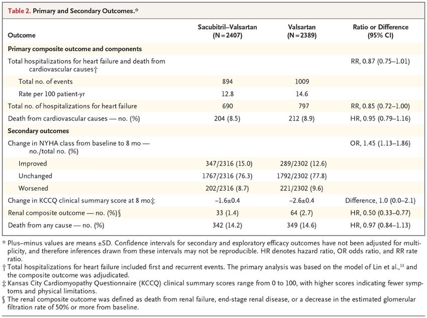
Angiotensin–Neprilysin Inhibition in Heart Failure with Preserved Ejection Fraction

The angiotensin receptor–neprilysin inhibitor sacubitril–
valsartan led to a reduced risk of hospitalization for
heart failure or death from cardiovascular causes
among patients with heart failure and reduced ejection
fraction. The effect of angiotensin receptor–neprilysin
inhibition in patients with heart failure with preserved
ejection fraction is unclear. We randomly assigned 4822
patients with New York Heart Association (NYHA) class
II to IV heart failure, ejection fraction of 45% or higher,
elevated level of natriuretic peptides, and structural
heart disease to receive sacubitril–valsartan (target
dose, 97 mg of sacubitril with 103 mg of valsartan twice
daily) or valsartan (target dose, 160 mg twice daily).
The primary outcome was a composite of total
hospitalizations for heart failure and death from
cardiovascular causes. Primary outcome components,
secondary outcomes (including NYHA class change,
worsening renal function, and change in Kansas City
Cardiomyopathy Questionnaire [KCCQ] clinical
summary score [scale, 0 to 100, with higher scores
indicating fewer symptoms and physical limitations]),
and safety were also assessed.
Time-to-Event Curves for Primary
Composite Outcome and Its Components.
Panel A shows Ghosh–Lin curves for the
primary composite outcome of total
hospitalizations for heart failure and death
from cardiovascular causes, Panel B
Ghosh–Lin curves for total hospitalizations
for heart failure, and Panel C Kaplan–Meier
curves for death from cardiovascular
causes. Insets show the same data on an
enlarged y axis.
Primary Outcome in Prespecified
Subgroups. The primary outcome was a
composite of total hospitalizations for
heart failure and death from
cardiovascular causes. Race was
reported by the patient. New York Heart
Association (NYHA) class may have
changed between screening and
randomization. The diamond indicates the
overall effect, the size of the boxes is
proportional to the number of patients in
the subgroup, and arrows indicate that
the upper or lower boundary of the
confidence interval is off the scale. ACE
denotes angiotensin-converting enzyme,
GFR glomerular filtration rate, and NT-
proBNP N-terminal pro–B-type natriuretic
peptide.
Discussion
In this trial involving patients with heart failure and preserved left ventricular ejection fraction,
we compared treatment with sacubitril–valsartan with treatment with valsartan alone. The
primary composite outcome of total hospitalizations for heart failure and death from
cardiovascular causes did not differ significantly between the two groups. The findings from
nine prespecified supportive and sensitivity analyses were consistent with those from the
primary analysis.
There were fewer primary outcome events with sacubitril–valsartan than with valsartan, and an
analysis of investigator-reported primary outcomes suggested a benefit of this therapy. There
was a modest, although statistically nonsignificant, lower rate of hospitalizations for heart failure
with sacubitril–valsartan than with valsartan and no significant difference in the risk of death
from cardiovascular causes. Of four prespecified secondary outcomes, which were considered
to be exploratory, the change in the NYHA class from baseline to month 8 and the occurrence
of a decline in renal function favored sacubitril–valsartan over valsartan. Sacubitril–valsartan
was associated with a higher incidence of hypotension and angioedema but a lower incidence
of elevated serum creatinine or potassium levels than valsartan. We did not find a significant
benefit of sacubitril–valsartan in patients with heart failure with preserved ejection fraction with
respect to the primary composite outcome of total hospitalizations for heart failure and death
from cardiovascular causes. In the context of known benefit of this treatment in patients with
heart failure and left ventricular systolic dysfunction, and with the suggestion of a differential
effect of sacubitril–valsartan in our trial in relation to left ventricular ejection fraction, future
research should focus on the potential role of angiotensin receptor–neprilysin inhibition in
patients with heart failure and ejection fraction that is below normal but not frankly reduced.
P2Y12 is a chemoreceptor for adenosine
diphosphate (ADP) that belongs to the Gi class
of a group of G protein-coupled (GPCR)
purinergic receptors. This P2Y receptor family
has several receptor subtypes with different
pharmacological selectivity, which overlaps in
some cases, for various adenosine and uridine
nucleotides. The P2Y12 receptor is involved in
platelet aggregation and is thus a biological
target for the treatment of thromboembolisms
and other clotting disorders. Two transcript
variants encoding the same isoform have been
identified for this gene.
In the field of purinergic signaling, the P2Y12
protein is found mainly but not exclusively on the
surface of blood platelets, and is an important
regulator in blood clotting. The drugs clopidogrel
(Plavix), prasugrel (Efient, Effient), ticagrelor
(Brilinta), and cangrelor (Kengreal) bind to this
receptor and are marketed as antiplatelet
agents.
A network meta-analysis of 37 studies involving
88,402 STEMI patients and 5,077 major adverse
cardiac events (MACE) patients found that use
of prasugrel was associated with lower mortality
and MACE than other drugs in this class
(clopidogrel and ticagrelor).
Cytochrome P450 2C19 (abbreviated CYP2C19) is an enzyme.
This protein, a member of the cytochrome P450 mixed-function oxidase system, is involved in the metabolism of
xenobiotics, including many proton pump inhibitors and antiepileptics. In humans, the CYP2C19 protein is
encoded by the CYP2C19 gene. CYP2C19 is a liver enzyme that acts on at least 10% of drugs in current clinical
use, most notably the antiplatelet treatment clopidogrel (Plavix) also drugs that treat pain associated with ulcers,
such as omeprazole, antiseizure drugs such as mephenytoin, the antimalarial proguanil, and the anxiolytic
diazepam. Genetic polymorphism (mainly CYP2C19*2, CYP2C19*3 and CYP2C19*17) exists for CYP2C19
expression, with approximately 3–5% of European and 15–20% of Asian populations being poor metabolizers
with no CYP2C19 function. This may reduce the efficacy of clopidogrel (Plavix). The basis for this reduced effect
of clopidogrel in patients who have a gene of reduced activity may seem somewhat paradoxical, but can be
understood as follows. Clopidogrel is administered as a “prodrug;” that is, a drug that is inactive when taken, and
then depends on the action of an enzyme in the body in order to be activated. In patients who have a gene of
reduced activity, clopidogrel may not be metabolized to its active form.
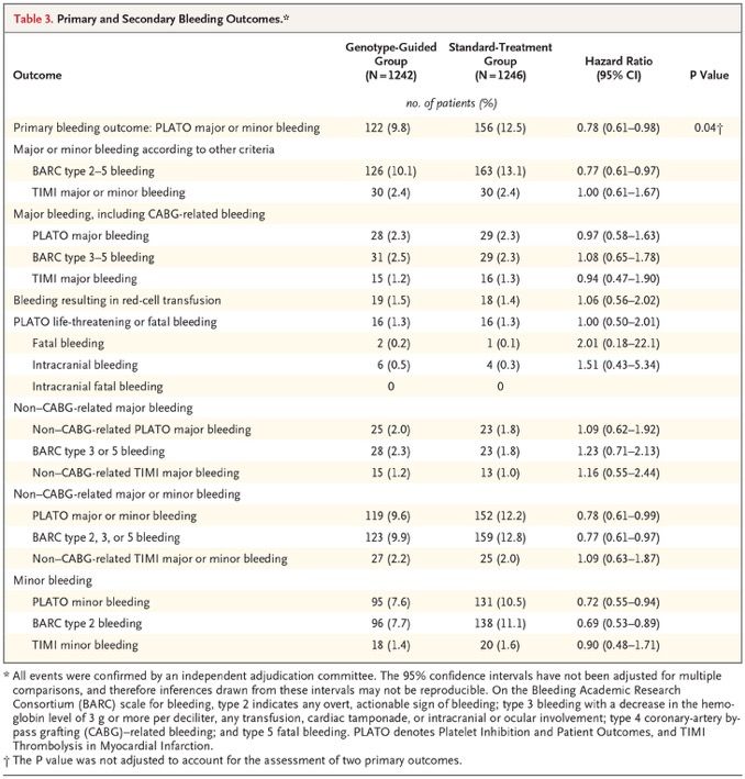
A Genotype-Guided Strategy for Oral P2Y12 Inhibitors in Primary PCI
It is unknown whether patients undergoing primary percutaneous coronary intervention (PCI) benefit from
genotype-guided selection of oral P2Y12 inhibitors. We conducted a randomized, open-label, assessor-blinded
trial in which patients undergoing primary PCI with stent implantation were assigned in a 1:1 ratio to receive
either a P2Y12 inhibitor on the basis of early CYP2C19 genetic testing (genotype-guided group) or standard
treatment with either ticagrelor or prasugrel (standard-treatment group) for 12 months. In the genotype-guided
group, carriers of CYP2C19*2 or CYP2C19*3 loss-of-function alleles received ticagrelor or prasugrel, and
noncarriers received clopidogrel. The two primary outcomes were net adverse clinical events — defined as
death from any cause, myocardial infarction, definite stent thrombosis, stroke, or major bleeding defined
according to Platelet Inhibition and Patient Outcomes (PLATO) criteria — at 12 months (primary combined
outcome; tested for noninferiority, with a noninferiority margin of 2 percentage points for the absolute
difference) and PLATO major or minor bleeding at 12 months (primary bleeding outcome).
The initial plan was for the trial to compare a genotype-guided strategy for selecting oral P2Y12 inhibitors with
a standard-treatment strategy for which clopidogrel was recommended. However, the 2011 European Society
of Cardiology (ESC) guideline for patients with acute coronary syndrome without ST-segment elevation
recommended use of ticagrelor or prasugrel over clopidogrel. In anticipation that the same recommendation
would be made for patients with ST-segment elevation, we changed the treatment in the standard-treatment
group from clopidogrel to ticagrelor or prasugrel. Because we were now testing guided de-escalation of
therapy instead of guided escalation of therapy, there were major changes in the primary outcomes and
hypotheses of the trial.
Incidence Curves for the Primary
Outcomes. Panel A shows the
cumulative incidence of the primary
combined thrombotic and bleeding
outcome, consisting of death from
any cause, myocardial infarction,
definite stent thrombosis, stroke, or
major bleeding defined according to
Platelet Inhibition and Patient
Outcomes (PLATO) criteria. Panel B
shows the primary bleeding outcome
of PLATO major or minor bleeding.
The inset in each panel shows the
same data on an enlarged y axis.
PCI denotes percutaneous coronary
intervention.
Discussion
In this trial, we investigated the possible clinical benefit of CYP2C19 genotype–guided antiplatelet
therapy in patients with STEMI undergoing primary PCI. There are two key findings from this trial.
First, the use of a genotype-guided strategy, in which patients without a CYP2C19 loss-of-function
allele received clopidogrel, was not associated with a higher risk of combined death from any
cause, myocardial infarction, definite stent thrombosis, stroke, or major bleeding 12 months after
primary PCI than standard treatment with the more potent P2Y12 inhibitors ticagrelor and
prasugrel. Second, the use of clopidogrel in the genotype-guided group resulted in a lower risk of
(mostly minor) bleeding than standard treatment. However, the incidence of bleeding was not
significantly lower in the de-escalation group than in the prasugrel group. Furthermore, this
method of guiding therapy requires patients to switch between P2Y12 inhibitors multiple times if
they have high platelet reactivity during treatment with clopidogrel, and it requires patients to
revisit the clinic to perform platelet-function testing. On the basis of the TROPICAL-ACS trial, the
latest ESC guidelines give a class IIb recommendation to use platelet-function testing for guided
de-escalation, especially in patients who are not deemed to be candidates for 12 months of potent
antiplatelet therapy. In conclusion, in patients with STEMI undergoing primary PCI, a CYP2C19
genotype–guided strategy for selection of oral P2Y12 inhibitor therapy was noninferior to standard
treatment with ticagrelor or prasugrel at 12 months with respect to thrombotic events and resulted
in a lower incidence of bleeding.
Editorial (Dan Roden)
The POPular Genetics trial provides strong support for a genotype-guided approach to clopidogrel
prescribing in patients of European ancestry, in whom the contribution of CYP2C19 variants was
first defined; a minority of patients of European ancestry carry loss-of-function variants, and very
few are poor metabolizers. The result has even greater implications for parts of the world where
these variants are much more common. Professional societies, which increasingly view
atherosclerosis as a worldwide epidemic, must now rethink their stance with respect to genotyping
to improve the effectiveness of clopidogrel therapy.
Lancet review on colon cancer last week
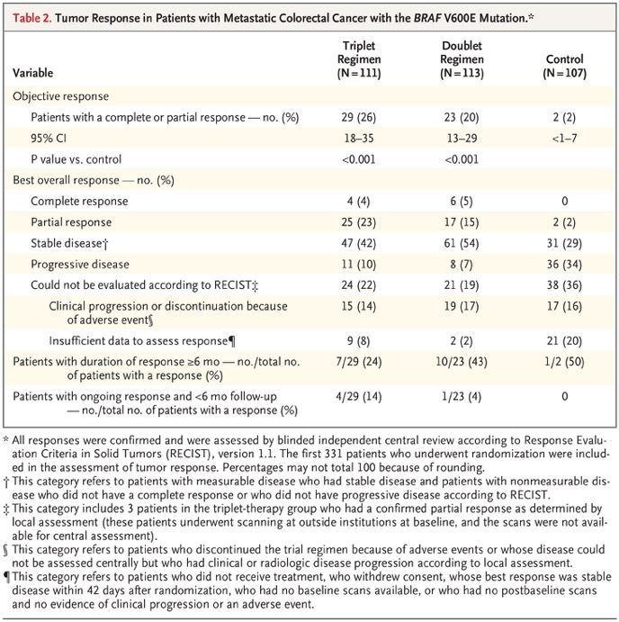
Encorafenib, Binimetinib, and Cetuximab in BRAF V600E–Mutated Colorectal
Cancer
Patients with metastatic colorectal cancer with the
BRAF V600E mutation have a poor prognosis, with a
median overall survival of 4 to 6 months after failure of
initial therapy. Inhibition of BRAF alone has limited
activity because of pathway reactivation through
epidermal growth factor receptor signaling. In this open-
label, phase 3 trial, we enrolled 665 patients with BRAF
V600E–mutated metastatic colorectal cancer who had
had disease progression after one or two previous
regimens. Patients were randomly assigned in a 1:1:1
ratio to receive encorafenib, binimetinib, and cetuximab
(triplet-therapy group); encorafenib and cetuximab
(doublet-therapy group); or the investigators’ choice of
either cetuximab and irinotecan or cetuximab and
FOLFIRI (folinic acid, fluorouracil, and irinotecan)
(control group). The primary end points were overall
survival and objective response rate in the triplet-
therapy group as compared with the control group. A
secondary end point was overall survival in the doublet-
therapy group as compared with the control group. We
report here the results of a prespecified interim
analysis.
Overall Survival.

Panel A shows the Kaplan–Meier analysis
of the probability of survival in the triplet-
therapy group as compared with the control
group, and Panel B the probability of
survival in the doublet-therapy group as
compared with the control group. Panel C
shows the results of the subgroup analysis
of overall survival in the triplet-therapy
group as compared with the control group.
Eastern Cooperative Oncology Group
(ECOG) performance-status scores range
from 0 to 5, with higher scores indicating
greater disability.
Best Percentage Change in Size of
Target Lesions. Shown are the best
percentage changes from baseline in the
sum of the diameters of the target
lesions in each patient in the three
groups, as determined by central review.
The dashed lines at 20% and −30%
indicate progressive disease and partial
response, respectively, according to
Response Evaluation Criteria in Solid
Tumors, version 1.1. The asterisks
indicate patients who had a complete
response, partial response, or stable
disease with respect to target lesions but
who had a new lesion, a progressing
nontarget lesion, or both.
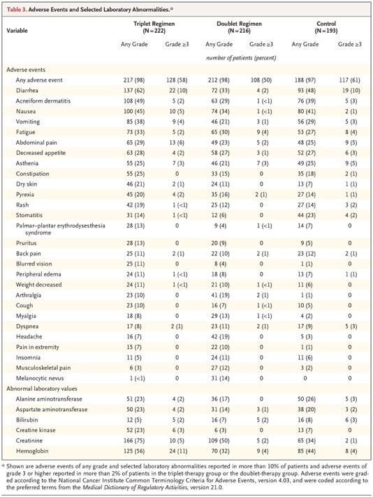
The most common adverse events in the
triplet-therapy group were gastrointestinal-
related and skin-related events, including
diarrhea, nausea, vomiting, and acneiform
dermatitis. Low hemoglobin level or anemia
was a common laboratory abnormality. Table
S3 lists selected adverse events that occurred
during the trial, grouped according to clinically
similar events that are commonly associated
with BRAF and MEK inhibitors. Class-related
toxic effects of MEK inhibitors, including
serous retinopathy and left ventricular
dysfunction, occurred at rates similar to those
described previously and were managed with
treatment interruptions with or without
subsequent dose reduction. Adverse events of
grade 3 or higher were observed in 58% of
patients in the triplet-therapy group, in 50% in
the doublet-therapy group, and in 61% in the
control group. Discontinuation of therapy
primarily because of an adverse event was
seen in 7% of patients in the triplet-therapy
group, in 8% in the doublet-therapy group, and
in 11% in the control group. Fatal adverse
events occurred in 4%, 3%, and 4% of the
patients, respectively. Three of the deaths
were determined by the investigators to be
related to treatment: one death (in the triplet-
therapy group) was from colonic perforation,
one (in the control group) was from
anaphylaxis, and one (in the control group)
was from respiratory failure.
Discussion
This initial analysis of the BEACON CRC trial shows that the triplet regimen of encorafenib,
binimetinib, and cetuximab resulted in longer overall survival and a higher objective response rate
than a control regimen of cetuximab plus the investigators’ choice of irinotecan-based
chemotherapy in patients with BRAF V600E–mutated metastatic colorectal cancer who had had
disease progression after one or two previous regimens. The doublet regimen of encorafenib and
cetuximab also resulted in significantly longer overall survival and a higher objective response rate
than were seen in the control group. The results in the control group were as expected on the
basis of recently reported prospective data in a similar population and several published
retrospective analyses. The triplet regimen was designed to be a combination of agents that
would provide the most effective inhibition of the MAPK pathway. Preclinical and clinical studies
showed that the lack of efficacy of single-agent BRAF or dual BRAF and MEK inhibition in BRAF
V600E–mutated colorectal cancer is related to EGFR-mediated adaptive feedback — a finding
that led to the development of a combination of BRAF, MEK, and EGFR inhibition through a series
of iterative studies.
This initial analysis in patients with BRAF V600E–mutated metastatic colorectal cancer who had
had disease progression after one or two previous regimens showed that a triplet regimen of
encorafenib, binimetinib, and cetuximab or a doublet regimen of encorafenib and cetuximab, as
compared with current standard therapy, resulted in a significant and clinically relevant benefit
with respect to overall survival and objective response rate. The side-effect profiles of both
combination regimens allowed maintenance of high dose intensity for the majority of patients and
are consistent with the known profile of each agent. Further follow-up is needed to better define
the relative benefits of the triplet and doublet regimens.
Die Spielmeyer-Vogt-Krankheit, auch Batten-
Syndrom ist eine sehr seltene angeborene
neurodegenerative Erkrankung mit den
Hauptmerkmalen ubiquitärer
Lipopigmentablagerung in den Lysosomen.
Diese zerebellare Form einer Heredoataxie ist
die klassische juvenile Form der Neuronalen
Ceroid-Lipofuszinosen als CLN3. Die Häufigkeit
beträgt in Deutschland bei Geburt etwa 1 zu
143.000, in Schweden etwa 1 zu 45.000. Die
Vererbung erfolgt autosomal-rezessiv.
Klinische Kriterien sind:
Manifestation im 6.–7. Lebensjahr
Sehstörung in 70 %, zunehmende Erblindung
mit Optikusatrophie, Makuladegeneration und
Pigmentablagerungen
Verlust erworbener intellektueller und
motorischer Funktionen
Haltungsschwäche, Gangunsicherheit, Athetose
Später Krampfanfälle
Überlebensdauer 6–20 Jahre.
Im Blutausstrich finden sich Lymphozyten mit
großen Vakuolen, eine Diagnosesicherung
erfolgt durch Humangenetik.
Als Spleißen bzw. Splicing (englisch splice ‚miteinander
verbinden‘, ‚zusammenkleben‘) wird ein wichtiger Schritt der
Weiterverarbeitung (Prozessierung) der Ribonukleinsäure
(RNA) bezeichnet, der im Zellkern von Eukaryoten
stattfindet und bei dem aus der prä-mRNA die reife mRNA
entsteht. Die zunächst in der Transkription gebildete prä-
mRNA enthält noch Introns und Exons. Durch das Splicing
werden die Introns entfernt und die angrenzenden Exons
miteinander zur fertigen mRNA verknüpft.
Splicing findet zusammen mit der Polyadenylierung (Tailing)
des 3'-Endes nach der Transkription statt, ist also ein
posttranskriptioneller Vorgang. Im Unterschied dazu ist das
Capping des 5'-Endes ein cotranskriptioneller Vorgang.
Einige RNAs können Introns ohne die Hilfe eines großen
Spliceosoms (siehe unten) entfernen. Die chemische
Aktivität dazu besitzen sie selbst, d. h. es handelt sich um
Ribozyme, die nur in einigen Fällen (Gruppe II Introns) die
Hilfe von Proteinen für eine korrekte Faltung benötigen.
Auch bei einigen Krankheitsbildern spielt das Splicing eine
große Rolle. Mutationen in introns haben keinen direkten
Effekt auf die Sequenz des Proteins, das durch ein Gen
codiert wird. In einigen Fällen jedoch betreffen Mutationen
Sequenzen, die für das Splicing wichtig sind und führen so
zu einem falschen Prozessieren der prä-mRNA. Die so
entstehenden RNAs codieren für unfunktionelle oder sogar
schädliche Proteine und führen damit zu erblichen
Erkrankungen. In den letzten Jahren zeigte sich immer
deutlicher, dass Transkription, Prozessierung der RNA (also
Splicing, Capping und Tailing), RNA-Export in das
Zytoplasma, RNA-Lokalisierung, Translation und RNA-
Abbau einander beeinflussen und regulieren.
Patient-Customized Oligonucleotide Therapy for a Rare Genetic Disease
Genome sequencing is often pivotal in the diagnosis of rare
diseases, but many of these conditions lack specific
treatments. We describe how molecular diagnosis of a rare,
fatal neurodegenerative condition led to the rational design,
testing, and manufacture of milasen, a splice-modulating
antisense oligonucleotide drug tailored to a particular patient.
Proof-of-concept experiments in cell lines from the patient
served as the basis for launching an “N-of-1” study of milasen
within 1 year after first contact with the patient. There were no
serious adverse events, and treatment was associated with
objective reduction in seizures (determined by
electroencephalography and parental reporting). This study
offers a possible template for the rapid development of
patient-customized treatments. (Funded by Mila’s Miracle
Foundation and others.) A 6-year-old girl presented with the
insidious onset of blindness, ataxia, seizures, and
developmental regression. The parents’ first concerns dated
back to when the girl was 3 years of age, when her right foot
began to turn inward. When she was 4 years of age, her
family noticed her pulling books close to her face at bedtime.
At 5 years of age, she came to medical attention because of
modest language and social regression, as well as increased
clumsiness and stumbling. In the months before she turned 6
years of age, the progression of symptoms accelerated, and
she was hospitalized after the rapid development of loss of
vision, frequent falls, dysarthria, and dysphagia. Magnetic
resonance imaging (MRI) of the head revealed mild cerebral
and cerebellar atrophy, and 24-hour electroencephalography
(EEG) revealed several subclinical generalized seizures.
Mutation Identification
Patient was heterozygous for c.1102G->C, a known mutation. Whole-genome sequencing was undertaken to
search for the missing second mutation, which was hypothesized to be a noncoding mutation or a structural
variant missed by standard clinical sequencing. Careful inspection of sequencing data revealed a cluster of
chimeric reads deep in MFSD8 intron 6 (Fig. 1B), detected in both the proband and the mother (Fig. S2A).
Chimeric reads in this cluster were fused either to unmapped poly-T sequences on one end or to unmapped
hexameric repeat sequences (AGAGGG) on the other end, which suggested the presence of a DNA insertion
beginning and ending with these motifs. Chimeric breakpoints were offset by 14 bp, suggesting duplication of
an endogenous target sequence (Fig. 1B). We deduced that these features were consistent with the insertion
of an SVA (SINE–VNTR–Alu) retrotransposon. Indeed, amplification by polymerase chain reaction (PCR)
across the breakpoints revealed an approximately 2-kb insertion in the proband and the mother that was
confirmed by RepeatMasker analysis to be an SVA retrotransposon.
Because SVA insertions have been reported to modulate splicing of nearby genes, we examined splicing
patterns in the patient’s family. RNA sequencing (RNA-seq) and reverse-transcriptase–PCR analyses revealed
missplicing of exon 6 into a cryptic splice-acceptor site (i6.SA) in MFSD8 intron 6, in a location 119 bp
upstream from the SVA insertion site, in blood samples and lymphoblasts from the proband and mother (but
not the father or an unaffected sibling). This missplicing precisely segregated with the SVA insertion (Fig. 1C)
and Fig. S5) and was predicted to lead to premature translational termination, which supported the
pathogenicity of the insertion.
Development of an Antisense Oligonucleotide Drug
Nusinersen is an FDA-approved antisense oligonucleotide drug for spinal muscular atrophy that changes the
splicing pattern of the SMN2 RNA. Reasoning that an antisense oligonucleotide might be similarly used to
correct missplicing and restore MFSD8 expression in our patient, we designed antisense oligonucleotides to
target the i6.SA cryptic splice-acceptor site and nearby splicing enhancers. When we tested these antisense
oligonucleotides in patient fibroblasts, we identified three that boosted normal:mutant splicing ratios by a factor
of 2.5 to 3. TY777 was the most efficacious (Figure 2B) and became our lead candidate; we dubbed it
“milasen.”
This report shows a path to personalized
treatment for patients with orphan diseases. It
describes the identification of a novel mutation in a
child with neuronal ceroid lipofuscinosis 7 (CLN7,
a form of Batten’s disease), a rare and fatal
neurodegenerative disease. Identification of the
mutation was followed by the development and
clinical deployment, within 1 year, of a tailored
drug to treat the patient.

Study Timeline and Genetic Diagnosis. Panel A
shows the timeline from the initial clinical diagnosis
to the initiation of treatment. ASO denotes
antisense oligonucleotide. Panel B shows an IGV
(Integrative Genomics Viewer) image of the
patient’s whole-genome sequencing (WGS) read
alignments near MFSD8 intron 6. Characters in
the mapped sequencing reads indicate
mismatched or unaligned (i.e., soft-clipped) bases.
Vertical dashed lines indicate chimeric read
breakpoints. Panel C shows splicing and
translational effects of the SVA (SINE–VNTR–Alu)
insertion in MFSD8. The abbreviation “i6” indicates
the upstream region of the SVA insertion site in Mutations in the MFSD8 gene
intron 6 that is misspliced with exon 6 as a result have been the cause of neuronal
of the activation of the i6.SA cryptic splice site by ceroid lipofuscinosis.
the SVA insertion.
Antisense Oligonucleotide Drug Development. Panel A
shows the location and chemistry of the ASOs that were
designed to block the i6.SA splice acceptor site or exonic
splicing enhancer (ESE) elements. The ESE elements were
predicted with RESCUE-ESE and ESEfinder. 2′-MOE denotes
2′-O-methoxyethyl, and 2′-OMe 2′-O-methyl. Panel B shows
the ratio of the normal exon 6–exon 7 (E6–E7) splicing to the
abnormal exon 6–intron 6 (E6–i6) splicing (normalized to a no-
transfection control), measured in patient fibroblasts that were
transfected (for 24 hours at 100 nmol per liter) as indicated. To
measure splice isoform-specific levels, multiplex reverse-
transcriptase polymerase chain reactions were conducted with
isoform-specific primer sets, and then the intensity of the
isoform-specific bands was quantified by gel electrophoresis
(Fig. S6). “Scrambled” indicates a nontargeting oligonucleotide
(TY772). bars indicate 95% confidence intervals of the
means. P values were calculated by two-sided t-test. Panel C
shows RNA sequencing (RNA-seq) analysis validation of the
splice-correcting effect of milasen (TY777). For the calculation
of the fraction of normal splicing (exon 6–exon 7), three other
splicing events that are mutually exclusive with the normal
splicing were considered. Splicing events supported by only
one read are not shown. P values were calculated by Fisher’s
exact test. Panel D shows intracellular vacuoles, visualized by
electron microscopy, in control fibroblasts (MFSD8 wild-type
human foreskin fibroblast; BJ cell line) and in patient
fibroblasts that are either untreated or transfected with the
indicated oligonucleotide. Scoring was performed on a scale of
0 to 5, with 0 representing the lowest and 5 representing the
highest level of vacuole accumulation.
N-of-1 Clinical Study. Panel A shows the dosing schedule.
 (Additional details are provided in Fig. S14A.) Panel B
 shows the concentration of milasen in cerebrospinal fluid
 (CSF) before each administration (trough). An additional
 measurement of the concentration in CSF was obtained at
 day 174 (without concurrent dose administration). bars
 indicate the minimum and maximum values of duplicate
 measurements. Trough levels rose steadily in a dose-
 proportional fashion until day 40, at which point they
 dropped to 1.7 ng per milliliter and then resumed their rise
 with repeated dosing up to a plateau of 18 to 27 ng per
 milliliter. The dip at day 40 may have been due to a CSF
 leak, given its coincident timing with a post–lumbar
 puncture headache after the previous dose. A similar
 plateauing of CSF trough levels was observed in a
 previous study of intrathecally delivered nusinersen (9 to
 11 ng per milliliter after four repeated doses of 12 mg).8
 Panel C shows the trends in seizure frequency and
 duration as reported in a seizure diary recorded by the
 parents. Seizures were all of the same type: sudden
 startle followed by uncontrollable, untriggered laughter
 that was different from the patient’s natural laugh, at times
 accompanied by an increase in the nonspecific repetitive
 hand movements she had at baseline. Panel D shows the
 trends in seizure activity as detected by
 electroencephalography (EEG). In a comparison of the
 means of the initial two recordings and the subsequent
 three recordings, the daily seizure count, seizure duration,
Drug given intrathecally every 14 days and percent cumulative time spent in seizure decreased
 by 63% (from 31.5 to 11.7 per day), 52% (from 108
 seconds to 52 seconds), and 85% (from 3.9% to 0.6%),
 respectively.
Discussion
Over the course of treatment to date, milasen appears to have had an acceptable side-effect
profile, with no safety concerns. These findings are consistent with those of previous clinical
studies of intrathecal delivery of an FDA-approved antisense oligonucleotide for spinal muscular
atrophy and investigational antisense oligonucleotides for amyotrophic lateral sclerosis and
Huntington’s disease, which had similarly acceptable side-effect profiles. Treatment has also
been accompanied by objective reductions in the frequency and duration of seizures. Milasen
itself remains an investigational drug, and it is not suited to the treatment of other patients with
Batten’s disease because its design is customized to our patient’s specific mutation.
Nonetheless, this experience indicates that antisense oligonucleotides may deserve
consideration as a platform for the rapid delivery of individualized treatments. Further exploration
of this approach will continue to require careful case-by-case consideration of a number of
scientific, clinical, and ethical issues, and expectations need to be tempered by the fact that this
approach bears substantial risks. At this time, it should be contemplated only in the context of
exceptionally serious or life-threatening circumstances. It should also be recognized that only a
minority of patients are likely to have mutations that are amenable to the “mRNA splice-
switching” strategy deployed here. More generally, this approach is at present probably scalable
to only a limited number of patients, given current limitations on systemic and infrastructural
issues (e.g., regulatory burden, manufacturing capacity, cost, and reimbursement).
This study illustrates the ability to rationally design, test, and deploy a novel therapeutic agent
for a patient with a rare disease on the basis of an understanding of her specific pathogenic
mutation. It is an example of individualized genomic medicine.
A 77-year-old right-handed woman with limited cutaneous systemic sclerosis presented to
the rheumatology clinic with a 2-year history of slowly progressive, painful swelling of her
fingertips. On physical examination, the patient had telangiectases on her face and thorax
and thickening of the skin on her right hand, both forearms, and face, as well as soft-tissue
swelling of the tips of the fingers of both hands (Panels A and B). Findings on capillaroscopy
were notable for disorganization of capillaries, avascular areas, and the presence of enlarged
capillaries. A radiograph of the hands showed calcifications near the distal phalanx of the
first, second, and third fingers of the right hand and the first finger of the left hand (Panel C),
which were consistent with calcinosis, a manifestation of limited cutaneous systemic
sclerosis. These calcium deposits may grow, ulcerate, and become infected. Despite trials of
multiple medications, the patient had progressive pain and numbness in her fingers, with
increasing difficulty handling objects, and was referred for surgical evaluation regarding
excision of the lesions.
A 60-year-old woman who was a current smoker presented to the emergency department with
acute chest pain. The troponin I level was 51 ng per liter on a high-sensitivity assay (reference
value,
Der Von-Willebrand-Faktor (VWF) oder von-Willebrand-Faktor
(vWF) ist ein Protein, das als Trägerprotein des
Blutgerinnungsfaktors VIII eine wichtige Rolle bei der
Blutstillung spielt und nach Erik Adolf von Willebrand (1870–
1949), einem finnischen Internisten, benannt wurde.
Der Von-Willebrand-Faktor wird sowohl von Megakaryozyten
als auch von den Endothelzellen gebildet, die die Innenwand
eines Blutgefäßes (die Intima) bilden. In den Blutplättchen
(Thrombozyten) als Abschnürungen der Megakaryozyten ist er
in den α-Granula gespeichert, das Gefäßendothel gibt ihn zur
Speicherung an die subendotheliale Matrix ab. Kommt es zu
einem Riss der Innenwand des Endothels, werden die darunter
liegenden Proteine der Gefäßwand freigelegt u. a. Kollagene.
An diese kann der Von-Willebrand-Faktor binden. Bestimmte
zelluläre Elemente des Blutes, die Blutplättchen
(Thrombozyten), verfügen auf ihrer Oberfläche über eine
Andockstelle, an die der Von-Willebrand-Faktor binden kann.
Diese wird als Von-Willebrand-Rezeptor oder Glykoprotein
Ib/V/IX bezeichnet. Der Von-Willebrand-Faktor schafft also eine
Brücke zwischen den Blutplättchen und der verletzten
Gefäßwand. An bereits adhärenten Thrombozyten kann der
vWF mittels Glykoprotein IIb bzw. IIIa binden. Der Von-
Willebrand-Faktor hat somit eine direkte Wirkung bei der
zellulären Blutstillung. Bei der plasmatischen Blutgerinnung ist
er zwar nicht direkt beteiligt, da er zur Bildung des Fibrins nicht
erforderlich ist. Durch seine Funktion als Träger- und
Schutzprotein für den Gerinnungsfaktor VIII führt ein Mangel an
Von-Willebrand-Faktor oder ein Defekt seiner Proteinstruktur
durch diese unmittelbare Wechselwirkung aber dennoch zur
Beeinträchtigung der plasmatischen Hämostase (s. Willebrand-
Jürgens-Syndrom und von-Willebrand-Krankheit).
N Engl J Med
2019 earlier
Pathophysiology of Severe ADAMTS13 Deficiency. In an
arteriole of a healthy person (Panel A), von Willebrand
factor is synthesized by endothelial cells and stored in
Weibel–Palade bodies as long strings of repeating
subunits. After stimulation of endothelial cells, these
ultralarge von Willebrand factor multimers are secreted
and some of these molecules remain anchored to the
endothelial-cell surface; others are released into the
circulation. Flowing blood unfolds ultralarge von
Willebrand factor multimers, exposing the platelet
binding and the ADAMTS13 cleavage sites. After
ADAMTS13 cleavage, von Willebrand factor molecules
again adopt a coiled, condensed shape without exposed
platelet binding or ADAMTS13 cleavage sites. In the
absence of ADAMTS13 (Panel B), many platelets attach
to the exposed platelet-binding sites on the ultralarge
von Willebrand factor multimer strings. Some of these
multimers remain attached to the endothelial surface,
coating the vessel wall. Other ultralarge von Willebrand
factor strings detach from the endothelial-cell surface
and enter the circulation, where they become coated
with adherent platelets. As these platelet-loaded
ultralarge von Willebrand factor multimers approach
small vessels and bifurcations with sharp turns, blood
flow accelerates, increasing shear stress. Increased
shear stress causes further unfolding of the ultralarge
von Willebrand factor molecules, exposing more
platelet-binding sites. In conditions involving increased
von Willebrand factor synthesis and release into plasma
(e.g., during infection, inflammation, and pregnancy), the
circulating ultralarge von Willebrand factor molecules
with their many attached platelets form webs at the sites
of increased shear stress, obstructing blood flow and
causing thrombosis
Hereditary Thrombotic Thrombocytopenic Purpura
Hereditary thrombotic
thrombocytopenic purpura (TTP), also
known as Upshaw–Schulman
syndrome (Online Mendelian
Inheritance in Man number, 274150), is
a rare autosomal recessive disorder
caused by ADAMTS13 mutations that
result in the absence or severe
deficiency of the plasma
metalloprotease ADAMTS13.
ADAMTS13 is required for cleavage of
newly synthesized von Willebrand
factor multimers. Decreased
ADAMTS13 activity is associated with
an increased size of von Willebrand
factor multimers and an increased risk
of microvascular thrombosis. Patients
with hereditary TTP may appear to be
healthy, but their increased risk of
critical thrombosis is always present.
This review describes the history,
pathogenesis, prevalence, clinical
features, and current management of
hereditary TTP, as well as potential
future treatments. The only known
substrate of ADAMTS13 is von
Willebrand factor, which is exclusively
synthesized by endothelial cells and
megakaryocytes.
ADAMTS13 Mutations in Hereditary TTP
More than 200 ADAMTS13 mutations, spread over all
ADAMTS13 protein domains, have been identified in
patients with hereditary TTP. In addition, there are at
least 10 missense ADAMTS13 single-nucleotide
polymorphisms, some of which are in strong linkage
disequilibrium with specific ADAMTS13 mutations and
influence their molecular effects.

 Clinical Features
 Some patients with hereditary TTP may have symptoms
 that manifest at birth, whereas others remain
 asymptomatic for decades. When symptoms occur, the
 clinical features may be indistinguishable from an acute
 episode of acquired TTP or they may differ
 substantially.

 Typical Triggers of Disease Manifestations and
 Heterogeneous Clinical Presentations in Hereditary
 TTP. Shown are four patients with hereditary TTP who
 present at times of risk (i.e., the neonatal period, after
 heavy alcohol intake, and during pregnancy) or with
 manifestations that occur spontaneously. All the
 patients have common clinical features that are distinct
 from those that are characteristic of patients with
 acquired TTP. Some patient data are courtesy of Eric
 Avery, M.D., Lincoln, Nebraska. CT denotes computed
 tomography, and MRI magnetic resonance imaging.
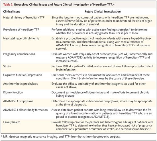
Distinguishing Hereditary TTP from Acquired TTP
The patient’s age may help to distinguish between hereditary TTP and acquired TTP. Acquired TTP is
much less common in young children than in adults, whereas hereditary TTP is commonly diagnosed in
children. In the Japanese cohort, the disease first manifested in 33 of the 43 patients (77%) before 10
years of age. The presence of a functional ADAMTS13 inhibitor or an increased anti–ADAMTS13 IgG
antibody titer argues against the diagnosis of hereditary TTP. An exception is the occurrence of severe
hemolysis with a high plasma hemoglobin concentration (≥2 g per deciliter), which can cause false positive
results in functional ADAMTS13 inhibitor assays.58 Although the absence of a functional ADAMTS13
inhibitor is characteristic of hereditary TTP, it does not rule out the diagnosis of acquired TTP. Among
patients with acquired TTP in the Oklahoma Thrombotic Thrombocytopenic Purpura–Hemolytic Uremic
Syndrome Registry, a functional ADAMTS13 inhibitor was not initially identified in 15 of 86 patients (17%).
Long-Term Outcomes
Data on long-term outcomes in patients with hereditary TTP are lacking. In the Japanese cohort of 43
patients, only 13 patients (30%) were followed after 35 years of age, and only 6 (14%) were followed after
45 years of age. Three of the 43 patients (7%) died at the ages of 38 to 79 years — 2 from chronic kidney
disease and 1 from stroke. In the Norwegian cohort of 21 patients (including 3 siblings who were probably
affected), 5 (24%) died. Two of these patients died before 1 year of age, and the other 3 died at 40 to 60
years of age. The causes of death were not reported. In the cohort in the United Kingdom, 4 of the 73
patients (5%) died from stroke; the patients’ ages were not reported. Five of the 120 patients (4%) in the
International Hereditary Thrombotic Thrombocytopenic Purpura Registry died; their ages and causes of
death were not reported.
Current Management
Plasma infusion is usually sufficient to treat acute episodes in patients with hereditary TTP, although for
severe manifestations (e.g., in pregnancy) therapeutic plasma exchange may be appropriate. For patients
who have recurrent symptoms, regular lifetime prophylactic plasma infusions are appropriate, although the
decision to begin prophylaxis is difficult, especially in a child. The half-life of ADAMTS13 activity in patients
receiving regular prophylactic plasma infusions is 2.5 to 3.5 days. The development of inhibitory
ADAMTS13 alloantibodies in patients receiving plasma therapy has been reported twice.
Future Management of Hereditary TTP
The availability of rhADAMTS13 may revolutionize the management of hereditary TTP.
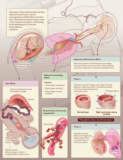
A 35-Year-Old Woman with Cardiopulmonary Arrest during Cesarean Section
At 6 weeks 4 days of gestation, the patient had been seen at
a community health center because of a positive pregnancy
test. She was gravida 4, 1-0-2-1. Her first pregnancy, 5 years
earlier, had resulted in the need for a cesarean section
because the fetus was in the breech presentation; the baby
was delivered at full term without complications. The second
and third pregnancies had been electively terminated. The
patient reported that her husband had human
immunodeficiency virus type 1 (HIV-1) infection and that he
had been taking antiretroviral therapy; he had had
undetectable plasma levels of HIV-1 RNA during the 5
months before this visit. Blood testing in the patient for HIV-1
and HIV-2 antibodies and HIV-1 p24 antigen with the use of a
fourth-generation combination assay was negative.
Preexposure prophylaxis with a fixed-dose combination of
emtricitabine and tenofovir disoproxil fumarate was
prescribed.
Twenty-five days before presentation, at 33 weeks of
gestation, vaginal bleeding developed, and the patient was
brought by ambulance to the obstetrical unit of this hospital.
She reported that on awakening that morning, she had
passed a large volume of bright red, liquid blood and three
large clots, each measuring approximately 1.5 cm in
diameter, through the vagina. She felt occasional
contractions; fetal movement was normal. She reported no
recent trauma, fall, or sexual intercourse. Ultrasonographic
examination was performed; the placenta was positioned
anteriorly along the midline of the uterus, completely
overlying the cervical os, and there was no evidence of
placenta accreta.
The patient underwent cesarean section under
combined spinal–epidural anesthesia. Cefazolin
was administered intravenously before incision of
the skin. During the surgery, a low transverse
uterine incision was made through the anterior
placenta. The membranes were ruptured bluntly,
yielding clear amniotic fluid. The uterine incision
was extended, and the infant was delivered 37
minutes after injection of the spinal anesthetic
agent, which was administered during a combined
spinal–epidural procedure; the opaque surgical
drape was lowered, allowing the patient to see the
infant through the clear drape. The 1-minute and
5-minute Apgar scores were 8 and 10,
respectively. The placenta was removed with
gentle traction.
Three minutes after delivery of the infant, while
clot and debris were being cleared from the uterine
cavity, the exteriorized uterus became blanched
with minimal bleeding. Concurrently, the patient
was noted to be unresponsive, with sporadic
respirations; carotid and aortic pulses were
present. Manual ventilation was begun. Two
minutes later, the patient became pulseless, with
normal sinus rhythm seen on the patient monitor of
the anesthesia machine. Cardiopulmonary
resuscitation was begun, the trachea was
intubated, and mechanical ventilation was initiated.
Pulmonary Embolism
Did this patient have a pulmonary embolism that led to cardiovascular collapse? In support of this diagnosis
was the acute nature of the event and the fact that the patient had several risk factors for pulmonary
embolism, including hypercoagulability, pregnancy, advanced maternal age, and obesity.
Hemorrhage
The patient had multiple risk factors for hemorrhage, and this was the adverse outcome for which we were
most prepared. Given that she had had a previous cesarean section and that she had a complete anterior
placenta previa in this pregnancy, we had concerns about a potential placenta accreta.
Venous Air Embolism
Venous air embolism has been reported to occur at the time of cesarean section. This diagnosis should be
considered any time the operative site is positioned above the patient’s heart, which occurred in this patient
when the uterus was exteriorized. However, as is the case with pulmonary embolism, these events are often
preceded by wheezing, dyspnea, chest pain, or gasping.
Anaphylaxis
Anaphylaxis was also a consideration in this case, given that many medications were administered before
and during the procedure. However, the patient did not have any known drug allergies, and the timing for an
anaphylactic event did not fit well, since cardiovascular collapse occurred more than 30 minutes after these
treatment exposures.
High Cephalad Spread of Neuraxial Anesthetic Agent
High cephalad spread of a neuraxial anesthetic agent can cause sudden loss of consciousness and cardiac
arrest, but in this case, the event occurred more than 30 minutes after injection of the spinal anesthetic
agent, which was administered during a combined spinal–epidural procedure.
Peripartum Cardiomyopathy and Myocardial Infarction
The patient had risk factors for peripartum cardiomyopathy, including being in the late stage of pregnancy,
being of advanced maternal age, and being of African descent.
Eclampsia
Eclampsia should be considered in any pregnant woman who has an acute change in mental status.
However, this patient was normotensive and did not report headaches, vision changes, or pain in the right
upper quadrant. Also, she did not show any signs of seizure activity.
Sie können auch lesen