Max Delbrück Center for Molecular Medicine
←
→
Transkription von Seiteninhalten
Wenn Ihr Browser die Seite nicht korrekt rendert, bitte, lesen Sie den Inhalt der Seite unten

https://www.mdc-berlin.de/de/veroeffentlichungstypen/clinical-
journal-club

  Als gemeinsame Einrichtung von MDC und Charité fördert das Experimental and Clinical Research Center
  die Zusammenarbeit zwischen Grundlagenwissenschaftlern und klinischen Forschern. Hier werden neue
  Ansätze für Diagnose, Prävention und Therapie von Herz-Kreislauf- und Stoffwechselerkrankungen, Krebs
  sowie neurologischen Erkrankungen entwickelt und zeitnah am Patienten eingesetzt. Sie sind eingelanden,
  um uns beizutreten. Bewerben Sie sich!
A baby boy born at 39 weeks of gestation had bilious
emesis, failure to pass meconium, and abdominal
distention within 24 hours after birth. A water-soluble
contrast enema showed a uniformly distended and
shortened colon. What is the most likely diagnosis?

Hirschsprung’s disease

Congenital syphilis

Pyloric stenosis

Duodenal atresia

Necrotizing enterocolitis

                              The correct answer is Hirschsprung’s disease. The
                              patient was taken to the operating room where biopsy
                              specimens of the colon revealed the absence of
                              ganglion cells. Abundant ganglion cells were found at
                              the level of the terminal ileum, and an end ileostomy
                              was performed followed by colectomy with stapled side-
                              to-side anastomosis of the terminal ileum next to the
                              rectal remnant 6 months later.
Beim kongenitalen Megakolon (Synonym Megacolon
congenitum, angeborenes Megakolon,
aganglionotisches Megakolon, Hirschsprung-Krankheit,
Morbus Hirschsprung) handelt es sich um eine
angeborene Erkrankung des Dickdarms aus der
Gruppe der Aganglionosen. Dabei fehlen in einem
Abschnitt des Dickdarms Nervenzellen, wodurch sich
die dortige Muskulatur zusammenzieht und den Darm
verengt. Der Stuhl staut sich vor der Engstelle, wodurch
sich der davorliegende Teil des Darms erweitert
(dilatiert). Systematischer Erstbeschreiber (1886) ist der
dänische Pädiater Harald Hirschsprung (1830–1916).
Im Durchschnitt tritt diese Fehlbildung bei 1 von 5000
Kindern auf, wobei Jungen im Vergleich deutlich
häufiger betroffen sind als Mädchen. Bei ca. 12 % der
Säuglinge mit einem Down-Syndrom (Trisomie 21) ist
Morbus Hirschsprung nachweisbar. Kombinationen mit
anderen Fehlbildungen (z. B. Mukoviszidose,
Brachydaktylie) kommen vor, sind aber selten.
In 80 % der Fälle sind bei der Aganglionose nur das
Rektum und/oder Sigma betroffen (Short-Segment-
Aganglionose). Etwa 5 % gehören zur Long-Segment-
Aganglionose, bei der der krankhaft veränderte
Dickdarmabschnitt insgesamt 40 cm und mehr
ausgedehnt ist. In weniger als 5 % der Fälle fehlen die
Nervenzellen im gesamten Abschnitt des Dickdarmes,
man spricht in diesem Fall von einem Jirásek-Zuelzer-
Wilson-Syndrom. In einigen Fällen fehlen die
Nervenzellen bis in den Dünndarm.
Das kongenitale Megakolon kann auch im Rahmen von
Syndromen auftreten, siehe Mowat-Wilson-Syndrom.
Ein Mangel an Ganglienzellen (Aganglionose) im Bereich des Plexus submucosus (Meißner-Plexus) bzw.
myentericus (Auerbach-Plexus) führt zu einer Hyperplasie (übermäßige Zellbildung) der vorgeschalteten
parasympathischen Nervenfasern mit vermehrter Acetylcholin-Ausschüttung. Durch diese permanente Stimulation
der Ringmuskulatur kommt es zu einem dauerhaften Zusammenziehen des betroffenen Darmabschnittes. Das
übermäßig gebildete Acetylcholin wird durch eine als Ausgleich vermehrt produzierte Acetylcholinesterase
abgebaut.
Späte Defekte der Neuroblasteneinwanderung, Reifungsstörungen eingewanderter Neuroblasten, zeitweilige
Ischämien (Minderdurchblutung) des Darms oder virale Infektionen beim Embryo kommen als Ursache in Frage.
Bei Untersuchungen zum Morbus Hirschsprung wurden Mutationen im sogenannten Ret-Protoonkogen
(autosomal dominante Form), im Endothelin-3-Gen (EDN3) und im Endothelinrezeptor-Gen (EDNRB) (autosomal
rezessive Form) belegt.
Among the patients in our study, Hirschsprung's disease arose from common noncoding variants, rare coding
variants, and copy-number variants affecting genes involved in enteric neural-crest cell fate that exacerbate the
widespread genetic susceptibility associated with RET. For individual patients, the genotype-specific odds ratios
varied by a factor of approximately 67, which provides a basis for risk stratification and genetic counseling.
Die Pylorusstenose beschreibt eine Verengung im
Bereich des Magenausganges. Diese kann angeboren
oder erworben sein. Sie führt zu einer gestörten
Fortleitung des Mageninhalts in den Zwölffingerdarm
und somit zu unstillbarem Erbrechen. Die Behandlung
besteht in der Regel in einer operativen Korrektur der
Engstelle. Die Erkrankung ist bereits mit der Geburt
angelegt und kommt familiär gehäuft vor (evtl. erblich).
Die Ursachen sind bislang ungeklärt. Zu finden ist die
Krankheit vor allem bei West- und Nordeuropäern mit
einer Häufigkeit von 1:300, selten bei Asiaten und fast
nie bei Afrikanern. Der Erkrankungsgipfel liegt bei drei
Wochen nach der Geburt. Die Krankheit tritt besonders
bei den erstgeborenen Jungen auf (Verhältnis Jungen :
Mädchen: 4-5 : 1). Der Säugling erbricht (nicht gallig)
etwa eine halbe Stunde nach der Mahlzeit die Nahrung
schwallartig teilweise oder vollständig. Durch die
Magenreizung können sich im Erbrochenen Blutfäden
befinden. Danach sucht er wieder nach Nahrung. Direkt
nach einer Mahlzeit können gesteigerte
Magenbewegungen (Peristaltik) auf der
Bauchoberfläche im Oberbauch beobachtet werden.
Teilweise ist der vergrößerte Pylorus tastbar. Die
betroffenen Kinder sind durch die behinderte
Nahrungspassage unterernährt, untergewichtig, ständig
hungrig und entsprechend unzufrieden. Sie setzen
Hungerstühle von geringer Masse in hoher Frequenz
ab.
Die Duodenalatresie, auch als Duodenojejunale Atresie
bezeichnet, ist eine angeborene Entwicklungsstörung,
bei der das Lumen des Zwölffingerdarmes nicht
durchgängig ist. Dies ist entweder durch ein Fehlen
eines kurzen oder längeren Darmanteiles (eine echte
Atresie), eine Membran im Darmlumen oder eine
Fehlanlage der Bauchspeicheldrüse bedingt
(ringförmige Fehlanlage der Bauchspeicheldrüse
Pancreas anulare), wobei in diesem Fall meist nur eine
Duodenalobstruktion vorliegt, s. Duodenalstenose. Die
fehlende Durchgängigkeit bewirkt, dass sich vor der
Geburt Magen und Darmanteil vor der Atresie stark
aufweiten und der übrige Darm klein bleibt, da das
geschluckte Fruchtwasser diesen Darmanteil nicht
erreicht. Ferner liegt ein Polyhydramnion vor.

                                                         Abdomenübersicht im Liegen Duodenalatresia bei
                                                         einem Neugeborenen mit typischen Double-Bubble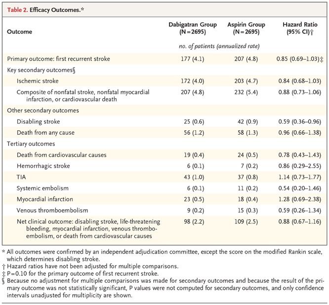
Antibacterial Envelope to Prevent Cardiac Implantable Device Infection
Infections after placement of cardiac implantable
electronic devices (CIEDs) are associated with
substantial morbidity and mortality. There is limited
evidence on prophylactic strategies, other than the use
of preoperative antibiotics, to prevent such infections.
We conducted a randomized, controlled clinical trial to
assess the safety and efficacy of an absorbable,
antibiotic-eluting envelope in reducing the incidence of
infection associated with CIED implantations. Patients
who were undergoing a CIED pocket revision,
generator replacement, or system upgrade or an initial
implantation of a cardiac resynchronization therapy
defibrillator were randomly assigned, in a 1:1 ratio, to
receive the envelope or not. Standard-of-care strategies
to prevent infection were used in all patients. The
primary end point was infection resulting in system
extraction or revision, long-term antibiotic therapy with
infection recurrence, or death, within 12 months after
the CIED implantation procedure. The secondary end
point for safety was procedure-related or system-
related complications within 12 months. The TYRX
envelope is an absorbable single-use envelope
designed to hold a CIED when the device is implanted
in the body. The envelope is constructed from a
multifilament knitted mesh and coated with an
absorbable polymer mixed with minocycline and
rifampin, which elutes the antibiotics into the local
tissue for a minimum of 7 days. The envelope is fully
absorbed in approximately 9 weeks.
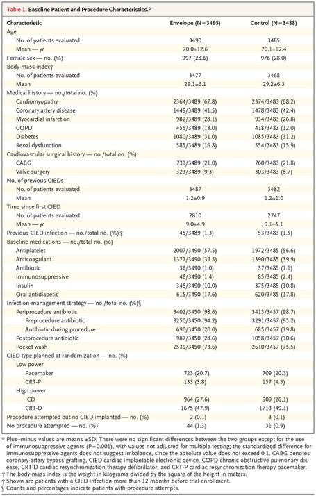
Procedure Characteristics
Procedure characteristics were balanced between the
two groups. A total of 3429 patients who were assigned
to the control group did not receive the envelope, and
3371 patients who were assigned to the envelope
group received the envelope at the time of the CIED
procedure. In the envelope group, the envelope was not
successfully implanted in 10 procedures owing to
limited pocket space; thus, the envelope was
successfully implanted in 99.7% of procedure attempts
(3371 of 3381 attempts) by 646 implanting physicians.
The mean procedure time did not differ significantly
between the two groups (56.3±46.2 minutes in the
envelope group and 55.0±48.0 minutes in the control
group; between-group difference, 1.3 minutes; 95%
confidence interval [CI], −0.9 to 3.5).
Kaplan–Meier Curves for First Major CIED Infection.
Results are for the overall randomized cohort through 12
months (Panel A) and through all follow-up (Panel B) and
were not adjusted for multiple comparisons. Hazard ratios
are derived from Cox regressions, with stratification
according to device class, and indicate the relative
(envelope vs. control) risk of CIED infection. The insets
show the same data on an enlarged y axis.

Major or minor CIED infections within 12 months occurred
in 50 patients in the envelope group and 75 patients in the
control group (12-month Kaplan–Meier estimated event
rate, 1.5% and 2.2%, respectively; hazard ratio, 0.67;
95% CI, 0.47 to 0.96). Through the entire follow-up
period, major CIED infections occurred in 32 patients in
the envelope group and 51 patients in the control group
(36-month Kaplan–Meier estimated event rate, 1.3% and
1.9%, respectively; hazard ratio, 0.63; 95% CI, 0.40 to
0.98)
Subgroup Analysis of Major CIED
Infections through 12 Months. The P
values are for the interaction between the
randomization group and the subgroup
variable. The body-mass index is the
weight in kilograms divided by the square
of the height in meters. The CIED type is
the type planned at randomization: low
power (pacemaker or cardiac
resynchronization therapy pacemaker) or
high power (implantable cardioverter–
defibrillator or cardiac resynchronization
therapy defibrillator). COPD denotes
chronic obstructive pulmonary disease.
Discussion
WRAP-IT was a randomized, controlled clinical trial to assess the safety and efficacy of the
TYRX absorbable antibiotic-eluting envelope. In a population of patients who were at
increased risk for CIED pocket infection, the envelope was significantly more effective at
preventing infection than standard infection-control strategies alone. The efficacy objective was
met, with 40% fewer patients in the envelope group than in the control group having a major
infection through 12 months of follow-up. The envelope was successfully implanted in 99.7% of
procedure attempts, and the safety objective was met, because the envelope group did not
have a higher incidence of CIED procedure-related or system-related complications than the
control group.
Some limitations should be considered when interpreting the results of this trial. First,
consecutive patients were not enrolled because this trial was limited to patients receiving
generators from one device manufacturer (leads from other manufacturers were permitted).
Second, the envelope was commercially available at the time of the trial, which may have
influenced participation in the trial. Third, the use of immunosuppressive agents was not
balanced between the trial groups; however, among these patients, there was only one major
infection within the first 12 months, and therefore this is unlikely to influence interpretation of
the results. Fourth, the lack of data on antibiotic susceptibility limits our ability to address the
unknown risk of the development of antibiotic resistance. Fifth, periprocedure and
postprocedure infection-prevention strategies including antibiotic use were not controlled;
however, there is not yet clear evidence that a particular strategy influences the infection rate.
In conclusion, adjunctive use of an antibacterial envelope resulted in a 40% lower incidence of
major CIED infection than standard-of-care infection-prevention strategies alone. Patients who
received the envelope did not have more procedure-related or system-related complications
than those who did not receive it.
Dabigatran-etexilat ist ein Arzneistoff aus der Gruppe
der Gerinnungshemmer (Antikoagulanzien). Die
Substanz ist ein Prodrug, das nach Umwandlung in das
pharmakologisch aktive Dabigatran den
Blutgerinnungsfaktor IIa (Thrombin) direkt hemmt. Nach
der Einnahme wird der Stoff in der Niere aus dem Blut
gefiltert und über den Urin entsorgt, weshalb
Dabigatranetexilat als Gerinnungshemmer für Patienten
mit Niereninsuffizienz nicht geeignet ist.
Dabigatranetexilat ist für die perorale Verabreichung
geeignet und in dieser Form (Handelsname Pradaxa;
Hersteller Boehringer Ingelheim) in der EU zugelassen
zur Vorbeugung gegen die Bildung von Blutgerinnseln
in den Venen nach elektivem chirurgischen Knie- oder
Hüftgelenksersatz, seit 2011 außerdem zur
Schlaganfallvorbeugung bei Patienten mit
Vorhofflimmern und Schlaganfallrisiko.
Dabigatran bindet kompetitiv und reversibel direkt an
Thrombin und blockiert dessen Wirkung, so dass die
Umwandlung von Fibrinogen zu Fibrin und damit eine
Gerinnselentstehung unterbleibt. Dabigatran verhindert
somit auch die Thrombin-induzierte Verklumpung der
Blutplättchen (Thrombozytenaggregation). Die
gerinnungshemmende (antikoagulatorische) Wirkung
des Dabigatran korreliert mit seiner Konzentration im
Plasma: die therapeutische Konzentration liegt bei
0,05–0,1 mg/l, messbar als 10–20 % der Norm im
neuen F10a/F2a Gerinnungstest EXCA (extrinsic
coagulation activity assay).

                                                         Plättchen
Dabigatran for Prevention of Stroke after Embolic Stroke of Undetermined
Source
Cryptogenic strokes constitute 20 to 30% of
ischemic strokes, and most cryptogenic strokes are
considered to be embolic and of undetermined
source. An earlier randomized trial showed that
rivaroxaban is no more effective than aspirin in
preventing recurrent stroke after a presumed
embolic stroke from an undetermined source.
Whether dabigatran would be effective in
preventing recurrent strokes after this type of
stroke was unclear.
We conducted a multicenter, randomized, double-
blind trial of dabigatran at a dose of 150 mg or 110
mg twice daily as compared with aspirin at a dose
of 100 mg once daily in patients who had had an
embolic stroke of undetermined source. The
primary outcome was recurrent stroke. The primary
safety outcome was major bleeding.
Trial Population
Patients 60 years of age or older were eligible for
enrollment if they had had an embolic stroke of
undetermined source within the previous 3 months
or, if they had at least one vascular risk factor,
within the previous 6 months; patients 18 to 59
years of age were eligible if they had had a
qualifying stroke within the previous 3 months and
had at least one additional vascular risk factorDiscussion The RE-SPECT ESUS trial showed no significant difference between the effect of dabigatran and that of aspirin on the risk of recurrent stroke among patients with embolic stroke of undetermined source. The rate of recurrent stroke was 4.1% per year among patients in the dabigatran group and 4.8% per year among patients in the aspirin group. Dabigatran was associated with major bleeding in 1.7% of the patients per year, and aspirin was associated with major bleeding in 1.4% of the patients per year. The percentages were similar in the two groups in all subcategories of major bleeding, but more patients in the dabigatran group than in the aspirin group had clinically relevant nonmajor bleeding. Our hypothesis was that dabigatran would be more effective than aspirin for stroke prevention in patients with embolic stroke of undetermined source because many of these patients might have had an unrecognized source of cardiac embolism, including atrial fibrillation. Post hoc analysis suggested that dabigatran may have had an effect on stroke recurrence after 1 year, but no inferences can be made because of the post hoc nature of the analysis. A possible explanation for this temporal pattern might be a progressive increase in the occurrence of asymptomatic, undetected atrial fibrillation and other cardiac sources of embolism over time.
Es geht um genomische Kollision
LIM and senescent cell antigen-like-containing domain
protein 1 is a protein that in humans is encoded by
the LIMS1 gene. The protein encoded by this gene is
an adaptor protein which contains five LIM domains, or
double zinc fingers. The protein is likely involved in
integrin signaling through its LIM domain-mediated
interaction with integrin-linked kinase, found in focal
adhesion plaques. It is also thought to act as a bridge
linking integrin-linked kinase to NCK adaptor protein 2,
which is involved in growth factor receptor kinase
signaling pathways. Its localization to the periphery of
spreading cells also suggests that this protein may play   LIMS1 mRNA gene expression and cis-eQTL effect of rs893403
a role in integrin-mediated cell adhesion or spreading.
LIMS1 has been shown to interact with Integrin-linked
kinase and NCK2.

GRIP and coiled-coil domain-containing protein 2 is
a protein that in humans is encoded by the GCC2 gene.
The protein encoded by this gene is a
peripheral membrane protein localized to the trans-
Golgi network. It is sensitive to brefeldin A. This
encoded protein contains a GRIP domain which is
thought to be used in targeting. Two alternatively
spliced transcript variants encoding
different isoforms have been described for this gene.

                                                            GCC2mRNA gene expression and cis-eQTL effect of
                                                            rs893403
We used a publicly available catalogue of known copy-number variants generated with the use of 2.1 M NimbleGen comparative genome hybridization arrays. From this data set, a total of 3266 copy-number variants were mapped to the human reference genome hg18 (accessed July 2010). To optimize the power of this study, we selected only copy-number polymorphisms that had a global minor allele frequency of more than 10%, corresponding to the expected homozygosity rates of more than 1%. For the 50 genotyped SNPs, we performed strict quality-control analysis of genotypes that included per-SNP and per- recipient genotyping rates of more than 95%, elimination of monomorphic SNPs, and elimination of markers that significantly deviated from the Hardy–Weinberg equilibrium within each ethnic group. On the basis of the HapMap3 data, we found a single nucleotide polymorphism (SNP) that was an informative tag (r2>0.8) for 50 (57%) of the identified deletions. In the Kaplan–Meier analysis, we observed that a single SNP (rs893403) surpassed a Bonferroni- corrected significance threshold and reached a false discovery rate of 0.3%. Kidney transplant recipients who were homozygous for the deletion- tagging allele had an approximately 84% higher risk of rejection than those who did not have this genotype.
Genomic Mismatch at LIMS1 Locus and Kidney Allograft Rejection
In the context of kidney transplantation, genomic incompatibilities between donor and recipient
may lead to allosensitization against new antigens. We hypothesized that recessive inheritance
of gene-disrupting variants may represent a risk factor for allograft rejection. We performed a
two-stage genetic association study of kidney allograft rejection. In the first stage, we performed
a recessive association screen of 50 common gene-intersecting deletion polymorphisms in a
cohort of kidney transplant recipients. In the second stage, we replicated our findings in three
independent cohorts of donor–recipient pairs. We defined genomic collision as a specific donor–
recipient genotype combination in which a recipient who was homozygous for a gene-
intersecting deletion received a transplant from a nonhomozygous donor. Identification of
alloantibodies was performed with the use of protein arrays, enzyme-linked immunosorbent
assays, and Western blot analyses. In the discovery cohort, which included 705 recipients, we
found a significant association with allograft rejection at the LIMS1 locus represented by
rs893403 (hazard ratio with the risk genotype vs. nonrisk genotypes, 1.84; 95% confidence
interval. We found that the LIMS1 locus appeared to encode a minor histocompatibility antigen.
Genomic collision at this locus was associated with rejection of the kidney allograft and with
production of anti-LIMS1 IgG2 and IgG3.
Discovery Phase. Panel A shows our strategy for selecting high-priority deletions for tagging and typing in the
discovery cohort. A total of 44 of 50 deletions were successfully tagged and genotyped in the discovery cohort; 6
of 50 deletion-tagging single-nucleotide polymorphisms (SNPs) were either monomorphic or failed our genotype
quality-control analysis. Annotations were based on the human reference genome hg18 (accessed in July 2010).
Copy-number polymorphisms (CNPs) were common copy-number variants (CNVs with an allele frequency of
>1%). MAF denotes minor allele frequency. Panel B shows the probability–probability plot for the genetic screen
for rejection in the discovery cohort of 705 recipients under a recessive model. The blue dots represent P values
for 44 successfully typed common deletions; the red dotted lines represent significance thresholds of 0.05
(unadjusted analysis) and 0.0011 (Bonferroni-corrected for 44 independent tests). The blue dotted line indicates
the expectation under the null hypothesis, and the shaded area corresponds to a 95% confidence interval for the
null hypothesis of no association. The top SNP (rs893403) represents a near-perfect tag (r2=0.98) for a common
1.5-kb deletion (CNVR915.1) on chromosome 2q12.3. Panel C shows the genomic characteristics of the 44 CNP-
tagging SNPs that were tested in the discovery phase. Plus–minus values are means ±SD.
Effects of rs893403 on Rejection-free Allograft
Survival in Study Cohorts. Panel A shows the
results in the discovery phase (involving 705
kidney transplant recipients [the Columbia cohort]
who had either a nonrisk genotype [blue] or a risk
genotype [red]). Tick marks indicate censored
data. Panel B shows the results in the replication
phase, which involved a stratified analysis of three
other cohorts (Belfast, TransplantLines, and
Torino) that included a total of 2004 donor–
recipient pairs. The P values correspond to the
minimally adjusted model, with adjustment for
cohort only (if applicable). Panel C shows the
results in all the cohorts combined, which involved
a stratified analysis of the four cohorts (i.e., 2709
kidney transplants [in 705 recipients from the
discovery cohort and 2004 donor–recipient pairs
from the replication cohorts]). Panel D shows the
estimated hazard ratios (with 95% confidence
intervals) of rejection in each of the four cohorts
individually, in all the replication cohorts, and in all
the cohorts combined. The effects were estimated
before (blue [recipient only]) and after (red [donor–
recipient pairs]) accounting for donor compatibility
in order to show that the inclusion of genetic
information from the donors resulted in
consistently improved hazard ratio estimates.
Detection of Anti-LIMS1 Antibodies in Kidney Transplant
Recipients at Genetic Risk for Rejection. Panel A shows
the change in intensity (x axis) as compared with the −log
P value (y axis) for the top-ranking proteins on the basis
of the mean signal intensity in a protein array; the change
is calculated as a ratio of the mean normalized intensity in
the high-risk rejection group to the mean normalized
intensity of all other groups (termed “fold change”). The
findings suggest the presence of anti-LIMS1 reactivity in
high-risk recipients with rejection. Panel B shows the
normalized intensity levels for LIMS1 on the protein array
for the comparison between the high-risk rejection group
and all other groups (P=0.002); the horizontal lines
represent the group means. Panel C shows the results of
anti-LIMS1 total IgG seroreactivity studies with the use of
an enzyme-linked immunosorbent assay that were
performed in 318 persons across seven genotype- and
phenotype-discordant groups. The results are shown as
the change in the optical density (OD), defined as a ratio
of the measured OD for each sample to the mean OD of
the same 5 normalization controls (serum samples
obtained from healthy persons) that were used on each
plate. These studies included 52 controls who had not
undergone transplantation (Control), 37 recipients who
were homozygous for the risk allele and did not have
rejection (Risk-NR), 31 recipients who were homozygous
for the risk allele and had rejection (Risk-R; in red), 50
recipients who were heterozygous for the risk allele and
did not have rejection (Het-NR), 50 recipients who were
heterozygous for the risk allele and had rejection (Het-R),
63 recipients who were homozygous for the non-risk–
associated allele and did not have rejection (Hom-NR),
and 35 recipients who were homozygous for the non-risk–
associated allele and had rejection (Hom-R). Total IgG
seroreactivity was detected only in recipients with a high-
risk genotype who had rejection.
Discussion
In this study, we examined a genomic-collision scenario in which an allograft recipient was
homozygous for a deletion polymorphism and received a kidney allograft from a donor who had at
least one normal allele. In the analysis of four large kidney transplant cohorts, we found that the
genomic collision at chromosome 2q12.3 led to a risk of rejection that was nearly 60% higher than
the risk among donor–recipient pairs with noncollision genotypes. The risk associated with the
collision genotype is equivalent to a mismatch of three of six HLA alleles, which is both clinically
significant and potentially modifiable by genetic testing and matching. The genomic collision at
chromosome 2q12.3 would be expected to occur in approximately 12 to 15% of transplants from
unrelated donors among persons of European and African ancestry but would be very rare among
persons of East Asian ancestry.
Taken together, our results provide support for genomic collision at chromosome 2q12.3
contributing to the risk of allograft rejection and point to LIMS1 as a potential minor
histocompatibility antigen encoded by this locus. In addition, we found that the LIMS1 protein was
expressed in other commonly transplanted tissues, such as the heart and lung, but follow-up
studies will be useful in determining whether our findings are generalizable to other organs.
Phosphoinositid-3-Kinasen, kurz PI3K, sind Enzyme aus der Gruppe der Transferasen, die in
sämtlichen eukaryotischen Zellen auffindbar sind. Sie gehören nach der EC-Klassifikation zur Klasse 2.7. Die
PI3-Kinase ist für die Bildung von Phosphatidylinositol-3,4,5-trisphosphat (PIP3) aus Phosphatidylinositol-4,5-
bisphosphat PIP2 zuständig. Das PIP3 ist u.a. für die Aktivierung einer weiteren Kinase, der Proteinkinase
B (PKB) verantwortlich. Die PKB fördert das Zellwachstum und verhindert v.a. die Apoptose. Außerdem
reguliert sie auch metabolische Reaktionen. Phosphoinositid-3-Kinasen katalysieren
einen Phosphorylierungsvorgang der 3'-OH Position am Inositolring von
einigen Phospholipiden (Phosphatidylinositole). Diese dienen als Andockstelle für weitere Proteine (z.B.
Proteinkinase B), sodass sie einen wichtigen Signalweg, den PI3K/Akt-Signalweg, aktivieren. Phosphoinositid-
3-Kinasen sind in eine Vielzahl von zellulären Schlüsselfunktionen wie etwa
Zellwachstum, Proliferation, Migration, Differenzierung, Überleben (Apoptose über PKB)
und Zelladhäsion involviert. Fehlfunktionen können die Entstehung
von Krankheiten (z.B. Allergien, Herzkrankheiten, Entzündungsprozesse oder Krebs) begünstigen.
Was ist PIK3CA?
The phosphatidylinositol-4,5-bisphosphate 3-kinase,
catalytic subunit alpha (the HUGO-approved official
symbol = PIK3CA; HGNC ID, HGNC:8975), also called
p110α protein, is a class I PI 3-kinase catalytic subunit.
The human p110α protein is encoded by
the PIK3CAgene.
Phosphatidylinositol-4,5-bisphosphate 3-kinase (also
called phosphatidylinositol 3-kinase (PI3K)) is
composed of an 85 kDa regulatory subunit and a 110
kDa catalytic subunit. The protein encoded by this gene
represents the catalytic subunit, which uses ATP to
phosphorylate phosphatidylinositols (PtdIns), PtdIns4P
and PtdIns(4,5)P2. The involvement of p110α in human
cancer has been hypothesized since 1995. Support for
this hypothesis came from genetic and functional
studies, including the discovery of common activating Bestimmung des PIK3CA-Mutations-Status
PIK3CA missense mutations in common human tumors. Die Bestimmung des PIK3CA-Mutations-
PIK3CA mutations are present in over one-third of Status ist u. a. wichtig bei der Behandlung
breast cancers, with enrichment in the luminal and in von Patienten mit kolorektalem Karzinom
human epidermal growth factor receptor 2-positive (CRC), Mammakarzinom und
subtypes (HER2 +). The three hotspot mutation Lungenkarzinom. Hierbei wird untersucht,
positions (GLU542, GLU545, and HIS1047) have been ob es im PIK3CA-Gen (Abk. für
widely reported till date. PIK3CA participates in a „Phosphatidylinositol-4,5-Bisphosphate 3-
complex interaction within the tumor Kinase Catalytic Subunit Alpha“) im Laufe
microenvironment in this phenomenon. der Entstehung des Tumors zu onkogenen
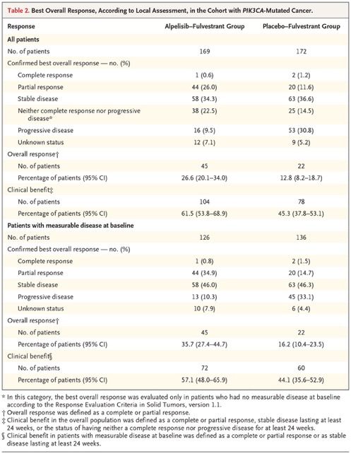
Mutationen gekommen ist oder nicht.Alpelisib for PIK3CA-Mutated, Hormone Receptor–Positive Advanced Breast Cancer PIK3CA mutations occur in approximately 40% of patients with hormone receptor (HR)–positive, human epidermal growth factor receptor 2 (HER2)–negative breast cancer. The PI3Kα-specific inhibitor alpelisib has shown antitumor activity in early studies. In a randomized, phase 3 trial, we compared alpelisib (at a dose of 300 mg per day) plus fulvestrant (at a dose of 500 mg every 28 days and once on day 15) with placebo plus fulvestrant in patients with HR- positive, HER2-negative advanced breast cancer who had received endocrine therapy previously. Patients were enrolled into two cohorts on the basis of tumor-tissue PIK3CA mutation status. The primary end point was progression- free survival, as assessed by the investigator, in the cohort with PIK3CA-mutated cancer; progression-free survival was also analyzed in the cohort without PIK3CA-mutated cancer. Secondary end points included overall response and safety.
Kaplan–Meier Analysis of
Progression-free Survival. In the
cohort of patients with PIK3CA-
mutated cancer, the median
progression-free survival was 11.0
months in the alpelisib–fulvestrant
group and 5.7 months in the
placebo–fulvestrant group (Panel
A). The primary end point crossed
the prespecified Haybittle–Peto
boundary (one-sided P≤0.0199)
(Panel A). Symbols indicate
censored data. In the cohort
without PIK3CA-mutated cancer,
the median progression-free
survival was 7.4 months in the
alpelisib–fulvestrant group and 5.6
months in the placebo–fulvestrant
group (Panel B). The gene PIK3CA
encodes for the alpha isoform of
phosphatidylinositol 3-kinase
(PI3Kα).
Subgroup Analysis of
Progression-free Survival in
the Cohort with PIK3CA-
Mutated Cancer. Confidence
intervals have not been
adjusted for multiplicity.
Inferences drawn from the
confidence intervals may not
be reproducible. The previous
chemotherapy subgroup was
based on the last line of
chemotherapy received.
Patients may have received
chemotherapy in the context of
both neoadjuvant and adjuvant
therapy. Patients may have
had more than one PIK3CA
mutation. There were multiple
subtypes of E545 and H1047
mutations. CDK denotes
cyclin-dependent kinase.
Proof-of-concept criteria were not met in the
cohort of patients without PIK3CA-mutated
cancer at the final efficacy analysis. The
median progression-free survival was 7.4
months (95% CI, 5.4 to 9.3) in the alpelisib–
fulvestrant group and 5.6 months (95% CI,
3.9 to 9.1) in the placebo–fulvestrant group
(hazard ratio for progression or death, 0.85;
95% CI, 0.58 to 1.25; posterior probability of
true hazard ratio
Safety
The total safety population included 284
patients who received alpelisib–
fulvestrant and 287 who received
placebo–fulvestrant. The adverse events
of any grade that occurred in at least
35% of the patients in either group were
hyperglycemia (in 63.7% of the patients
who received alpelisib–fulvestrant and
9.8% of those who received placebo–
fulvestrant), diarrhea (in 57.7% and
15.7%, respectively), nausea (in 44.7%
and 22.3%), decreased appetite (in
35.6% and 10.5%), and rash (in 35.6%
and 5.9%) or maculopapular rash (in
14.1% and 1.7%)
Discussion
These results show improvements in patients’ outcomes with the addition of an α-specific
PI3K inhibitor to standard treatment for PIK3CA-mutated, HR-positive, HER2-negative
advanced breast cancer, findings that validate PIK3CA as an important treatment target in this
population. Patients with PIK3CA-mutated, HR-positive, HER2-negative advanced breast
cancer that had progressed during or after the receipt of endocrine therapy had significantly
longer progression-free survival when they received alpelisib–fulvestrant than when they
received placebo–fulvestrant, with an estimated 35% lower risk of progression or death. A
clinically relevant treatment benefit was not observed for alpelisib–fulvestrant in the cohort
without PIK3CA-mutated cancer. In the cohort with PIK3CA-mutated cancer, alpelisib–
fulvestrant was also associated with significantly higher percentages of patients with tumor
response than was placebo–fulvestrant, a finding that is consistent with observations from the
phase 1b study. Progression-free survival was similar in the placebo groups in the two cohorts
defined according to PIK3CA mutation status.
Alpelisib has activity in patients with PIK3CA-mutated, HR-positive, HER2-negative advanced
breast cancer that has progressed during or after treatment with an aromatase inhibitor.
Therefore, the integration of genomic testing for PIK3CA mutation into routine clinical practice
may be useful in the selection of therapy; validated diagnostic testing procedures are not yet
available. This trial shows that treatment with alpelisib–fulvestrant can provide an extension of
progression-free survival among patients with PIK3CA-mutated disease. This effect was
observed across various subgroups. Preliminary analysis of progression-free survival on the
basis of ctDNA results shows a similar effect.
In conclusion, this phase 3 trial showed a significant prolongation of progression-free survival
and greater overall response with alpelisib–fulvestrant than with placebo–fulvestrant among
patients with PIK3CA-mutated, HR-positive, HER2-negative advanced breast cancer who had
disease that had relapsed or progressed during or after the receipt of previous endocrine
therapy.
Muco-Obstructive Lung Diseases
A spectrum of lung diseases that affect the airways, including chronic obstructive pulmonary disease
(COPD), cystic fibrosis, primary ciliary dyskinesia, and non–cystic fibrosis bronchiectasis, can be
characterized as muco-obstructive diseases. These diseases have the clinical features of cough, sputum
production, and episodic exacerbations that are often associated with a diagnosis of chronic bronchitis.
However, neither “chronic bronchitis” nor “hypersecretory diseases” adequately describes the diffuse
mucus obstruction, airway-wall ectasia, chronic inflammation, and bacterial infection that are typical of
these conditions; therefore, “muco-obstructive” may be a preferred descriptive term. Although asthma can
also be associated with diffuse airway mucus obstruction, its distinct pathophysiological mechanisms
preclude discussion in this grouping.
In healthy persons, a well-hydrated mucus layer is transported rapidly (at a rate of approximately 50 μm
per second) from the distal airways toward the trachea. In muco-obstructive diseases, epithelial defects in
ion–fluid transport, mucin secretion, or a combination of these lead to hyperconcentrated (dehydrated)
mucus, failed mucus transport, and mucus adhesion to airway surfaces. Mucus that is accumulated in the
trachea can be expectorated by cough as phlegm or sputum. Mucus in the small airways cannot be
cleared by cough and accumulates, forming the nidus for airflow obstruction, infection, and inflammation.
Biochemical and Biophysical Properties of Mucus Relative to Airway Function
Human airway mucus is a hydrogel composed of approximately 98% water, 0.9% salt, 0.8% globular
proteins, and 0.3% high-molecular-weight mucin polymers.12 The hydration (concentration) status of mucus
is measured as the wet-to-dry content of mucus (i.e., the percentage of a given volume that consists of
nonvolatile solids) or as the mucin concentration determined by light-scattering techniques.The correlation
between the two measurements (in healthy persons or those with muco-obstructive disease) is high, which
allows both measures to be used to describe this mucus property.
The two major synthesized and secreted respiratory mucins, MUC5B and MUC5AC, are physically very
large, spanning 0.2 to 10 μm in length for single polymers. The secreted mucin polymers interweave to
form mesh-like gels with mesh sizes that are concentration dependent (i.e., higher concentrations are
associated with smaller mesh sizes). MUC5B and MUC5AC share many features, including their
multimeric organization and high carbohydrate content (approximately 75% of total weight).
Muco-Obstructive Disease Pathogenesis, Mucin Species,
and Total Mucin Concentrations in Health and Disease.
Panel A shows the progression from normal to muco-
obstructed airways. In healthy persons (left), well-balanced
epithelial sodium (Na+) absorption and secretion of chloride
anions (Cl−) hydrates airway surfaces and promotes efficient
mucociliary clearance (MCC). In persons with muco-
obstructive lung disease (middle), an imbalance of ion
transport coupled with mucin hypersecretion increases
mucin concentrations in the mucus layer, osmotically
compresses the periciliary layer (PCL), and abolishes MCC.
The adherent mucus may be expelled as sputum by cough
(upper right). Mucus that cannot be expelled by cough
accumulates, concentrates, obstructs airflow, and becomes
the nidus for infection (lower right). CFTR denotes cystic
fibrosis transmembrane conductance regulator, and ENaC
the epithelial sodium channel. Panel B shows domain
structures and relative sizes of the secreted mucins
(MUC5AC and MUC5B) and tethered mucins (MUC1,
MUC4, and MUC16). For reference, the globular protein
albumin (ALB) is shown. The secreted mucins are
composed of monomers with N terminals (MUC5AC, green;
MUC5B, light blue), glycosylated domains, and C terminals
(MUC5AC, yellow; MUC5B, purple). Both the C–C terminal
dimers and N–N terminal multimers are linked by S–S
bonds. The glycosylated domains contain sugar side chains
from 2 to 15 sugar molecules in length, often terminally
capped with sialic acid or sulfate, which gives mucins a
negative charge. The glycosylation domains provide mucin
hydration, reflecting the avidity of sugar molecules for water,
and a combinatorial library of binding sites capable of
trapping most inhaled materials with a low but sufficient
binding affinity to mediate clearance. The tethered mucins
have cytoplasmic N-terminal (dark gray), transmembrane
(light gray), and heavily glycosylated extracellular domains.
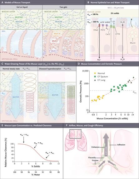
Two-Gel Model of Mucus Transport. Panel A, left, shows the
classic “gel-on-liquid” model showing a mucus layer (MUC5AC
and MUC5B) and a “sol” layer as liquid surrounding cilia. Panel
A, right, shows the “two-gel” model in which the mucus layer
remains the same, but a periciliary layer (gel) is formed by
tethered macromolecules, including MUC1, MUC4, and MUC16.
Panel B shows epithelial ion and water transport in normal
human airway epithelia. The percentage of solids in the normal
mucus layer and osmotic pressures (π) of the normal mucus
layer (πML) and PCL (πPCL) in pascals (Pa) are noted. An ENaC
in the apical membrane mediates sodium and liquid absorption.21
In parallel, the epithelium secretes chloride and bicarbonate
anions through CFTR and calcium-activated chloride channels
(CaCC). The balance between active sodium absorption and
secretion of chloride and bicarbonate anions, and hence fluid
flow, is regulated in part by lumenal concentrations of
extracellular ATP, interacting through P2Y2 receptors, and
adenosine (ADO), interacting through A2b receptors. Panel C
describes the relative water-drawing powers of the mucus layer
(πML) and PCL (πPCL) interfaced to epithelial cell–mediated fluid
absorption or secretion (blue arrows). The length of the Hookean
springs (to the right of the blue arrows) denotes the height of the
PCL (purple) or mucus layer (green), and the spring diameter is
inversely proportional to osmotic pressure. In a normal state
(left), πML is lower than πPCL, represented by a green spring (πML)
with a diameter larger than the purple spring (πPCL). In a
dehydrated state (right), persistent or abnormal absorption
initially removes fluid from the lower-osmotic-pressure mucus
layer but ultimately coordinately removes fluid from both the
mucus layer and the PCL. The osmotic pressures of both layers
are increased and equalized (smaller spring diameters) and
volume depleted (springs shortened). This state osmotically
compresses the cilia and produces mucus stasis. Panel D shows
the relationship between mucus concentration (i.e., percent
solids) and osmotic pressure (Pa).
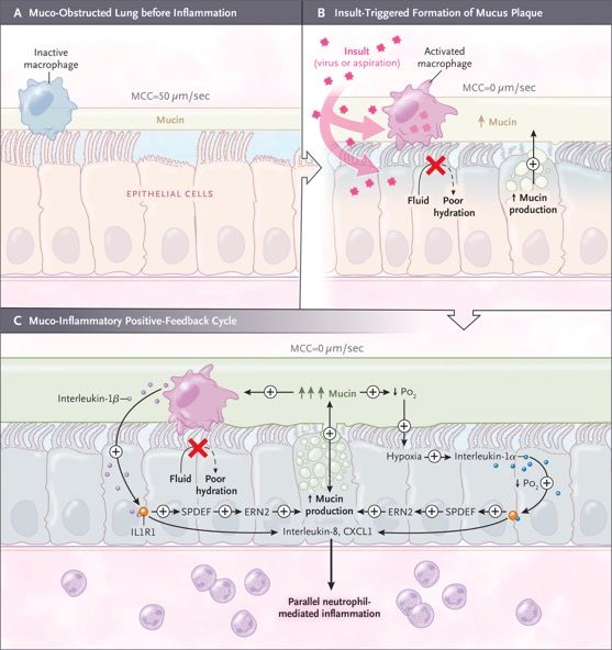
Muco-Inflammatory Positive-Feedback
(“Vicious”) Cycles in Muco-Obstructive Disease.
Panel A shows the basal muco-obstructive
disease state in which surface hydration and
mucus concentration may be relatively normal
but vulnerable to muco-obstruction with insults
(e.g., aspiration or viral infection). Panel B shows
insult-triggered formation of abnormal mucus
plaque. Insults increase mucin secretion not
accompanied by adequate hydration owing to
abnormal fluid-secretory responses.
Hyperconcentrated mucus activates resident
macrophages (pink) and produces hypoxia in
subjacent airway epithelia (blue). Panel C shows
the generation of persistent, muco-inflammatory
positive-feedback cycles. Activated resident
macrophages release interleukin-1β (left), and
hypoxic necrotic epithelia (blue) release
interleukin-1α (right). Interleukin-1α and -1β
activate epithelial interleukin-1 receptors (IL1R1)
to induce mucin biosynthesis mediated by
SPDEF and ERN2 and expression of
proinflammatory cytokines and chemokines
(e.g., IL8 and CXCL1). Accelerated mucin
secretion without proper hydration worsens
mucus hyperconcentration on airway surfaces
and stimulates hypoxic macrophages and
epithelia to release additional interleukin-1α and
-1β in positive-feedback loops (denoted by a
plus sign). Secretion of proinflammatory
mediators induces parallel neutrophil-mediated
inflammation; parallel neutrophil-mediated,
protease-induced mucin secretion; and other
positive-feedback cycles (not shown). Po2
denotes partial pressure of oxygen.
Diagnosis
Disease-specific criteria assist in the diagnosis of each muco-obstructive lung disease: for cystic fibrosis,
levels of chloride anions in sweat and genetic testing; for COPD, exposure history and spirometry; for primary
ciliary dyskinesia, nasal nitric-oxide measurements, cilia waveform analyses, and genetic testing; and for
non–cystic fibrosis bronchiectasis, CT scanning. The tools to make a general diagnosis of muco-obstructive
disease are also available.
Therapies for Muco-Obstructive Diseases
Disease-specific therapies for one muco-obstructive disease, cystic fibrosis, are now available. Ivacaftor (VX-
770) is a potentiator of residual CFTR function that has been approved for patients with cystic fibrosis with
gating and some splicing CFTR mutations. Ivacaftor provided a model for the development of therapies for
muco-obstruction, as evidenced by impressive associations between improved airway-surface hydration in
vitro, peripheral and central mucociliary clearance in vivo, and clinically relevant outcomes (e.g., forced
expiratory volume in 1 second [FEV1]).
Hydrators
Perhaps the most direct approach in the treatment of muco-obstructive lung diseases is to reduce the
concentration of pathologic mucus — that is, rehydrate it. The currently available approach to achieve this
goal is inhalation of osmotically active aerosols (e.g., hypertonic saline or mannitol). In clinical development
are modulators of ion transport that may redirect airway epithelial ion transport from net absorptive to
secretory directions, providing a mechanism for epithelial restoration of airway-surface hydration.
Mucolytics
Two recent observations suggest that attacking abnormal mesh and gel properties of mucus in addition to
reducing concentration may be therapeutic. First, mucus viscosity is a key variable governing the mucus
cohesive and adhesive properties relevant to cough efficiency, and reductions in viscosity with mucin S–S
bond–reducing or surfactant agents decrease mucus adhesion and cohesion independent of concentration.
Second, the mucus plaques or flakes that are recovered from young patients with cystic fibrosis during
bronchoalveolar lavage are unable to swell and dissolve in excess solvent. Inhaled acetylcysteine has not
proved to have the mucin-reductive activity required for a therapeutic effect.
Conclusions
Muco-obstructive diseases are characterized by mucus hyperconcentration. The four muco-obstructive
diseases differ with respect to the epithelial abnormalities that produce mucus hyperconcentration but follow a
final common path of mucus concentration–dependent formation of mucus plaques and plugs.
A 60-year-old man presented to the otorhinolaryngology
clinic with a feeling of nasal obstruction and postnasal
drip that had developed 10 years earlier and had
worsened over the past month. He also had a sensation
of a foreign body in his throat. Symptoms worsened
when he was lying flat. On physical examination, no
deviation of the nasal septum or hypertrophy of the
inferior turbinates was detected. Nasopharyngoscopy
revealed a smooth, pink, cystic mass in the midline of
his nasopharynx, with no obstruction of the openings to
the eustachian tubes (the image shows the view
through the right naris). Computed tomography of the
head revealed a 2.5 cm by 1.3 cm by 1.4 cm cystic
tumor in the midline of the nasopharynx without
intracranial extension. The findings were consistent with
Tornwaldt’s cyst, a benign cyst that arises between the
roof of the nasopharynx and the remnant of the
notochord. Because the patient was symptomatic, he
underwent surgical marsupialization of the cyst. On
follow-up examination 3 months after surgery, he no
longer had the sensation of a foreign body in his throat,
his nasal symptoms were reduced, and there was no
recurrence of the cyst.                                     Eine Tornwaldt-Zyste (lat.: Bursa pharyngea(lis)),
                                                            (gelegentlich auch in den Schreibweisen Thornwaldt- oder
                                                            Thornwald-Zyste) ist eine benigne Zyste (eine gutartige,
                                                            flüssigkeitsgefüllte Raumforderung), die im oberen hinteren
                                                            Nasopharynx (Nasen-Rachenraum) lokalisiert ist.
                                                            Sie wird meist zufällig bei der Computertomografie (CT) oder
                                                            Magnetresonanztomografie (MRT) des Kopfes als in der
                                                            Mittellinie gelegene, gut abgegrenzte rundliche
                                                            Raumforderung diagnostiziert.
A 55-Year-Old Man with Jaundice A 55-year-old man with a history of opioid use disorder and hepatitis C virus (HCV) infection presented to this hospital with jaundice. Four months before the current presentation, the patient was released from prison after a 2-year incarceration. After he left prison, he resumed injecting heroin and had three episodes of overdose. He was evaluated at another hospital for symptoms of depression and was admitted for psychiatric treatment. During that admission, sublingual buprenorphine–naloxone therapy was initiated, and the patient was discharged. One day after discharge and 5 weeks before the current presentation, headache, body aches, sweats, diarrhea, and nausea developed. The patient presented to a clinic for substance use disorder that is affiliated with the other hospital and reported that he had been unable to obtain sublingual buprenorphine–naloxone from a pharmacy after discharge. The temperature was 36.6°C, the pulse 70 beats per minute, and the blood pressure 100/68 mm Hg. The weight was 72 kg. He appeared to be restless, but the remainder of the physical examination was normal. Urine toxicologic screening was positive for buprenorphine, norbuprenorphine, and norfentanyl; buprenorphine– naloxone therapy was resumed. Three weeks before the current presentation, dark urine and light-headedness developed and did not improve with increased fluid consumption. The patient also noticed slow thinking and arthralgias in the hands, wrists, and elbows. He was evaluated by a new primary care provider.
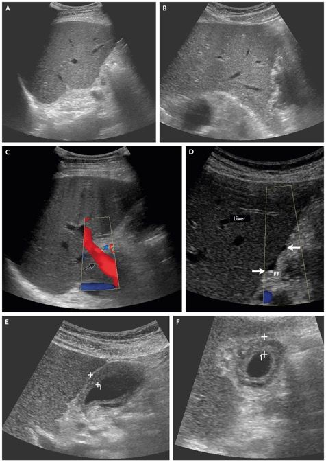
Limited ultrasonography of the right upper
quadrant revealed no bile-duct dilatation, a
patent main portal vein with hepatopetal flow,
and normal hepatic parenchymal echotexture.
Diffuse, hypoechoic gallbladder-wall thickening
was present, without gallbladder distention,
cholelithiasis, or pericholecystic fluid. Murphy’s
sign was negative. There was trace perihepatic
ascites.

Abdominal Ultrasound Images.
Grayscale images (Panels A, B, E, and F) and
color Doppler images (Panels C and D) of the
right upper quadrant show no intrahepatic or
extrahepatic bile-duct dilatation (Panels A and
B), a patent main portal vein with flow in the
correct direction (Panel C, arrow), trace
perihepatic ascites (Panel D, arrows), and a
collapsed gallbladder with diffuse, hypoechoic
wall thickening (Panels E and F, plus signs). FF
denotes free fluid.
A long-held dogma regarding the differential diagnosis of severe acute liver injury is that most cases are due to
vascular, viral, or toxic causes. A recent multicenter study examined the causes of severe acute liver injury in
patients with an aspartate aminotransferase or alanine aminotransferase level of more than 1000 U per liter and
confirmed the leading causes to be ischemic hepatitis, pancreaticobiliary disease, drug-induced liver injury, and
viral hepatitis.
Ischemic Hepatitis
Does this patient have a vascular or ischemic process that is causing acute liver injury? Ischemic hepatitis is
defined as a decrease in hepatic blood flow that results in hepatocyte death and necrosis. This condition is
commonly seen in critically ill patients, and in many cases, a clear hypotensive episode is not documented,
which suggests that the insulting event may be fleeting or subclinical. Recent data further suggest that elevated
central venous pressure and cardiac pressures on the right side are important predisposing factors.
Pancreaticobiliary Disease
The absence of bile-duct dilatation, gallstones, and pancreatic abnormality on ultrasonography provides
evidence against a pancreaticobiliary cause of this patient’s liver injury. The magnitude of hyperbilirubinemia
and the kinetics of elevation in aminotransferase levels are also not suggestive of biliary disease; the alanine
aminotransferase level tends to rise and fall rapidly, and it is uncommon to have a peak bilirubin level of more
than 10 mg per deciliter (171 μmol per liter).
Drug-Induced Liver Injury
Drug-induced liver injury must be considered in all cases of severe acute liver injury or failure; studies have
determined it to be the most common cause of acute liver failure in the United States and most of Europe.
In this case, the only new medication was buprenorphine–naloxone, which is an exceedingly rare cause of
drug-induced liver injury. One report identified buprenorphine–naloxone as the cause of severe liver injury in
two patients with chronic HCV infection; the drug had been injected at high doses, and symptomatic hepatitis
had occurred within 4 days after the injection.
Viral Hepatitis
This patient’s clinical presentation is strongly suggestive of acute viral hepatitis. The acute viral infections that
most commonly cause severe liver injury are HCV infection (in 8.0% of cases) and hepatitis B virus (HBV)
infection (in 2.1% of cases). Hepatitis A virus (HAV), hepatitis delta virus (HDV), Epstein–Barr virus, and
cytomegalovirus infections are much less common causes (in 0.3% of cases combined).
Acute HCV Infection
Symptoms associated with acute HCV infection occur approximately 9 weeks after exposure. There is a very
low level of viremia in the first 2 months after infection, followed by a brisk and exponential increase in viremia
over a period of 8 to 10 days. In the subsequent phase, the host immune system responds to the viremia: the
aminotransferase levels rise, the HCV RNA viral load plateaus, and symptoms develop. Could this patient
have acute HCV infection? Although he reportedly had a history of HCV infection, we have not seen an HCV
viral load obtained before this presentation. A positive test for HCV antibodies alone suggests previous
exposure but does not confirm a chronic infection. An estimated 25% of persons with exposure to HCV have
successful viral clearance; female sex, symptomatic infection, and favorable host genetic factors are
associated with spontaneous clearance.
Acute HBV Infection
The clinical presentation and outcome of acute HBV infection are strongly influenced by the patient’s age; in
neonates, viral clearance is exceedingly rare, occurring in less than 5% of cases, whereas in adults, the
infection is successfully controlled in more than 95% of cases. The virus itself is noncytopathic, and the
symptoms and liver injury are consequences of the host immune response. Liver failure is rare but occurs in
approximately 1% of cases. Even in the absence of liver failure, liver injury can be severe; in one case series,
the mean alanine aminotransferase level was 1419 U per liter, and the mean bilirubin level 6.5 mg per deciliter
(111 μmol per liter).
Acute HDV Infection
What other factors could explain this patient’s severe acute liver injury? HDV infection is dependent on and
exacerbates HBV infection. There are two clinical patterns of infection with HDV: coinfection, in which
exposure to HDV and to HBV are simultaneous; and superinfection, in which acute HDV infection occurs in a
person with established chronic HBV infection. In cases of coinfection, HDV infection relies on the successful
development of HBV infection, which is uncommon in adults, and thus chronic coinfection is rare.
HAV Infection
HAV infection is a classic cause of severe acute viral hepatitis. Similar to HBV infection, HAV infection has a
clinical presentation that varies according to the patient’s age; children are often asymptomatic, whereas more
than 70% of adults have symptoms, such as fever, malaise, nausea, vomiting, and abdominal discomfort, that
last 2 to 8 weeks.
Sie können auch lesen