Instructions For Use For professional use only - IVD - ScheBo ...
←
→
Transkription von Seiteninhalten
Wenn Ihr Browser die Seite nicht korrekt rendert, bitte, lesen Sie den Inhalt der Seite unten
ANTIGEN

 Gebrauchsanweisung
 Instructions For Use                              ��

 Nur für den professionellen Gebrauch
 For professional use only

      In-Vitro-Diagnostikum
IVD   For in vitro diagnostic use only

(Fluorescence Immunochromatography)
INHALTSVERZEICHNIS / CONTENT �. VERWENDUNGSZWECK � �. INTENDED USE �� �. TESTPRINZIP � �. TEST PRINCIPLE �� �. KIT BESTANDTEILE � �. KIT COMPONENTS �� �. WARNHINWEISE UND VORSICHTSMAßNAHMEN � �. WARNINGS AND PRECAUTIONS �� �. AUFBEWAHRUNG UND HALTBARKEIT � �. STORAGE CONDITIONS AND SHELF LIFE �� �. ERFORDERLICHE GERÄTE � �. APPLICABLE INSTRUMENTS �� �. PROBENMATERIAL � �. SAMPLE REQUIREMENTS �� �. BENÖTIGTE MATERIALIEN � �. MATERIALS REQUIRED BUT NOT PROVIDED �� �. GEWINNUNG DER ABSTRICHPROBEN � �. COLLECTION OF SWAB SAMPLES �� ��. TESTDURCHFÜHRUNG MIT ABSTRICHPROBEN � ��. TEST PROCEDURE FOR SWAB SAMPLES �� ��. TESTDURCHFÜHRUNG MIT SERUMPROBEN � ��. TEST PROCEDURE FOR SERUM SAMPLES �� ��. AUSWERTUNG DER TESTERGEBNISSE �� ��. INTERPRETATION OF THE RESULTS �� ��. EINSCHRÄNKUNGEN �� ��. LIMITATION OF THE PROCEDURES �� ��. LEISTUNGSMERKMALE �� ��. PERFORMANCE CHARACTERISTICS �� ��. VERFAHRENSANMERKUNGEN �� ��. PROCEDURAL NOTES �� ��. ERKLÄRUNG DER SYMBOLE �� ��. SYMBOLS ��
SARS-CoV-� Antigen                                                                                             Gebrauchsanweisung

�. VERWENDUNGSZWECK                                            �. TESTPRINZIP

   Das Genom des SARS-CoV-� codiert Spike-Protein, Hüllpro-      Dieser Schnelltest verwendet ein Fluoreszenz-immunchro-

   tein, Membranprotein und Nukleokapsid. Während des            matographisches-Verfahren zum Nachweis von SARS-CoV-�

   Zusammenbaus der Viren bindet das N-Protein an die virale     N-Antigen. Die zu testende Probe wird in das Probenfenster

   RNA und führt zur Bildung eines spiralförmigen Nukleokap-     der Testkassette aufgetragen. Das SARS-CoV-� N-Antigen in der

   sids. Das N-Protein ist ein hoch immunogenes Phospho-         Probe bildet einen Komplex mit dem, mit fluoreszierenden

   protein, das mit der Replikation des Virusgenoms und der      Mikrokügelchen markierten, Antikörper. Dieser Komplex

   Signalübertragung von Zellen zusammenhängt. Aufgrund          wandert entlang der Membran und erreicht die Testregion

   der konservierten Sequenz von N-Protein ist der Nachweis      (T-Linie), auf der ein zweiter Antikörper gegen das SARS-CoV-�

   von SARS-CoV-� N-Protein von großer klinischer Bedeutung.     N-Antigen aufgebracht ist. Ungebundene fluoreszierende

   Dieser Schnelltest wird zum qualitativen Nachweis von         Mikrokugeln wandern entlang der Membran zur Kontroll-

   SARS-CoV-�-Nukleokapsid-Proteinantigen (im Folgenden          region (C-Linie) und werden vom Antikörper der Kontroll-

   als SARS-CoV-� N-Antigen bezeichnet) in humanem Serum         region gebunden. Das Testergebnis im Testfenster wird mit

   und aus nasopharyngealen und/oder oropharyngealen Ab-         einer UV-Lampe mit einer Wellenlänge von ��� nm sichtbar

   strichproben extrahiertem SARS-CoV-� N-Antigen verwendet.     gemacht. Fluoreszieren sowohl die T-Linie als auch die C-Linie

                                                                 zeigt dies ein SARS-CoV-� N-Antigen positives Ergebnis an;

                                                                 fluoresziert nur die C-Linie und nicht die T-Linie ist der Test

                                                                 SARS-CoV-� N-Antigen negativ. Wird die C-Linie nicht ange-

                                                                 zeigt, ist das Testergebnis ungültig und die Probe muss

                                                                 erneut mit einer anderen Testkassette getestet werden.

/ ��                                                                                                                        / ��
SARS-CoV-� Antigen Gebrauchsanweisung
�. KIT BESTANDTEILE taminierten Materialien gemäß den örtlichen Anforderungen
sicher umgehen.
�� Testkassetten �� sterile Tupfer
�.�. Wischen und waschen Sie alle Probenspritzer mit einem
�� Pipetten � Extraktionspuffer (à � ml)
hochwirksamen Desinfektionsmittel ab. Spritzer und Aerosol-
� Gebrauchsanweisung �� Extraktionsröhrchen/Deckel
bildung vermeiden.
�.�. Verwenden Sie für jede Probe eine neue saubere Einweg-
�. WARNHINWEISE UND VORSICHTSMAßNAHMEN
pipettenspitze bzw. Extraktionsröhrchen, um eine Kreuzkon-
�.�. Bei dem Test handelt es sich um ein In-vitro-Diagnostikum
tamination zu vermeiden.
ausschließlich für den professionellen Gebrauch. Nicht nach
�.�. Schauen Sie nicht direkt in die UV-Lampe.
Ablauf des Verfallsdatums verwenden.
�.�. Entsorgen Sie alle Proben und potenziell kontaminierte
�.�. Die Proben sollten als potenziell infektiös angesehen
Materialien ‒ wie infektiöse Abfälle ‒ in einem Biogefährdungs-
werden. Die Anwender sollten Schutzkleidung, Masken und
abfallbehälter.
Handschuhe tragen und andere geeignete Sicherheitsvor-
�.��. Verwenden Sie die Testkassette so bald wie möglich,
kehrungen treffen, um das Infektionsrisiko zu vermeiden oder
nachdem die Testkassette aus dem Beutel genommen wurde,
zu verringern.
um eine Befeuchtung zu vermeiden. Die Testkassette ist feuch-
�.�. Dieser Test sollte bei �� ‒ �� °C durchgeführt werden.
tigkeits- und hitzeempfindlich.
Stellen Sie sicher, dass der Test und die Proben auf Raum-
�.��. Verwenden Sie die Testkassette nicht, wenn der Beutel
temperatur gebracht werden, bevor Sie den Test durchführen.
oder die Siegelnaht beschädigt ist.
�.�. Befolgen Sie die Gebrauchsanweisung sorgfältig. Die Zu-
�.��. Die Testkassette kann nicht wiederverwendet werden.
verlässigkeit der Testergebnisse kann nicht garantiert werden,
wenn die Gebrauchsanweisung nicht exakt befolgt wird.
�.�. Medizinisches Fachpersonal muss mit den potenziell kon-
/ �� / ��SARS-CoV-� Antigen Gebrauchsanweisung
�. AUFBEWAHRUNG UND HALTBARKEIT �.�. Serumproben können � Tage bei �‒ � °C gelagert werden.
Sofern eine Lagerung über diesen Zeitpunkt hinaus erforder-
Der Test kann ab Herstellungsdatum �� Monate bei �‒�� °C
lich ist, sollte diese bei ‒�� °C erfolgen. Serumproben können
gelagert werden. Die Testkassette muss innerhalb einer
maximal � x aufgetaut und wieder eingefroren werden.
Stunde nach dem Öffnen des Aluminiumfolienbeutels ver-
Bringen Sie das Serum auf Raumtemperatur und mischen
wendet werden.
Sie dieses vor dem Testen gut. Wenn sich sichtbare Partikel
im Serum befinden, sollte das Serum vor dem Testen zentri-
�. ERFORDERLICHE GERÄTE
fugiert werden, um den Niederschlag zu entfernen.
UV-Lampe mit einer Wellenlänge von ��� nm.
Wenn das Serum lipämisch (Triglyceridkonzentration über
�. PROBENMATERIAL ��mmol/l), hämolytisch oder trüb ist, verwenden Sie die
Probe bitte nicht, um eine Beeinträchtigung der Ergebnis-
�.�. Anwendbar für humanes Serum und für anterior nasale
interpretation zu vermeiden.
oder nasopharyngeale und/oder oropharyngeale Abstrich-
proben.
�. BENÖTIGTE MATERIALIEN
�.�. Es wird empfohlen die Probe zum Zeitpunkt der Proben-
nahme zu testen. Stoppuhr
�.�. Falls die Abstrichproben nicht unverzüglich getestet UV-Lampe mit einer Wellenlänge von ��� nm
werden, sollten sie in einem sauberen, trockenem Röhrchen
�. GEWINNUNG DER ABSTRICHPROBEN
aufbewahrt und fest verschlossen werden (stecken Sie die
Tupferspitze in ein Röhrchen und knicken/schneiden Sie �.�. Abstrich nach dem Standardverfahren einer anterior
das Applikatorstäbchen ab). Die Tupfer können für bis zu nasalen oder nasopharyngealen oder oropharyngealen
�� Stunden bei �‒� °C aufbewahrt werden. Abstrichprobe nehmen.
/ �� / ��SARS-CoV-� Antigen Gebrauchsanweisung
�.� Gewinnung einer anterior nasalen Abstrichprobe: ��. TESTDURCHFÜHRUNG MIT ABSTRICHPROBEN
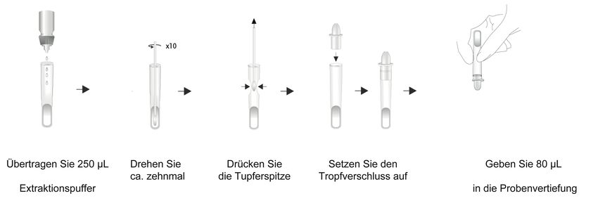
Führen Sie den Tupfer vorsichtig ca. � cm in ein Nasenloch Schritt �: Übertragen Sie ��� μl (� Tropfen) Extraktions-
ein und streichen Sie �- bis ��-mal rotierend entlang der puffer senkrecht in das Extraktionsröhrchen.
Nasenwand und ziehen den Tupfer vorsichtig aus der Schritt �: Stecken Sie den Tupfer, mit dem die Sekrete ge-
Nasenhöhle. Wiederholen Sie den Vorgang mit demselben sammelt wurden, in das mit Puffer gefüllte Extraktions-
Tupfer im anderen Nasenloch. röhrchen und schwenken Sie dieses ca. zehnmal im Kreis, um
�.�. Gewinnung einer nasopharyngealen Abstrichprobe: so viel wie möglich von der Probe in der Lösung freizusetzen.
Legen Sie den Kopf des Patienten um ��° nach hinten. Schritt �: Drücken Sie die
� Tupferspitze aus, indem Sie seit-
Führen Sie den Tupfer in das Nasenloch ein (der Tupfer lich an das Probenröhrchen drücken, um möglichst viel von
sollte eine dem Abstand zwischen den Nasenlöchern und der Flüssigkeit im Röhrchen zu behalten.
der Außenöffnung des Ohrs entsprechende Tiefe erreichen). Schritt �: Setzen Sie den Tropfverschluss auf.
Lassen Sie den Tupfer zur Sekretaufnahme mehrere Sekund- Schritt �: Reißen Sie den Aluminiumbeutel auf, entnehmen �
en dort. Entfernen Sie langsam den Tupfer, während Sie ihn Sie die Testkassette und legen Sie diese auf eine ebene Fläche.
gleichzeitig drehen. Schritt �: Schreiben Sie die Probennummer auf die Testkassette.
�.�. Gewinnung einer oropharyngealen Abstrichprobe: Schritt �: Geben Sie �� μl (� Tropfen) der aufbereiteten Probe
Führen Sie den Tupfer in den Bereich des hinteren Rachens in das Probenfenster. Vermeiden Sie die Bildung von Luftblasen.
und der Mandeln ein. Reiben Sie den Tupfer über beide Ton- Schritt �: Lesen Sie die Testergebnisse nach �� bis �� Minuten
sillen und den hinteren Mundrachenraum und vermeiden mit einer UV-Lampe (Wellenlänge ��� nm) innerhalb von
Sie es, die Zunge, Zähne und das Zahnfleisch zu berühren. �� Sekunden ab (eine Langzeitbelichtung mit der UV-Lampe
�.�. Es wird empfohlen, die Probe zum Zeitpunkt der Proben- führt zu einer Verringerung des Fluoreszenssignals, was sich
nahme zu testen. auf die Interpretation des Ergebnisses auswirken kann).
/ �� / ��SARS-CoV-� Antigen Gebrauchsanweisung
� � � � Schritt �: Pipettieren Sie �� μl (� Tropfen mit der mitgelie-
x �� ferten Pipette) des zu testenden Serums in das Probenfenster
der Testkassette. Vermeiden Sie die Bildung von Luftblasen.
Schritt �: Lesen Sie die Testergebnisse nach �� bis �� Minuten
� mit einer UV-Lampe (Wellenlänge ��� nm) innerhalb von
Übertragen Sie Drehen Sie Drücken Sie Setzen Sie den
��� µL (� Tropfen) ca. zehnmal die Tupferspitze Tropfverschluss auf �� Sekunden ab (eine Langzeitbelichtung mit der UV-Lampe
Extraktionspuffer
führt zu einer Verringerung des Fluoreszenzsignals, was sich
� � �
auf die Interpretation des Ergebnisses auswirken kann).
SARS-Cov-�
Ag
C
�
��
�
T
��
�� ��
� �
�� ��
�� ��
�� ��
��
Geben Sie �� µL Warten Sie Ergebnis innerhalb
(� Tropfen) in die ��‒�� Minuten von �� Sekunden
Probenvertiefung unter der UV-Lampe
ablesen
Pipettieren Sie �� µl Serum � Tropfen (=
^ �� µl)
��. TESTDURCHFÜHRUNG MIT SERUMPROBEN
SARS-Cov-�
Ag
� �
Schritt �: Bringen Sie die Testkassette und das Serum vor ��
��
�
C
T
der Testdurchführung auf Raumtemperatur.
�� ��
�� ��
�� ��
Schritt �: Reißen Sie den Aluminiumbeutel auf, entnehmen
�� ��
��
Sie die Testkassette und legen Sie diese auf eine ebene Fläche.
��‒�� Minuten warten Ergebnis innerhalb von �� Sekunden
Schritt �: Schreiben Sie die Probennummer auf die Testkassette. unter der UV-Lampe ablesen
/ �� / ��SARS-CoV-� Antigen Gebrauchsanweisung
��. AUSWERTUNG DER TESTERGEBNISSE ��.�. Aufgrund der komplexen Struktur bioaktiver Sub-
stanzen in Proben und des Unterschieds der Antigen-
��.�. Wenn unter der UV-Lampe gleichzeitig sichtbare rot
Antikörper-Spezifität kann die Möglichkeit falsch positiver
fluoreszierende Banden in der Testregion (T-Linie) und in der
Ergebnisse mit diesem Test nicht vollständig ausgeschlossen
Kontrollregion (C-Linie) auftreten, zeigt dies an, dass der Test
werden. Wenn die Testergebnisse nicht mit den klinischen
SARS-CoV-� N-Antigen positiv ist. Wenn die rote fluoreszie-
Indikationen übereinstimmen, sollten zur Bestätigung
rende Bande im Bereich der Kontrollregion (C-Linie) sichtbar
weitere geeignete Testmethoden verwendet werden.
erscheint und in der Testregion (T-Linie) keine sichtbare rote
��.�. Ist der Test SARS-CoV-� N-Antigen positiv, weist dies
fluoreszierende Bande vorhanden ist, weist dies darauf hin,
darauf hin, dass eine SARS-CoV-�-Infektion vorliegt. Ein
dass der Test auf das SARS-CoV-� N-Antigen negativ ist. Wenn
negatives Ergebnis des Tests auf das SARS-CoV-� N-Antigen
im Bereich der Kontrollregion (C-Linie) keine sichtbare rote
kann die Infektion mit SARS-CoV-� jedoch nicht vollständig
fluoreszierende Bande vorhanden ist, ist das Testergebnis
ausschließen. Dies kann vorkommen, wenn der Gehalt an
ungültig und die Probe muss erneut getestet werden.
SARS-CoV-� N-Antigen in der Probe unter der Nachweis-
SARS-Cov-� SARS-Cov-� SARS-Cov-� SARS-Cov-� grenze liegt oder ein Anti-N-Antigen-Antikörper im Serum
Ag Ag Ag Ag
vorliegt und dadurch der Gehalt an N-Antigen abnimmt.
��.�. Wie bei allen diagnostischen Tests müssen alle Ergeb-
C C C C
nisse im Zusammenhang mit anderen klinischen Informa-
T T T T
tionen und weiteren Ergebnissen, die durch andere Test-
methoden dem Arzt zur Verfügung stehen, interpretiert
werden.
positiv negativ ungültig ungültig
/ �� / ��SARS-CoV-� Antigen Gebrauchsanweisung
��. EINSCHRÄNKUNGEN N-Antigen zugesetzt wurde. Bei sechzigfacher Testung betrug
die LoD �,� pg/ml.
��.�. Lipämische, hämolytische und mit Mikroorganismen
��.�. Viruserkennungsgrenze:
kontaminierte Serumproben, mehr als dreimaliges wieder-
Die Stammlösung (�,� × ��� TCID��/ml) (IVCAS �,����) des
holtes Einfrieren und Auftauen oder Serumproben nach
SARS-CoV-� wurde auf ��� TCID��/ml, ��� TCID��/ml, ��
Hitzeinaktivierung können die Genauigkeit des Nachweises
TCID��/ml, �� TCID��/ml, �� TCID��/ml, � TCID��/ml Proben
beeinträchtigen und zu fehlerhaften Ergebnissen führen.
verdünnt. Jede Probe wurde dreimal getestet.
��.�. Ikterische Serumproben oder starke Verunreinigung
können zu falschen Ergebnissen führen. Bestimmung der Nachweisgrenze
��.�. Die Genauigkeit des Tests hängt vom Probennahme-
Konzentration Ergebnis Ergebnis
Testzyklen
Verfahren ab. Eine unsachgemäße Probennahme, unsach- (TCID��/mL) Serumproben Abstrichproben
gemäße Probenaufbewahrung oder wiederholtes Einfrieren ����� � �/� positiv �/� positiv
und Auftauen der Probe wirken sich auf das Testergebnis aus. ��� � �/� positiv �/� positiv
��� � �/� positiv �/� positiv
��. LEISTUNGSMERKMALE �� � �/� positiv �/� positiv
��.�. Nachweisgrenze �� �/� positiv �/� positiv
�
Die Nachweisgrenze für Serumproben des Tests wurde mit
�� � �/� positiv �/� positiv
negativen Serumproben untersucht, denen rekombinantes
� � �/� positiv �/� positiv
N-Antigen zugesetzt wurde. Bei sechzigfacher Testung betrug
die LoD �,� pg/ml. Die Nachweisgrenze für Serumproben liegt bei �� TCID��/ml
Die Nachweisgrenze für Abstrichproben wurde mit nega- und die Nachweisgrenze für Abstrichproben liegt bei
tiven Abstrichproben untersucht, denen rekombinantes �� TCID��/ml.
/ �� / ��SARS-CoV-� Antigen Gebrauchsanweisung
Kreuz-
��.�. Kreuzreaktivitätsstudien Substanz Konzentration
reaktivität
Kreuz-
Substanz Konzentration
Die Kreuzreaktivität des SARS-CoV-�-Antigen-Schnelltests reaktivität
Escherichia coli �.���� CFU/mL negativ
wurde mit verschiedenen mikrobiellen Substanzen, die mög- Chlamydia pneumoniae �.�×��� CFU/ml negativ
Hepatitis C Virus (HCV) �.���� TCID��/mL negativ
licherweise mit dem Nachweis des SARS-CoV-�-Antigens Mycoplasma pneumoniae �.���� CFU/ml negativ
Hepatitis B Virus (HBV) �.���� TCID��/mL negativ
kreuzreagieren könnten, in Serum- und Abstrichproben
Parainfluenza virus �.���� TCID��/ml negativ
getestet. Die Ergebnisse zeigten keine Kreuzreaktivität mit Influenza B �.�×���.�� TCID��/mL negativ
Respiratory syncytial virus �.���� TCID��/ml negativ
den in der Tabelle aufgeführten Substanzen. Influenza A �.�×���.�� TCID��/mL negativ
Adenovirus �.���� TCID��/ml negativ
Kreuz- Herpes Simplex Virus-� (HSV-�) �.���� TCID��/mL negativ
Substanz Konzentration
reaktivität
Cytomegalovirus (CMV) �.���� TCID��/ml negativ
Herpes Simplex Virus-� (HSV-�) �.���� TCID��/mL negativ
Escherichia coli �.���� CFU/ml negativ
Human Immunodeficiency
Epstein-Barr Virus (EBV) �.���� TCID��/ml negativ
�.���� TCID��/mL negativ
Virus ‒ � (HIV-�)
Hepatitis C Virus (HCV) �.���� TCID��/ml negativ
Varicella Zoster Virus (VZV) �.��� �
TCID ��/ml negativ
negativ
Enterovirus �.��� �
TCID ��/mL
Hepatitis B Virus (HBV) �.���� TCID��/ml negativ
Parvovirus
Staphylococcus B��
epidermidis �.���
�.���� TCID��/ml
CFU/mL
� negativ
negativ
Influenza B �.����.�� TCID��/ml negativ
Legionella pneumophila
Streptococcus pneumoniae �.���
�.��� � CFU/ml
CFU/mL
� negativ
negativ
Influenza A �.����.�� TCID��/ml negativ
Chlamydia pneumoniae
Streptococcus pyogenes �.��� CFU/ml negativ
negativ
�.��� �
CFU/mL
�
Herpes Simplex Virus-� (HSV-�) �.���� TCID��/ml negativ
Mycoplasma pneumoniae
Staphylococcus aureus �.���
�.���
�
CFU/mL
�
CFU/ml negativ
negativ
Herpes Simplex Virus-� (HSV-�) �.���� TCID��/ml negativ
Parainluenza
Humanes virus ���E
Coronavirus �.���
�.���TCID ��/mL
TCID Negative
negativ
��/ml
� �
Humanes Immundefizienz- �.���� TCID��/ml negativ
Virus-� (HIV-�)
Respiratory syncytial virus �.��� TCID ��/mL Negative
Humanes Coronavirus OC�� �.��� TCID ��/ml negativ
� �
Enterovirus �.���� TCID��/ml negativ
Adenovirus �.��� TCID ��/mL Negative
Humanes Coronavirus NL�� �.��� TCID ��/ml negativ
�
�
Staphylococcus epidermidis �.���� CFU/ml negativ
Cytomegalovirus (CMV) �.���� TCID��/mL Negative
negativ MERS �.���� TCID ��/ml negativ
Legionella pneumophila �.���� CFU/ml
Epstein-Barr Virus (EBV) �.���� TCID��/mL Negative
Chlamydia pneumoniae �.���� CFU/mL negativ
/ �� Varicella Zoster Virus (VZV) / ��
�.���� TCID��/mL Negative
Mycoplasma pneumoniae �.���� CFU/mL negativ
Parvovirus B�� �.���� TCID��/mL NegativeSARS-CoV-� Antigen Gebrauchsanweisung
��.�. Interferenzen ��.�.�. Mikrobielle Interferenzstudien
��.�.�. Studien zu endogenen Interferenzsubstanzen Die folgenden Pathogene hatten in der genannten Konzen-
Keine der folgenden endogenen Interferenzsubstanzen hatte tration keinen Einfluss auf das Testergebnis SARS-CoV-� N-
in der angegenenen Konzentration einen Einfluss auf das Antigen positiver Proben:
Testergebnis des SARS-CoV-� Antigen-Schnelltests.
Mikrobielle Interferenz Konzentration
.
Potentiell störende Substanz Konzentration Escherichia coli �.�×��� CFU/ml
Hepatitis C Virus (HCV) �.���� TCID��/ml
Bilirubin �.� mg/ml
Hepatitis B Virus (HBV) �.���� TCID��/ml
Triglyceride �� mmol/l
Influenza B �.����.�� TCID��/ml
Hämoglobin � mg/ml
Influenza A �.����.�� TCID��/ml
α - Interferon ���� IU/ml
Herpes Simplex Virus-� (HSV-�) �.���� TCID��/ml
Zanamivir ��� ng/ml
Herpes Simplex Virus-� (HSV-�) �.���� TCID��/ml
Ribavirin � μg/ml
Human Immunodeficiency
Oseltamivir �� μg/ml Virus ‒ � (HIV-�) �.�×��� TCID��/ml
Levofloxacin �� μg/ml Enterovirus �.�×��� TCID��/ml
Staphylococcus epidermidis �.���� CFU/ml
Ceftriaxone ��� μg/ml
Meropenem �.� mg/ml Legionella pneumophila �.���� CFU/ml
Tobramycin � μg/ml Chlamydia pneumoniae �.�×��� CFU/ml
HAMA ��� ng/ml Mycoplasma pneumoniae �.���� CFU/ml
/ �� / ��Chlamydia pneumoniae �.���� CFU/mL
Mycoplasma pneumoniae �.���� CFU/mL
SARS-CoV-� Antigen Gebrauchsanweisung
Parainluenza virus �.���� TCID��/mL
Respiratory syncytial virus �.���� TCID��/mL
��.�. Klinische Bewertung
Parainfluenza virus �.���� TCID��/ml
Die Sensitivität des Tests bei der Verwendung von anterior
Respiratory syncytial virus �.���� TCID��/ml
nasalen Abstrichproben wurde anhand von ��� PCR positi-
Adenovirus �.���� TCID��/ml
ven Proben mit einem Ct-Wert � �� bestimmt. Die Spezifität
Cytomegalovirus (CMV) �.���� TCID��/ml
wurde anhand von ��� PCR negativen Proben bestimmt.
Epstein-Barr Virus (EBV) �.�×��� TCID��/ml Die Sensitivität und Spezifität des Tests wurde mit auf dem
Varicella Zoster Virus (VZV) �.�×��� TCID��/ml Markt erhältlichen PCR Tests verglichen (Thermo Fisher
Parvovirus B�� �.���� TCID��/ml TaqPath COVID �� RT PCR Kit, MIKROGEN ampliCube Corona-
Streptococcus pneumoniae �.���� CFU/ml
virus assay).
Für den ScheBo®·SARS-CoV-� Quick™ Antigen wurde eine
Streptococcus pyogenes �.���� CFU/ml
Sensitivität von ��,� % und eine Spezifität von ��,� % er-
Staphylococcus aureus �.���� CFU/ml
mittelt.
Humanes Coronavirus ���E �.���� TCID��/ml
Humanes Coronavirus OC�� �.���� TCID��/ml PCR
Humanes Coronavirus NL�� �,���� TCID��/ml Positiv Negativ
MERS �,���� TCID��/ml Positiv ��� �
ScheBo®·SARS-
CoV-� Quick™ Negativ � ���
��.�. Hook-Effekt Antigen Gesamt ���
���
��.�.�. Verwendet wurde ��� ng/ml rekombinantes N-Anti-
Sensitivität ��,��% (��CI:��,��%‒��,�� %)
gen, das mit negativen Serum- und Abstrichproben herge-
Spezifität ��,��% (��CI:��,��%‒��,�� %)
stellt wurde. Es wurde kein Hook-Effekt festgestellt.
/ �� / ��SARS-CoV-� Antigen Gebrauchsanweisung
Die Sensitivität des Tests bei der Verwendung von oro- Die Sensitivität des Tests bei Verwendung von Serumproben
pharyngealen und nasopharyngealen Abstrichproben wurde anhand von ��� PCR positiven Proben mit einem
wurde anhand von ��� PCR positiven Proben mit einem Ct-Wert � �� bestimmt. Die Serumproben wurden parallel
Ct-Wert � �� bestimmt. Die Spezifität wurde anhand von zum Abstrich entnommen. Die Spezifität wurde anhand von
��� PCR negativen Proben bestimmt. ��� PCR negativen Proben bestimmt. Die Sensitivität und
Die Sensitivität und Spezifität des Tests wurde mit auf dem Spezifität des Tests wurde mit auf dem Markt erhältlichen
Markt erhältlichen PCR Tests verglichen (Thermo Fisher PCR Tests verglichen (Thermo Fisher TaqPath COVID �� RT
TaqPath COVID �� RT PCR Kit, MIKROGEN ampliCube Corona- PCR Kit, MIKROGEN ampliCube Coronavirus assay).
virus assay).
Für den ScheBo®·SARS-CoV-� Quick™ Antigen wurde eine Für den ScheBo®·SARS-CoV-� Quick™ Antigen wurde eine
Sensitivität von ��,� % und eine Spezifität von ��,� % er- Sensitivität von ��,� % und eine Spezifität von ��,� % er-
mittelt. mittelt.
PCR PCR
Positiv Negativ Positiv Negativ
Positiv ��� � Positiv ��� �
ScheBo®·SARS- ScheBo®·SARS-
CoV-� Quick™ Negativ � ��� CoV-� Quick™ Negativ � ���
Antigen Gesamt Antigen Gesamt
��� ��� ��� ���
Sensitivität ��,�� % (��CI:��,�� %‒��,�� %) Sensitivität ��,��% (��CI:��,� %‒��,�� %)
Spezifität ��,�� % (��CI:��,�� %‒��,�� %) Spezifität ��,��% (��CI:��,��%‒��,�� %)
/ �� / ��SARS-CoV-� Antigen Gebrauchsanweisung
��. VERFAHRENSANMERKUNGEN
��.�. Lesen Sie die Gebrauchsanweisung sorgfältig durch
bevor Sie den Test durchführen.
��.�. Die Tests müssen unter geeigneten Testbedingungen
durchgeführt werden. Alle Proben und Materialien im
Prüfverfahren sind gemäß der Vorgaben für Infektionskrank-
heiten zu behandeln.
��.�. Schützen Sie die Testkassette vor Feuchtigkeit.
��.�. Alle Reagenzien und Proben sollten vor Gebrauch auf
Raumtemperatur gebracht werden.
��.�. Verwenden Sie keine lipämischen Proben.
��.�. Verwenden Sie keine hämolytischen Proben.
��.�. Verwenden Sie keine trüben oder kontaminierten
Proben.
��.�. Lagern Sie diesen Test nicht in gefrorenem Zustand.
��.�. Die Interpretation der Testergebnisse muss in strikter
Übereinstimmung mit dieser Gebrauchsanweisung erfolgen.
Lorem ipsum
/ �� / ��SARS-CoV-� Antigen Instructions For Use
�. INTENDED USE �. TEST PRINCIPLE
The genome of SARS-CoV-� encodes spike protein, envelope This rapid test uses a fluorescence immunochromatographic
protein, membrane protein and nucleocapsid. In the process method to detect SARS-CoV-� N-antigen. The sample to be
of viral assembly N-protein binds to viral RNA and leads to tested is applied to the sample window of the test cassette.
the formation of spiral nucleocapsid. N-protein is a highly The SARS-CoV-� N-antigen in the sample forms a complex
immunogenic phosphoprotein which is related to viral with the antibody labeled with fluorescent microspheres.
genome replication and cell signalling. Because of the con- This complex migrates along the membrane and reaches the
served sequence of N-protein, detection of SARS-CoV-� test region (T-line) on which a second antibody against the
N-protein is of great clinical significance. SARS-CoV-� N-antigen is applied. Unbound fluorescent
This rapid kit is used for the qualitative detection of SARS-CoV-� microspheres migrate along the membrane to the control
nucleocapsid protein antigen (hereinafter referred to as region (C-line) and are bound by the control region anti-
SARS-CoV-� N-antigen) in human serum and nasopharyngeal body. The test result in the test window is made visible with
swab and/or oropharyngeal swab samples. a UV lamp with a wavelength of ��� nm. If both, the T-line
and the C-line fluoresce, the test result is SARS-CoV-� N-anti-
gen positive; if only the C-line fluoresces and no T-line
becomes visible, the test result is SARS-CoV-� N-antigen
negative. If no C-line becomes visible the test result is invalid
and the sample must be retested with a new test cassette.
/ �� / ��SARS-CoV-� Antigen Instructions For Use
�. KIT COMPONENTS �.�. Wipe and wash away any sample spills with highly effective
disinfectant. Avoid splashing and the formation of aerosols.
�� Test Cassettes �� Sterile Swabs
�.�. Use a new clean disposable pipette/extraction vial for
�� Pipettes � Extraction Buffer (à � ml)
each sample to avoid cross contamination.
� Instructions For Use �� Extraction Vials/Caps
�.�. Do not look into the UV light directly.
�.�. Dispose of all samples and potentially contaminated
�. WARNINGS AND PRECAUTIONS
materials as if they were infectious waste in a biohazard waste
�.�. For in vitro diagnostic professional use only. Do not use
container.
after the expiration date.
�.��. Once the test cassette is removed from the pouch per-
�.�. Samples should be considered as potentially infectious.
form the test as soon as possible to avoid being humidified.
Operators should wear protective clothing, masks, gloves and
The test cassette is sensitive to humidity as well as to heat.
are advised to take other appropriate safety precautions to
�.��. Do not use the test cassette if the pouch is damaged or if
avoid or reduce the risk of infection.
the seal is broken.
�.�. This test should be performed at ��‒�� °C. The test and
�.��. The test cassette cannot be reused.
samples must be brought to room temperature before the
test is performed.
�.�. Follow the instructions for use carefully. The accuracy of
the assay results cannot be guaranteed if there is any devia-
tion from the Instructions For Use.
�.�. Operators must handle the potentially contaminated
materials safely according to local requirements.
/ �� / ��SARS-CoV-� Antigen Instructions For Use
�. STORAGE CONDITIONS AND SHELF LIFE Let the serum reach room temperature and mix well before
testing. If there are visible particles in the serum, it should be
The test can be stored at �‒�� °C for �� months from the date
centrifuged in order to remove the precipitate.
of manufacture. The test cassette inside the foil bag shall be
If there is a lot of lipid (Triglyceride concentration over
used within � hour after opening.
�� mmol/L), hemolysis or turbidity in the serum, please do not
use the sample to avoid affecting the result interpretation.
�. APPLICABLE INSTRUMENTS
UV light with a wavelength of ��� nm
�. MATERIALS REQUIRED BUT NOT PROVIDED
�. SAMPLE REQUIREMENTS Timer
�.�. Applicable to human serum and to anterior nasal swab or UV light with a wavelength of ��� nm
nasopharyngeal swab and/or oropharyngeal swab samples.
�.�. It is recommended that the samples are tested at the time �. COLLECTION OF SWAB SAMPLES
of sample collection. �.�. The test can be performed according to the standard
�.�. If the swab samples are not tested immediately, they anterior nasal swab or nasopharyngeal swab or oropharyn-
should be stored in a dry and clean tube tightly sealed (place geal swab sample collection procedure.
tip of swab into a tube and snap/cut off the applicator stick). �.�. Anterior nasal swab sample collection: Insert the
The swabs can be stored at �‒� °C for up to �� hours. swab carefully about � cm into nostril and rotate the swab
�.�. Serum samples can be stored for � days at �‒� °C. For � to �� times against the nasal wall and then gently remove
long-term storage the sample should be stored at ‒�� °C. from the nostril. Using the same swab repeat the collection
Avoid repeated freezing and thawing of samples. The samples procedure with the second nostril.
can be subjected to a maximum of � freezing/thawing cycles. �.�. Nasopharyngeal swab sample collection: Tilt back the
/ �� / ��SARS-CoV-� Antigen Instructions For Use
head of the patient �� degrees. Insert swab into nostril (swab Step �: Write the sample number on the test cassette.
should reach depth equal to the distance from nostrils to outer Step �: Apply �� µl (� drops) of the sample extract into the
opening of the ear). Leave swab in place for several seconds sample hole of the test cassette. Ensure that there is no
to absorb secretions. Slowly remove swab while rotating it. bubble during the operation.
�.�. Oropharyngeal swab sample collection: Insert swab Step �: Read the test results within �� and �� minutes. Ob-
into the posterior pharynx and tonsillar areas. Rub swab over serve the test result immediately (within �� seconds) under
both tonsils and posterior oropharynx and avoid touching the UV light with a wavelength of ��� nm. (Longtime exposure
the tongue, teeth and gums. under UV light will cause the diminishing of the fluorescence
�.�. It is recommended that the sample is tested at the time signal which may affect the interpretation of the result.)
of sample collection. � � � �
x ��
��. TEST PROCEDURE FOR SWAB SAMPLES
Step �: Transfer ��� μl (� drops) extraction buffer to the
sample extraction vial. Transfer ��� µL Rotate about Squeeze the � Cover the extraction
(� drops) �� times swab tip vial with the cap
Step �: Insert the swab which has collected secretions into extraction buffer
the extraction buffer and rotate about �� times to dissolve
SARS-Cov-�
� �
Ag
� �
the sample in the buffer as much as possible.
Step �: Squeeze out the swab tip by pressing the side of the
C
�
��
�
T
��
�� ��
�� ��
extraction tube to keep as much liquid as possible in the tube.
�� ��
�� ��
��
Step �: Cover the vial with the cap.
�
Step �: Tear open the aluminum foil bag, take out the test Add �� µL (� drops) Wait �� ‒�� Minutes Read the result imme-
to the sample well diately (within �� seconds)
cassette and place it on a horizontal surface. under the UV flashlight
/ �� / ��SARS-CoV-� Antigen Instructions For Use
��. TEST PROCEDURE FOR SERUM SAMPLES � �
SARS-Cov-�
Ag
Step �: Take out the test cassette and sample to be tested ��
��
�
C
T
and let it reach room temperature.
�� ��
�� ��
�� ��
Step �: Tear open the aluminum foil bag, take out the test
�� ��
��
cassette and place it on a horizontal surface.
Wait �� ‒ �� Minutes Read the result
Step �: Write the sample number on the test cassette.
immediately (within �� seconds)
Step �: Pipette �� μl (� drops with the included pipette) of the under the UV flashlight
sample to be tested and apply it into the sample hole on the
��. INTERPRETATION OF THE RESULTS
test cassette. Ensure that there is no bubble during the operation.
Step �: Read the test results within �� and �� minutes. Ob- ��.� Under the UV flashlight; if a visible red fluorescent band
serve the test result immediately (within �� seconds) under appears in the detection area (T) and the control region (C) at
the UV light with a wavelength of ��� nm. (Long-time exposure the same time, the test is SARS-CoV-� N-antigen positive. If a
under UV light will cause the diminishing of the fluorescence red fluorescent band becomes visible in the control region (C)
signal which will affect the interpretation of the result.) and no visible red fluorescent band becomes visible in the
� � detection area (T), the test is SARS-CoV-� N-antigen negative.
If there is no visible red fluorescent band in the control
region (C), regardless of whether there is a red fluorescent
band visible in the detection area (T), the test result is invalid
and the sample needs to be tested again with a new test
Pipette the serum: ��μl � drops (=
^ �� µl) cassette.
/ �� / ��SARS-CoV-� Antigen Instructions For Use
SARS-Cov-� SARS-Cov-� SARS-Cov-� SARS-Cov-�
been produced and are present in the serum which decreas-
Ag Ag Ag Ag
es the N-antigen.
��.�. The test results of this kit are only used as the basis of
C C C C
auxiliary diagnosis. Clinical diagnosis should be combined
T T T T
with clinical information and other diagnostic methods.
��. LIMITATION OF THE PROCEDURES
��.�. Hyperlipidemia, hemolytic samples, serum samples
Positive result Negative result Invalid result Invalid result
contaminated with microorganisms, repeated freezing and
thawing more than � times or serum samples after heat
��.�. Due to the complex structure of bioactive substances in
inactivation may affect the accuracy of the detection and
samples and the difference of antigen antibody specificity,
may lead to erroneous results.
the possibility of false positive results cannot be completely
��.�. Serum samples with severe jaundice or serious contam-
ruled out when using this kit. If the test results are inconsis-
ination may lead to false results.
tent with the clinical indications, other appropriate test
��.�. The accuracy of the test depends on the sample
methods should be used for confirmation.
collection process. Improper sample collection, improper
��.�. If the SARS-CoV-� N-antigen is positive, it is an indicator
sample storage or repeated freezing and thawing of the
of a SARS-CoV-� infection. A negative result of SARS-CoV-�
sample may affect the test result.
N-antigen cannot completely rule out a SARS-CoV-� infec-
tion. A negative result can be caused if the sample is below
the detection limit or if the anti-N-antigen antibodies have
/ �� / ��SARS-CoV-� Antigen Instructions For Use
��. PERFORMANCE CHARACTERISTICS Determination of detection limit
��.�. Detection limit Test Concentration Test Result Test Result
Test cycles
(TCID��/mL) Serum Samples Swab Specimen
The detection limit (LoD) for serum samples was deter-
����� � �/� positive �/� positive
mined with negative serum samples added with recombi-
��� � �/� positive �/� positive
nant N-antigen; the test was repeated �� times, �.� pg/ml has
��� � �/� positive �/� positive
been determined as the LoD. The LoD for negative swab
samples was determined with swab samples added with �� � �/� positive �/� positive
recombinant N-antigen; the test was repeated �� times, �� � �/� positive �/� positive
�.� pg/ml has been determined as the LoD. �� � �/� positive �/� positive
� � �/� positive �/� positive
��.�. Virus Detection Limit
Novel coronavirus stock solution (�.���� TCID��/mL) (IVCAS The limit of detection for serum samples is determined at
�.����), that has been inactivated at �� °C for �� minutes has �� TCID��/mL and the limit of detection for swab samples
been diluted to ��� TCID��/mL, ��� TCID��/mL, �� TCID��/mL, was determined at �� TCID��/mL.
�� TCID��/mL, �� TCID��/mL, � TCID��/mL samples. Each
sample was tested � times.
/ �� / ��SARS-CoV-� Antigen Instructions For Use
��.�. Cross-reactivity Studies Cross-reactivity
Microbial Substance Test Concentration
Results
The cross-reactivity was evaluated by testing a panel of
Chlamydia pneumoniae �.���� CFU/mL Negative
microbials that could potentially cross-react with the
Mycoplasma pneumoniae �.���� CFU/mL Negative
SARS-CoV-� Antigen rapid test in serum and swab samples.
The results do not show any cross reactivity with the below Parainfluenza virus �.���� TCID��/mL Negative
listed microbial substances: Respiratory syncytial virus �.���� TCID��/mL Negative
Adenovirus �.���� TCID��/mL Negative
Cross-reactivity
Microbial Substance Test Concentration
Results
Cytomegalovirus (CMV) �.���� TCID��/mL Negative
Escherichia coli �.���� CFU/mL Negative
Epstein-Barr Virus (EBV) �.���� TCID��/mL Negative
Hepatitis C Virus (HCV) �.���� TCID��/mL Negative
Varicella Zoster Virus (VZV) �.���� TCID��/mL Negative
Hepatitis B Virus (HBV) �.���� TCID��/mL Negative
Parvovirus B�� �.���� TCID��/mL Negative
Influenza B �.����.�� TCID��/mL Negative
Streptococcus pneumoniae �.���� CFU/mL Negative
Influenza A �.����.�� TCID��/mL Negative
Streptococcus pyogenes �.���� CFU/mL Negative
Herpes Simplex Virus-� (HSV-�) �.���� TCID��/mL Negative
Staphylococcus aureus �.���� CFU/mL Negative
Herpes Simplex Virus-� (HSV-�) �.���� TCID��/mL Negative
Human Immunodeficiency Human coronavirus ���E �.���� TCID��/mL Negative
�.���� TCID��/mL Negative
Virus ‒ � (HIV-�)
Human coronavirus OC�� �.���� TCID��/mL Negative
Enterovirus �.���� TCID��/mL Negative
Human coronavirus NL�� �.���� TCID��/mL Negative
Staphylococcus epidermidis �.���� CFU/mL Negative
Legionella pneumophila MERS �.���� TCID��/mL Negative
�.���� CFU/mL Negative
Chlamydia pneumoniae �.���� CFU/mL Negative
/ �� / ��
Mycoplasma pneumoniae �.���� CFU/mL NegativeSARS-CoV-� Antigen Instructions For Use
��.�. Interference Studies ��.�.� Microbial Interference Studies:
��.�.� Endogenous Interference Substance Studies The following pathogens had no influence on the test results
The endogenous interference substances listed below do on SARS CoV-� N-antigen positive samples in the tested con-
not interfere with the test results of the SARS CoV- � antigen centration:
rapid test:
Microbial Interfering Substance Test Concentration
Interfering Substance Concentration Escherichia coli �.���� CFU/mL
Bilirubin �.� mg/mL Hepatitis C Virus (HCV) �.���� TCID��/mL
Triglyceride �� mmol/L Hepatitis B Virus (HBV) �.���� TCID��/mL
Hemoglobin � mg/mL Influenza B �.����.�� TCID��/mL
α - interferon ���� IU/mL Influenza A �.�×���.�� TCID��/mL
Zanamivir ��� ng/mL Herpes Simplex Virus-� (HSV-�) �.���� TCID��/mL
Ribavirin � μg/mL Herpes Simplex Virus-� (HSV-�) �.�×��� TCID��/mL
Human Immunodeficiency
Oseltamivir �� μg/mL Virus ‒ � (HIV-�) �.�×��� TCID��/mL
Levofloxacin �� μg/mL Enterovirus �.�×��� TCID��/mL
Ceftriaxone ��� μg/mL Staphylococcus epidermidis �.�×��� CFU/mL
Meropenem �.� mg/mL Legionella pneumophila �.���� CFU/mL
Tobramycin � μg/mL Chlamydia pneumoniae �.�×��� CFU/mL
HAMA ��� ng/mL Mycoplasma pneumoniae �.���� CFU/mL
Parainluenza virus �.���� TCID��/mL
/ �� / ��
Respiratory syncytial virus �.���� TCID��/mLStaphylococcus epidermidis �.���� CFU/mL
Legionella pneumophila �.���� CFU/mL
SARS-CoV-� Antigen Instructions For Use
Chlamydia pneumoniae �.���� CFU/mL
Mycoplasma pneumoniae �.���� CFU/mL
��.�. Clinical Evaluation
Parainfluenza virus �.���� TCID��/mL
The sensitivity of the test using anterior nasal swab samples
Respiratory syncytial virus �.���� TCID��/mL
was determined with ��� PCR confirmed positive swab
Adenovirus �.���� TCID��/mL
samples with a Ct value � ��. The specificity was determined
Cytomegalovirus (CMV) �.���� TCID��/mL
with ��� PCR confirmed negative swab samples. The
Epstein-Barr Virus (EBV) �.���� TCID��/mL
sensitivity and specificity of the test was compared to a
Varicella Zoster Virus (VZV) �.���� TCID��/mL
commercial PCR test (Thermo Fisher TaqPath COVID �� RT
Parvovirus B�� �.���� TCID��/mL PCR Kit, MIKROGEN ampliCube Coronavirus assay).
Streptococcus pneumoniae �.��� CFU/mL
�
A sensitivity of ��.� % and a specificity of ��.� % were
Streptococcus pyogenes �.��� CFU/mL
� determined for the ScheBo® · SARS-CoV-� Quick ™ Antigen.
Staphylococcus aureus �.���� CFU/mL
PCR
Human coronavirus ���E �.���� TCID��/mL
Positive Negative
Human coronavirus OC�� �.���� TCID��/mL
Positive ��� �
ScheBo®·SARS-
Human coronavirus NL �� �,���� TCID��/mL
CoV-� Quick™ Negative � ���
MERS �,���� TCID��/mL
Antigen Total ��� ���
��.�. Hook Effect Sensitivity ��,��% (��CI:��,�� %‒��,�� %)
��� ng/ml recombinant N-antigen has been prepared with Specificity ��,��% (��CI:��,�� %‒��,�� %)
negative serum- and negative swab samples. No hook effect
was observed.
/ �� / ��SARS-CoV-� Antigen Instructions For Use
The sensitivity of the test using oropharyngeal and The sensitivity of the test with serum samples was deter-
nasopharyngeal swab samples was determined with ��� mined with ��� PCR positive samples with a Ct value ���.
PCR confirmed positive swab samples with a Ct value � ��. The specificity was determined with ��� confirmed negative
The specificity was determined with ��� PCR confirmed serum samples. The serum samples were collected on the
negative swab samples. The sensitivity and specificity of the same day as the swab samples. The sensitivity and specificity
test was compared to a commercial PCR test (Thermo Fisher was compared to commercial PCR tests (Thermo Fisher
TaqPath COVID �� RT PCR Kit, MIKROGEN ampliCube Corona- TaqPath COVID �� RT PCR Kit, MIKROGEN ampliCube
virus assay). Coronavirus assay).
A sensitivity of ��.� % and a specificity of ��.� % were A sensitivity of ��.� % and a specificity of ��.� % were
determined for the ScheBo® · SARS-CoV-� Quick ™ Antigen. determined for the ScheBo® · SARS-CoV-� Quick ™ Antigen.
PCR PCR
Positive Negative Positive Negative
Positive ��� � Positive ��� �
ScheBo®·SARS- ScheBo®·SARS-
CoV-� Quick™ Negative � ��� CoV-� Quick™ Negative � ���
Antigen Total Antigen Total
��� ��� ��� ���
Sensitivity ��.�� % (��CI:��.�� %‒��.�� %) Sensitivity ��.��% (��CI:��.�%‒��.�� %)
Specificity ��.�� % (��CI:��.�� %‒��.�� %) Specificity ��.��% (��CI:��.��%‒��.�� %)
/ �� / ��SARS-CoV-� Antigen Instructions For Use ��. PROCEDURAL NOTES ��.�. Read this manual carefully before performing the test. ��.�. Testing needs to be performed under proper testing conditions. All samples and material shall be handled according to the local requirements for infectious diseases. ��.�. Protect the test cassette from moisture. ��.�. All reagents and samples should reach room temperature before use. ��.�. Do not use lipemic samples. ��.�. Do not use hemolytic samples. ��.�. Do not use turbid or contaminated samples. ��.�. Do not store this kit in a frozen condition. ��.�. The interpretation of the test results must be carried out in strict accordance with this manual. / �� / ��
SARS-CoV-� Antigen                                                                                      Gebrauchsanweisung / Instructions For Use

��. ERKLÄRUNG DER BENUTZTEN SYMBOLE/ SYMBOLS

                                                                                  Netanyastr. �
            IVD       In-vitro-Diagnostikum/ In vitro diagnostic medical device   ����� Gießen/Germany
                                                                                  Tel.: +�����/����-�
            REF       Bestellnummer/ Catalogue Number
                                                                                  Fax: +�����/����-��
        LOT           Chargenbezeichnung/ Batch Code                              E-Mail: schebo@schebo.com
                      Hersteller/ Manufacturer

                      Herstellungsdatum/ Date of Manufacture

                      Verwendbar bis/ Use by date

                      Bei beschädigter Verpackung nicht verwenden/
                      Do Not Use if Package is Damaged

                      Gebrauchsanweisung beachten/ Consult Instruction for Use

                      Lagerungstemperatur �‒�� °C/ Temperature Limit at �‒�� °C
                ��℃
       �℃

                      Inhalt ausreichend für �� Testungen/
              ��      Contents Sufficient for �� Tests

            �         Nicht wiederverwenden/ Do Not Re-use

                      Achtung/ Caution

                      Trocken aufbewahren/ Keep Dry

                                                                                                                                REV�_��-��-����

/ ��                                                                                                                                        / ��
Sie können auch lesen