Elektronen-mikroskopie - Deutsche Gesellschaft für ...
←
→
Transkription von Seiteninhalten
Wenn Ihr Browser die Seite nicht korrekt rendert, bitte, lesen Sie den Inhalt der Seite unten
Nummer 26 · August 2005                                        Nummer   26
                         ISSN 0936-6911

Elektronen-
mikroskopie
Mitteilungen der
Deutschen Gesellschaft für Elektronenmikroskopie e.V.

                                                                         http://www.dge-homepage.de

S. Hirzel Verlag · Wissenschaftliche Verlagsgesellschaft mbH Stuttgart
Postfach 10 10 61 · 70009 Stuttgart
                                                                                                 26
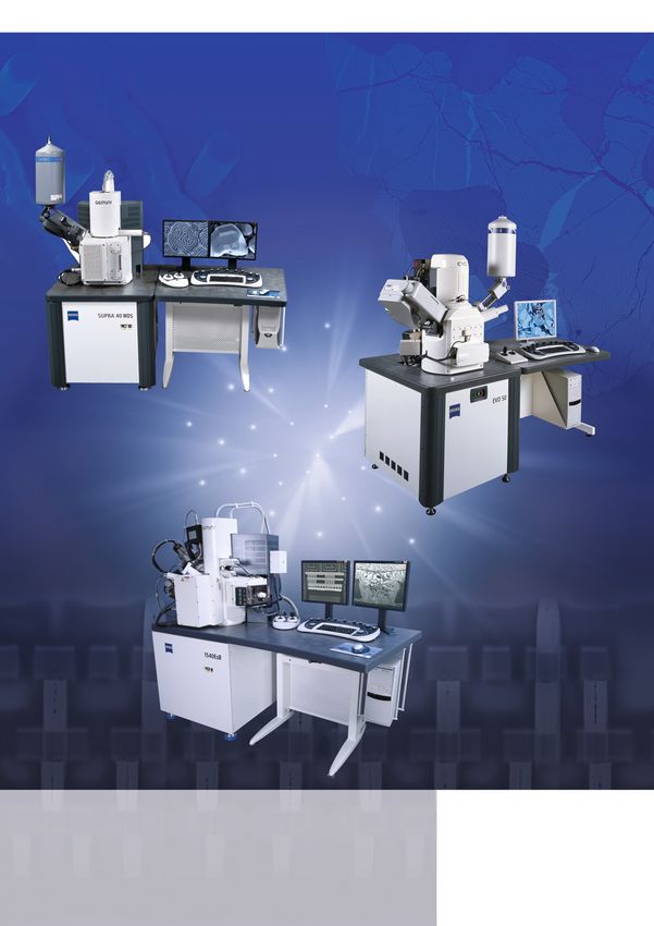
D I G I T A L   C A M E R A S            F O R          T E M                M I C R O S C O P Y

                               The Perfect Winning Hand -
                        TEM Camera Solutions
                                    by Soft Imaging System

                                                 The stakes are high. The competition tough.
                                                 Deal yourself the best available hand in the TEM
                                                 sector. Soft Imaging System's extensive range
                                                 of TEM hardware and software solutions places
                                                 the perfect set of tools at your fingertips. Meet
                                                 steadily rising demands in the transmission electron
                                                 microscopy business. Whether your application
                                                 is in the biomedical or materials analysis sector.
                                                 Our product range includes high-resolution CCD
                                                 cameras for wide-angle ports and bottom-mounted
                                                 positions on all standard TEMs. The cameras are
                                                 fiber or lens coupled, offer up to 11 MegaPixels
                                                 and provide dynamic ranges of up to 16 bits.
                                                 All cameras are cooled and highly sensitive with
                                                 fast readout rates, and use advanced techniques
                                                 to ensure optimal signal-to-noise ratio. The cameras
                                                 are easy to install, generally without any major
                                                 changes to your microscope. All cameras are
                                                 integrated with our TEM imaging software platform,
                                                 iTEM.
                                                 www.soft-imaging.net/tem

         TEM Cameras
                   Cantega2k, Morada,
                MegaView III, KeenView
                                                 For detailed information please contact:
                                                 Soft Imaging System
                                                 info.de@soft-imaging.net
                                                 www.soft-imaging.net/tem
                                                 North America: (888) FIND SIS +1 (303) 234-9270
                                                 Europe: +49 (251) 79800-0
                                                 Asia | Pacific: +60 (3) 8313-1400

                                                                                       An Olympus Company
Mitteilungen der Deutschen Gesellschaft für Elektronenmikroskopie e. V.
                                                           http://www.dge-homepage.de

             Nummer 26                                 Inhalt
                                                       Deutsche Gesellschaft für Elektronenmikroskopie e. V. ____________________________ 2
             August 2005                                 Geschäftsführung 2004–2005
                                                       Aktivitäten –––––––––––––––––––––––––––––––––––––––––––––––––––––––––––––––––––– 3
Herausgeber:                                            Wissenschaftliche Tagungen
Deutsche Gesellschaft für Elektronenmikroskopie e.V.    DGE-Laborkurse
                                                        DGE-Arbeitskreise
Redaktion:                                              Offizielle Publikationsorgane
Dr. Bernd Tesche
MPI für Kohlenforschung                                Geschäftsführung –––––––––––––––––––––––––––––––––––––––––––––––––––––––––––––– 4
Abt. Elektronenmikroskopie                              B.Tesche: Berichte des Schatzmeisters, Bilanzen der Geschäftsjahre 2003 und 2004
Kaiser-Wilhelm-Platz 1, 45470 Mülheim/Ruhr
Telefon (02 08) 3 06 22 60                             Wissenschaftliche Veranstaltungen –––––––––––––––––––––––––––––––––––––––––––––– 10
                                                        A. Nationale Tagungen
Anzeigen:
                                                            Microscopy Conference
                                                            6. Dreiländertagung 2005, Davos, Switzerland
Verlagsleitung Anzeigen: Kornelia Wind, Stuttgart              Informationen zur Tagung
Objektleitung Anzeigen:, Ilona Kern, Stuttgart
Telefon (07 11) 25 82-262, Telefax (07 11) 25 82-294
                                                               Mitgliederversammlung, Tagesordnung
S. HIRZEL                                                      Ernst-Ruska-Preis
Wissenschaftliche Verlagsgesellschaft                          Förderpreis der DGE
                                                            MC-2003, Dresden
Bezugsbedingungen:                                             31. DGE-Tagung
                                                               Wissenschaftliche Tagungsbeiträge der DGE-Geförderten:
Der Bezugspreis für Mitglieder der DGE ist durch
den Beitrag abgegolten. Jahresabonnement für                   Thomas R. Matzelle, Mi Young Park, Anne-Kathrina Rokahr
Nichtmitglieder (2 bis 3 Hefte) E 39,–, Einzelheft
                                                         B. Internationale Tagungen
E 23,–, jeweils zuzüglich Versandkosten. Die Bezugs-
dauer verlängert sich um 1 Jahr, wenn bis zum               EMC-2004, Antwerpen
30. September keine Abbestellung zum Jahresende             IMC 16-2006, Sapporo
beim Verlag erfolgt.
Alle Rechte, wie Nachdruck, auch von Abbildun-         DGE-Laborkurse 2005 –––––––––––––––––––––––––––––––––––––––––––––––––––––––––– 22
gen, Vervielfältigungen jeder Art, Vortrag, Funk,       Bericht zum DGE-Laborkurs 2004
Tonträger und Fernsehsendungen sowie Speiche-
rung in Datenverarbeitungsanlagen, auch auszugs-       Arbeitskreise der DGE –––––––––––––––––––––––––––––––––––––––––––––––––––––––––– 23
weise, sind vorbehalten.                                 Berichte aus den Arbeitskreisen
Mit Namen gekennzeichnete Beiträge geben nicht un-       EDO, PANS, EMED, HREM
bedingt die Meinung der Redaktion wieder. Der
Verlag haftet nicht für unverlangt eingereichte Ma-    Aus Forschung und Industrie –––––––––––––––––––––––––––––––––––––––––––––––––––– 29
nuskripte. Die der Redaktion angebotenen Ori-            Atomic Force
ginalbeiträge dürfen nicht gleichzeitig in anderen          Local Electrode Atom Probe Microscopy:
Publikationen veröffentlicht werden. Mit der An-            3D Imaging and Analysis with True Atomic Resolution
nahme zur Veröffentlichung überträgt der Autor           Ifg – Institut für Gerätebau GmbH
dem Verlag das ausschließliche Verlagsrecht für             Spurensuche im Elektronenmikroskop
die Zeit bis zum Ablauf des Urheberrechts. Einge-        Leica:
schlossen sind insbesondere auch das Recht zur
Herstellung elektronischer Versionen und zur Ein-
                                                            Weltweit einziges interaktives 3D-System für die Mikroskopie
speicherung in Datenbanken sowie das Recht zur           Carl Zeiss SMT
Verfielfältigung und Verbreitung online und offline         Carl Zeiss SMT sets new record milestone in sub-Ångström e-beam imaging
ohne zusätzliche Vergütung.                                 REM-Workshop bei Carl Zeiss SMT
                                                            Supra™ 40: Neues Feld Emissions Raster Elektronen Mikroskop

S. HIRZEL
                                                       Veranstaltungen außerhalb der DGE-Förderung: –––––––––––––––––––––––––––––––– 37
Wissenschaftliche Verlagsgesellschaft                    Fortbildungskurse von Leica
Birkenwaldstr. 44, D-70191 Stuttgart,
Telefon (07 11) 25 82-0
                                                       Personalien –––––––––––––––––––––––––––––––––––––––––––––––––––––––––––––––––––– 38
Telefax (07 11) 25 82-290                                Verstorben: H. Themann, Münster, E. Kellenberger, Schweiz, K. Mannweiler, Hamburg
Satz, Druck, Verarbeitung:                               Geburtstage/Festkolloquien:
Hofmann, D-73614 Schorndorf                                60. Geburtstag: Hannes Lichte, Dresden
© 2005 S. HIRZEL                                           70. Geburtstag: Harald Rose, Berkley
Wissenschaftliche Verlagsgesellschaft mbH,                 Beiträge zum Ehrenkolloquium Harald Rose von Max Haider und Hannes Lichte
Stuttgart
ISSN 0936-6911                                         Nationaler und Internationaler Tagungskalender –––––––––––––––––––––––––––––––––– 46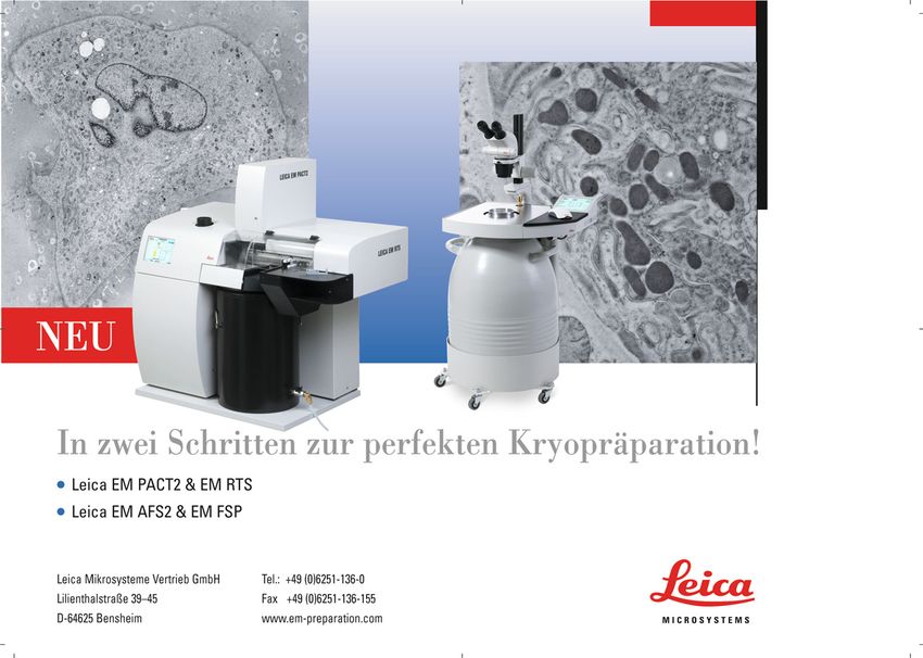
Deutsche Gesellschaft für Elektronenmikroskopie e. V.

    Geschäftsführung 2004–2005

1. Vorsitzender                        Beisitzer                           Rechnungsprüfer
Prof. Dr. Hannes Lichte                Prof. Dr. Helmut Kohl               und Ehrenbeisitzer
Institut für Angewandte Physik         Physikalisches Institut             Dr.-Ing. Gerhard Schimmel
TU Dresden                             Westfälische Wilhelms-Universität   Am Sonnenberg 17a
Triebenberglabor                       Münster                             64385 Reichelsheim
01062 Dresden                          Wilhelm-Klemm-Str. 10               Telefon: (0 61 64) 8 20
Telefon: (03 51) 21 50-89 10           48149 Münster                       Telefax: (0 61 64) 51 53 64
Telefax: (03 51) 21 50-89 20           Telefon: (02 51) 8 33-36 40         E-Mail: GeSchimmel@aol.com
E-Mail: hannes.lichte@triebenberg.de                    8 33-36 20
                                                                           Rechnungsprüfer
                                       Telefax: (02 51) 8 33-36 02
Stellvertretender Vorsitzender                                             Dr. Kurt Scheerschmidt
                                       E-Mail: kohl@nwz.uni-muenster.de
Prof. Dr. Paul Walther                                                     Max-Planck-Institut für
Zentrale Einrichtung Elektronen-       Prof. Dr. Joachim Mayer             Mikrostrukturphysik
mikroskopie der Universität Ulm        Gemeinschaftslabor                  Weinberg 2
Albert-Einstein-Allee 11               für Elektronenmikroskopie (GFE)     06120 Halle
89069 Ulm                              RWTH Aachen                         Telefon: (03 45) 5 58 29 10
Telefon: (07 31) 5 02 34 40            Aahornstraße 55                     Telefax: (03 45) 5 51 12 23
Telefax: (07 31) 5 03 33 83            52074 Aachen                        E-Mail: schee@mpi-halle.de
E-Mail: paul.walther@medizin.          Telefon: (02 41) 8 02 43 45
                                                                           Mitglied des Wahlausschusses
uni-ulm.de                             Telefax: (02 41) 8 88 83 13
                                                                           Prof. Dr. Rolf Goldberg
                                       E-Mail: mayer@gfe.rwth-aachen.de
Kom. Geschäftsführer,                                                      Institut für Angewandte
Schatzmeister                          Prof. Dr. Josef Zweck               Physik und Didaktik
Dr. Bernd Tesche                       Universität Regensburg              Technische Universität Dresden
Max-Planck-Institut für                NWF II – Physik                     Zellescher Weg 16
Kohlenforschung                        93040 Regensburg                    01062 Dresden
Kaiser-Wilhelm-Platz 1                 Telefon: (09 41) 9 43 25 90         Telefon: (03 51) 4 63-60 50
45470 Mülheim/Ruhr                                      9 43 41 88         Telefax: (03 51) 4 63-31 99
Telefon: (02 08) 3 06-22 60            Telefax: (09 41) 9 43 45 44         E-Mail: goldberg@physik.
                 3 06-22 16            E-Mail: josef.zweck@physik.         tu-dresden.de
                                       uni-regensburg.de
Telefax: (02 08) 3 06-29 80                                                Prof. Dr. Wolfgang Jäger
E-Mail: tesche@mpi-muelheim.           Berater für Biologie und Medizin    Mikrostrukturanalytik
mpg.de                                 PD Dr. Reinhard Rachel              Christian-Albrechts-Universität
                                       Lehrstuhl für Mikrobiologie der     Kaiserstraße 2
                                       Universität Regensburg              24143 Kiel
                                       Labor für Elektronenmikroskopie     Telefon: (04 31) 8 80 61 77
                                       Universitätsstraße 31               Telefax: (04 31) 8 80 61 78
                                       93053 Regensburg                    E-Mail: wj@tf.uni-kiel.de
                                       Telefon: (09 41) 9 43 45 34
                                       Telefax: (09 41) 9 43 18 24
                                       E-Mail: reinhard.rachel@biologie.
                                       uni-regensburg.de

Redaktionsschluss für Nummer 27: 15. November 2005

Redaktion: Dr. Bernd Tesche
Deutsche Gesellschaft für Elektronenmikroskopie
MPI für Kohlenforschung, Abt. Elektronenmikroskopie, Kaiser-Wilhelm-Platz 1, 45470 Mülheim/Ruhr

2                                                                          Elektronenmikroskopie · Nr. 26 · August 2005
Aktivitäten

Wissenschaftliche Tagungen                      (D) Energiefilterung und Elektronen-      E-Mail: gelderblomh@rki.de
                                                    Energieverlustspektroskopie           Konsiliarlabor: www.rki.de/
einschließlich der Mitgliederversammlungen
                                                    (EELS + EFTEM)                        INFECT/CONSUL/EM-DIAG.HTM
DGE-Laborkurse
DGE-Arbeitskreise                               Sprecher:                                 2. Sprecher:
                                                Dr. Klaus Leifer                          Dr. Dirk Soike
Offizielle Publikationsorgane                   Swiss Federal Institute of Technology     Landesamt für Verbraucherschutz und
                                                Lausanne (EPFL)                           Landwirtschaft
European Journal of Cell Biology
                                                Institute of Quantum Photonics and        Laborbereich Potsdam
Optik                                           Electronics                               Pappelallee 20
(Gustav Fischer Verlag GmbH & Co. KG            1015 Lausanne                             14469 Potsdam
D-07705 Jena, Germany                           Switzerland                               Telefon: (03 31) 5 68 82 73
E-Mail: office.j@gfischer.de                    Telefon: (0 21) 6 93 48 27                Telefax: (03 31) 5 68 82 57
                                                                6 93 70 31                E-Mail: dirk.soike@lvl.brandenburg.de
Ultramicroscopy
                                                Telefax: (0 21) 6 93 44 01
(North-Holland Physics Publishing)                                                        (G) Digitale Informationserfassung,
                                                E-Mail: klaus.leifer@epfl.ch
                                                                                                -verarbeitung und -archivierung
Arbeitskreise der DGE                           Stellvertretender Sprecher:                     (DIVA)
(A) Elektronenmikroskopische Direkt-            Dr. Gerald Kothleitner                    Sprecher:
       abbildung von Oberflächen (EDO)          FELMI                                     Dr. Werner Zuschratter
                                                Universität Graz                          Special Laboratory Electron &
Für weitere Informationen: EDO-HOMEPAGE
                                                Steyrergasse 17                           Laserscanning Microscopy
(Wuppertal)
                                                A-8010 Graz                               Leibniz-Institut für Neurobiologie
http://www.edo.uni-wuppertal.de/edo
                                                Austria                                   Universität Magdeburg
1. Sprecher:                                    Telefon: +43 (0) 3 16 8 73-83 36          Brenneckestraße 6
Prof. Dr. rer. nat. L. J. Balk                  Telefax: +43 (0) 3 16 81 15 96            39118 Magdeburg
Lehrstuhl für Elektronik,                       E-Mail: gerald.kothleitner@felmi-zfe.at   Telefon: (03 91) 6 26 33 29
Fachbereich Elektrotechnik                                                                Telefax: (03 91) 6 26 33 28
Informationstechnik, Medientechnik              Aktivitäten: Jährliches Kolloquium        E-Mail: zuschratter@ifn-magdeburg.de
Bergische Universität Wuppertal                 (E)   Präparation und Abbildung Nativer
Rainer-Gruenter-Straße 21                                                                 Stellvertretender Sprecher:
                                                      Systeme (PANS)                      Dr. Josef A. Schroeder
42119 Wuppertal
Telefon: +49 (0) 2 02-4 39-15 72                1. Sprecher:                              Zentrales EM-Labor
Telefax: +49 (0) 2 02-4 39-18 04                Dr. Stefan S. Biel                        Universitätsklinikum Regensburg
E-Mail: balk@uni-wuppertal.de                   Beiersdorf AG                             Institut für Pathologie
                                                Analytical Microscopy                     Franz-Josef-Strauß-Allee 11
(B)    Analytische Elektronenmikroskopie        Unnastraße 48                             93042 Regensburg
       in Biologie und Medizin (AEMB)           20245 Hamburg                             Telefon: (09 41) 9 44 66 36
1. Sprecher:                                    Telefon: (040) 49 09 66 71
                                                                                          Weitere Informationen:
Dr. Michael Laue                                Telefax: (040) 49 09 38 55
                                                                                          http://www.physik.uni-regensburg.de/
Institut für Anatomie und Zellbiologie          E-Mail: BielS@hamburg.beiersdorf.com
                                                                                          forschung/zweck/dge/diva/DIVA.html
Universität des Saarlandes
66421 Homburg                                   2. Sprecher:                              (H) Hochauflösende Transmissions-
Telefon: (0 68 41) 1 62 68 13                   Dr. Thomas Müller-Reichert                      Elektronenmikroskopie (HRTEM)
Telefax: (0 68 41) 1 62 62 93                   Max-Planck-Institut für Molekulare
                                                Zellbiologie und Genetik                  Sprecher:
E-Mail: michael.laue@uniklinik-saarland.de                                                Kurt Scheerschmidt
                                                EM-Facility
2. Sprecher:                                    Pfotenhauerstraße 108                     Dr. Kurt Scheerschmidt
Prof. Dr. Ludwig Jonas                          01307 Dresden                             MPI für Mikrostrukturphysik
Elektronenmikroskopisches Zentrum am            Telefon: (03 51) 2 10 17 63               Weinberg 2
Institut für Pathologie                         Telefax: (03 51) 2 10 20 00               06120 Halle
Universität Rostock                             E-Mail: mueller-reichert@mpi-cbg.de       Telefon: (03 45) 55 82-9 10
18055 Rostock                                                                                              55 82-9 17
Telefon: (03 81) 4 94 58 50                     Aktivitäten: Laborkurse, Symposium.       Telefax: (03 45) 5 51 12 23
E-Mail: ludwig.jonas@medizin.uni-rostock.de                                               E-Mail: schee@mpi-halle.de
                                                (F)   Elektronenmikroskopische
Aktivitäten: Laborkurse; Symposium 2005               Erregerdiagnostik (EMED)            Stellvertretender Sprecher:
                                                                                          Dr. Michael Seibt
(C) Rastersondenmikroskopie (SPM)               1. Sprecher:                              Physik. Institut Universität Göttingen
Die Aktivitäten auf dem Gebiet der Raster-      Dr. Hans Gelderblom                       Friedrich-Hund-Platz 1
sondenmikroskopie wurden von der DGE seit       Robert-Koch-Institut                      37077 Göttingen
Verfügbarkeit bzw. frühen Verbreitung der Me-   Nordufer 20                               Telefon: (05 51) 39-45 53
thode mit besonderem Interesse gefördert.       13353 Berlin                              E-Mail: seibt@ph4.physik.uni-goettingen.de
                                                Telefon: 00 49 (0) 1 88 87 54 23 37
Zurzeit keine Aktivitäten im Arbeitskreis.      Telefax: 00 49 (0) 1 88 87 54 23 34       Aktivitäten: Jährlich zweimaliges Kolloquium

Elektronenmikroskopie · Nr. 26 · August 2005                                                                                           3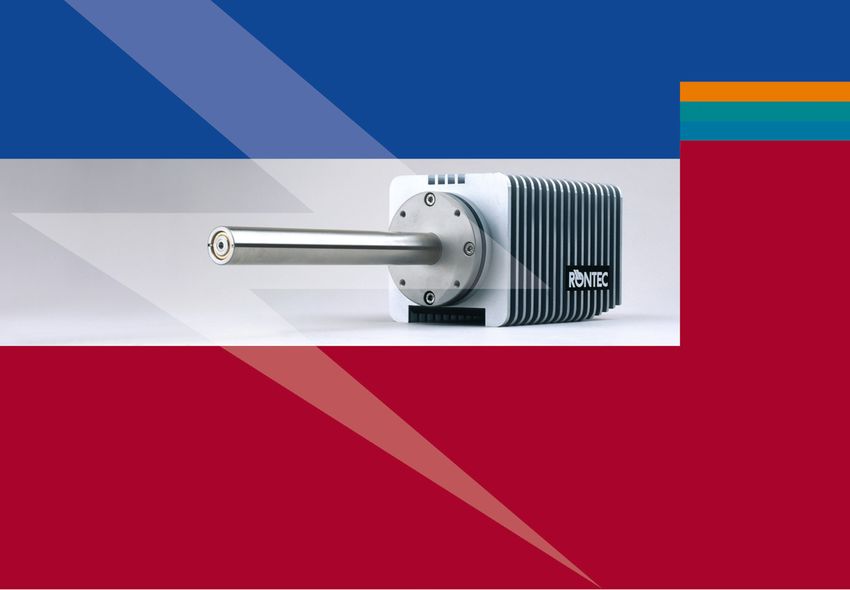
Geschäftsführung
    B. Tesche: Berichte des Schatzmeisters
               Bilanzen der Geschäftsjahre 2003 und 2004

4                                                     Elektronenmikroskopie · Nr. 26 · August 2005
Elektronenmikroskopie · Nr. 26 · August 2005 5
6 Elektronenmikroskopie · Nr. 26 · August 2005
Elektronenmikroskopie · Nr. 26 · August 2005 7
8 Elektronenmikroskopie · Nr. 26 · August 2005
Elektronenmikroskopie · Nr. 26 · August 2005 9
Wissenschaftliche Veranstaltungen

     Wissenschaftliche Veranstaltungen

                                                                                      most recent developments in light,
 A. Nationale Tagungen                                                                electron, and scanning probe micro-
                                                                                      scopes, together with specimen prepa-
                                                                                      ration equipment and image analysis
                                                                                      systems for both the life and materials
                                                                                      sciences.
Microscopy Conference,                     Ladies and gentlemen,                      We have made a special effort to keep
6. Dreiländertagung 2005                   dear micro-scopists,                       conference fees low, i.e. 65 Euros for
Die Microscopy Conference, 6. Drei-        Not only is it a great pleasure but a      students, in order to encourage young
ländertagung 2005, mit dem Status          honour for me to invite you on behalf      scientists to present their most recent
einer European Microscopy Society          of the Organizing Committee to con-        findings to the more established in-
(EMS) Extension, findet vom 28. Au-        tribute to and take part in the Micro-     vestigators in the community.
gust bis 2. September 2005 in Davos,       scopy Conference (MC) 2005, which          No doubt, the world-class Davos Con-
Switzerland, statt. Zu den 3 Confer-       will be held from August 28 to Sep-        gress Center will provide an excellent
ence Topics: Life Sciences (LS), Instru-   tember 2 in Davos, Switzerland. What       home for our Microscopy Conference
mentation & Methodology (IM), und          has started in 1985 in Konstanz, Ger-      2005. It offers a good selection of
Material Sciences wurden viele Bei-        many, as a convention of the three         meeting and conference rooms, all
träge eingereicht. Die Tagung wird be-     German-speaking countries Germany,         equipped with state-of-the-art audio-
gleitet von einer umfangreichen Ge-        Austria and Switzerland, called “Drei-     visual facilities. The professional staff
räteausstellung mit über 40 Firmen,        landertagung”, will take place this        from “Davos Tourismus” will help
die die neuesten Entwicklungen auf         year for the 6th time. Slowly but defi-    you with any questions or request you
dem Gebiet der Präparation, Geräte-        nitely, the “Dreilandertagung” has         might have with regard to lodging,
entwicklung und Bild- und Signalver-       become a multinational event with          meals and travel during the meeting.
arbeitung präsentieren.                    English as the conference language         Davos, one of the largest mountain
                                           and, for the first time, will have the     resorts in Europe, offers unforgettable
                                           status of a European Microscopy            encounters with nature and a broad
                                           Society (EMS) Extension.                   range of delights. And anyone favou-
                                           The Program Committee has put to-          ring the quiet idyll over the pulsing life
                                           gether a well balanced selection of        in the thriving city, will find peace and
                                           Plenary Talks, Special Interest Lectu-     tranquillity in the wide Davos out-
                                           res, and Regular Scientific Sessions.      doors.
                                           In addition, the program will provide      We are all looking forward to greeting
                                           plenty of room for discussing more         you in Switzerland and wish you al-
                                           specialized issues during Workshops.       ready now an interesting conference
                                           An International Scientific Advisory       and a pleasant stay.
                                           Board was actively involved in suggest-    Welcome to Davos for the Microscopy
                                           ing speakers and topics, as well as or-    Conference 2005.
                                           ganizing and chairing sessions. Last       Kurt Pulfer
                                           but not least, the Poster Sessions re-     Chairman MC 2005 Davos
                                           present an essential part of the confer-
                                           ence where scientific results from
                                           more specific investigations will be
                                           presented. There will be ample time
                                           for discussion with the authors at the
                                           posters.
                                           Furthermore, the Davos Congress
                                           Center provides an optimal platform
                                           for housing a large Commercial Exhi-
                                           bition. There will be on display the

10 Elektronenmikroskopie · Nr. 26 · August 2005Ý«Ài]Ê
ÃVÛiÀ]Ê Õ`°
/`>Þ½ÃÊ>`Û>Vi`Ê`ÃVÛiÀÞÊ>`Ê«À`ÕVÌÊ
`iÛi«iÌÊÀiµÕÀiÃÊÕ«i`i`ÊÎ Ê>VViÃÃÊÌÊ
Ì iÊ>ÃV>iÊÜÌ ÊÌ iÊLiÃÌÊÀiÃÕÌÊ«ÃÃLi°Ê
½ÃÊ/ÃÊvÀÊ >ÌiV ÒÊÃÕ««ÀÌÊÌ iÊÃÕVViÃÃÊvÊ
ÞÕÀÊÃÌÊ`i>`}Ê«ÀiVÌÃÊLÞÊ}Û}ÊÞÕÊÌ iÊ
«ÜiÀÊÞÕÊii`ÊÌÊÀi>V Êv>ÀÊÌÊÌ iÊ>ÃV>i°Ê
7ÊqÊ/Ì>ÒÊÎääÊ-®/ ÊqÊ/ iÊ>iÜÊ
/Ì>Ê-®/ ÊLÀi>ÃÊÌ iÊ}ÃÌÀÊL>ÀÀiÀÊÜÌ Ê
-ÕL}ÃÌÀÊ/ Ê>}i
>ÌVÃV>iÊÀiÃÕÌ°Ê/ ÃÊ`i`V>Ìi`ÊÊ
«>ÌvÀÊvÀÊ>LiÀÀ>ÌÊVÀÀiVÌi`ÊÃV>}®Ê
iiVÌÀÊVÀÃV«ÞÊ`iÛiÀÃÊÌ iÊ } iÃÌÊ
ÀiÃÕÌÊ>Û>>LiÊÊ>ÊViÀV>ÞÊ>Û>>LiÊ
-®/ ÊÃÞÃÌi°Ê
/iV>ÒÊÓÊ-«ÀÌÊ/ ÊqÊ iÃ}i`ÊvÀÊ
>`Û>Vi`Ê>}}Ê>`Ê>>ÞÃÃÊÃÌÕ`iÃÊÊviÊ
/Ì>ÊÎääÊ-®/
ÃViViÊ>`ÊÃvÌÊ>ÌÌiÀÊÀiÃi>ÀV ]ÊÌ iÊ/iV>Ê
-«ÀÌÊ«À`ÕViÃÊÕÃÕÀ«>ÃÃi`ÊÎ Ê } ÀiÃÕÌÊ
>}iðÊ`Û>Vi`ÊVÀÞÊ/ ÊÃÌÕ`iÃÊ>ÀiÊi>ÃÞÊ
>V iÛi`ÊÕÃ}Ê ½ÃÊ6ÌÀLÌÒÊvÀÊVÀÞÊÃ>«iÊ
«Ài«>À>ÌÊÜÌ ÊÌ iÊ/iV>Ê-«ÀÌ°Ê
Û>ÒÊ >
>LÊ>`Ê+Õ>Ì>ÒÊÎ ÊqÊ ½ÃÊ
À>}iÊvÊ>ÀiÌi>`}Ê Õ> i>Ê É
- ®ÊÃÞÃÌiÃÊ>ÀiÊ«Ìâi`ÊÌÊ}ÛiÊÞÕÊÌ iÊ
Û>Ê >>
>L «iÀvÀ>ViÊÞÕÊ`i>`ÊvÀÊÞÕÀÊëiVvVÊ
>««V>ÌÃÊÀ>}i°Ê/ iÊ Û>Ê >
>LÊ`iÛiÀÃÊ
} ÞÊ>VVÕÀ>ÌiÊ Ê}ÊÜÌ Ê } ÊÀiÃÕÌÊ
- Ê>}}ÊvÀÊ>ÃV>iÊ>V }Ê>`Ê
«ÀÌÌÞ«}]Ê>`Ê«Ài«>À>ÌÊvÊÕÌÀ>Ì Ê
/ ÊÃ>«iÃ°Ê ½ÃÊ+Õ>Ì>ÊÎ ÊÀi>ÃÊÌ iÊ
}`ÊÃÌ>`>À`ÊvÀÊ } Û>V]ÊÜÛ>VÊ>`Ê
iÛÀiÌ>ÊÎ Ê- Ê>««V>ÌðÊ
Û>Ê >- 7ÊqÊ Û>Ê >- ÊqÊ Ý«iÀiViÊÌ iÊ
ÜÀ`½ÃÊÞÊÌÀÕiÊ } ÀiÃÕÌ]ÊÜÛ>VÕÕÊ
- ÊvÀÊV >À>VÌiÀâ>ÌÊvÊV >À}}Ê>`ÉÀÊ
VÌ>>Ì}Ê>>ÌiÀ>ÃÊÀÊqÊ`iÛVið
7Ì Ê1«>À>ii`ÊVViÃÃÊ ÊÌ iÊÀ>ViÊvÀÊ>ÃV>iÊ`ÃVÛiÀÞÊ>`Ê
ViÀV>â>ÌÊÞÕÊV>½ÌÊÜ>ÌÊÕÌÊÌÀÀÜÊ
ÌÊÌ iÊ >ÃV>i vÀÊÌ iÊÌÃÊÞÕÊii`ÊÌ`>Þ°Ê ]ÊÌ iÊÜÀ`½ÃÊ
i>`iÀÊÊ/ÃÊvÀÊ >ÌiV ]Ê}ÛiÃÊÞÕÊÀiÊ
>VViÃÃÊÌÊÌ iÊ>ÃV>iÊÃÊÞÕÊV>ÊiÝ«Ài]Ê
`ÃVÛiÀÊ>`ÊLÕ`ÊiÊÞÕ½ÛiÊiÛiÀÊ>}i`°
ÜÜÜ°viV«>Þ°VÊ
Ã>iÃJviV°V
^ÓääxÊ Ê «>Þ°ÊÊÌÀ>`i>ÀÃÊ>ÀiÊÌ iÊ«À«iÀÌÞÊvÊÌ iÀÊÀiëiVÌÛiÊÜiÀðWissenschaftliche Veranstaltungen
International Scientific – Rudolf Reichelt LS3 Secretion and Endocytosis
Advisory Board – Paul Walther M. Pavelka, Wien
– S. Daglioglu, President of the Turk- – Josef Zweck L. Huber, Innsbruck
ish Society for Electron Microsco- – Klaus Leifer LS4 Microscopy in Vascular Biology
py – Jens Gobrecht A. Lametschwandtner, Salzburg
– M. de Lourdes Pereira, Department U. M. Spornitz, Basel
IT/Publications LS5 Microscopy of Stem Cells,
of Biology, University of Aveiro – Daniel Mathys
(Portugal) Cell Cycle and Programmed
– Stefan Dürrenberger Cell Death
– B. M. Humbel, Utrecht University. – URZ Basel
Electron Microscopy Unit, Dept. of W. Klepal, Wien
Mol. Cell Biol (Netherlands) General Organisation Th. Cremer, München
– M. Luisa No, Departamento Física – Kurt Pulfer LS6 Architecture of the Cell Nucle-
Aplicada II, Facultad de Ciencias – Markus Dürrenberger us and the Cytoskeleton
(Spain) V. Small, Wien
– P. Hozak, President Czech Micro- Exhibition/Sponsoring I. Raska, Prague
scopy Society – Patrick Schwarb LS7 Microscopy of Biomaterials and
– A. Czyrska-Filemonowicz, Presi- – Peter Eggli the Extracellular Matrix
dent of the Polish Society for G. Richards, Davos
Social Events Wl Workshop: EM Diagnostics of
Microscopy – Geoff Richards
– I. Ronchetti, Dept. Biomedical Infectious Deseases
Sciences, University of Modena Accommodation/Registration W2 Workshop: Visualisation of Cel-
and Reggio Emilia (Italy) – Davos Tourismus lular Dynamics
Instrumentation &
Conference Topics Methodology (IM)
Organizing Commitee
IM l Electron Optics
Finances Life Sciences (LS) M. Haider, Heidelberg
– Marcel Düggelin LS1 3D and Cryo Electron Micro- E. Plies, Tübingen
scopy: From the Molecular to IM2 Advances in TEM Contrast
Scientific Board the Cellular Level
– Ueli Aebi Analysis and Simulation
H. Stark, Gottingen P. Stadelmann, Lausanne
– Heinz Gross S. Nickell, Martinsried M. Lehmann, Dresden
Scientific Board Representatives LS2 Localization of Intracellular IM3 Electron Holography
– Ferdinand Hofer Elements and Macromolecules H. Lichte, Dresden
– Peter Karnthaler In Memoriam Karl Zierold G. Pozzi, Bologna
– Margit Pavelka H. Schwarz, Tübingen IM4 SEM and LEEM
– Reinhard Rieger R. Wepf, Hamburg R. Reichelt, Münster
– Hannes Lichte L. Frank, Brno
Sun, Aug 28 Mon, Aug 29 Tue, Aug 30 Wed, Aug 31 Thu, Sep 1 Fri, Sep 2
08:30–10:00 Opening Ceremony Plenary Lectures Plenary Lectures Plenary Lectures Plenary Lectures
Ruska Lectures
EMS lectures
10:00–10:30 Coffee Break Coffee Break Coffee Break Coffee Break Coffee Break
10:30–12:30 3 parallel Sessions 3 parallel Sessions 3 parallel Sessions 3 parallel Sessions 3 parallel Sessions
IM1/LS7/MS1 IM3/LS4/MS5 IM5/LS1/MS2 IM7/LS5/MS4 IM6/LS2/MS7
12:30–14:00 Lunch Break Lunch Break Lunch Break Lunch Break End of Conference
14:00–16:00 Poster Poster General Assemblies Poster
16:00–16:30 Registration Coffee Break Coffee Break Train Trip Coffee Break
Get Together Party
16:30–18:30 Exhibitor’s Presen- 3 parallel Sessions Conference Dinner 3 parallel Sessions
tation and Reception IM4/LS3/MS3 IM2/LS6/MS6
Welcome “Land-
schaft Davos”
18:30–20:00 Break Break
20:00–22:00 Workshops Workshop W1 Workshop W2/W3
12 Elektronenmikroskopie · Nr. 26 · August 2005Erfolg ist präparierbar
Ion Mill
Ionendünnung von Proben, optional
Plasma Cleaner mit Flüssigstickstoffkühlung
Gleichzeitiges schonen-
des Entfernen von
organischen Verunreini-
gungen auf Proben und
Probenhalter
Automated Sample Preparation System
Vollautomatische SEM-Probenpräparation
mit Plasmareinigung, Ionenstrahlätzen,
reaktivem Ionenätzen und Ionenstrahl-
unterstützter Beschichtung
Nano Mill
Bearbeiten von TEM-Proben und
FIB-Nachbearbeitung mit niederenergetischem
fokussiertem Raster-Ionenstrahl und stickstoff-
gekühlter Probe
Weitere Produkte für das Labor:
Q-Control: Aktive
Molecular Cantileverdämpfung AFM-Cantilever XP-2 Stylus-
Force Probe – oder -entdämpfung für Rasterkraft- Profiler zur
AFM Systeme für oszillatorische mikroskopie Stufenhöhen-
AFM-Modi messung
Rufen Sie uns an: Atomic Force F&E GmbH · Hauptstrasse 161 · 68259 Mannheim/Germany
Telefon +49 (0)621 762117-0 · Fax +49 (0)621 762117-11· info@atomicforce.de · www.atomicforce.deWissenschaftliche Veranstaltungen
IM5 X-Ray Optics/Microscopy
C. David, Villingen
R. Fink, Erlangen
IM6 Analytical TEM
P. Schattschneider, Wien
H. Kohl, Münster
IM7 Advances in Specimen Prepara-
tion and Nano-Structuring
H. Cerva, München
J. Mayer, Aachen
W3 Workshop:
Specimen Preparation
Material Sciences (MS)
MS 1 Spectroscopy in
Material Sciences
F. Hofer, Graz
W. Sigle, Stuttgart
MS2 Nanoparticles
P. Hoffmann, Lausanne
R. Schlögl, Berlin
MS3 Nanostructured Materials
D. Gerthsen, Karlsruhe
Th. Waitz, Wien
MS4 Alloys and Intermetallics
H. P. Karnthaler, Wien
W. Neumann, Berlin
MS5 Semiconductors
H. Strunk, Erlangen
P. Werner, Halle
MS6 Solid State Chemistry
F. Krumeich, Zürich
D. Hesse, Halle
MS7 Magnetic and
Dielectric Materials
J. Zweck, Regensburg
H. Hug, Basel
Informationen zur Tagung:
Website:
http://www.davos2005.unibas.ch
Contact Mitgliederversammlung am Ernst-Ruska-Preis
Markus Dürrenberger 31. August 2005 in Davos Seit seiner satzungsmäßigen Um-
Universität Basel struktierung (Neugründung) wird der
Der Vorstand der DGE lädt hiermit zur
Zentrum für Mikroskopie (ZMB) Ernst-Ruska-Preis erstmals als DGE-
ordentlichen Mitgliederversammlung
Klingelbergstrasse 50 Preis im Rahmen der Eröffnungssit-
ein. Die Versammlung findet am 31.
CH-4056 Basel zung der 6. Dreiländertagung verlie-
August von 14.00 bis 16.00 Uhr im
Tel. +41(0)61-267 14 04 hen.
Kongresszentrum Davos statt. Tages-
Fax +41(0)61-267 14 10
ordnung siehe Einladungsschreiben
E-Mail:
des kom. Geschäftsführers.
duerrenberger@davos2005.unibas.ch
Anmerkungen: Förderpreis der DGE
Die Jahresrechnungen 2003 und 2004 Dieser an junge Wissenschaftler zu
sind in diesen Mitteilungen veröffent- vergebene Förderpreis wird im Rah-
licht. men der Mitgliederversammlung der
Die Kandidaten für den Vorstand DGE verliehen.
haben für den Fall ihrer Wahl ihr Ein- Die Ausschreibungen beider Preise
verständnis erklärt. wurden auf der Homepage der DGE
veröffentlicht.
14 Elektronenmikroskopie · Nr. 26 · August 2005DETECTORS
XRD
XRF
EDX
RÖNTEC AG
sales@roentec.de
RÖNTEC USA, Inc.
sales@rontecusa.com
RÖNTEC France
roentecfrance@free.fr
www.roentec.com
The future Microanalysis at lightning speed Ask for a live
demonstration
of the world’s fastest
■ 3rd generation of LN2-free XFlash® Detectors (SDD)
of EDX ■ Best energy resolution of 127eV at MnKα 1000 cps
EDX analyzer!
■ Light element quantification
■ Mapping up to 10 times faster
■ New L- and M-line data for precise IDWissenschaftliche Veranstaltungen
MC 2003 Dresden metry of the precipitates in the ele-
31. DGE-Tagung mental map is nearly a circle. It is ne-
Die DGE hat 3 jungen Wissenschaft- cessary to compare the experimental
lern die Kongressteilnahme durch eine results with simulated images. There-
Reisebeihilfe ermöglicht. Wie bisher fore we made a simulation of a spheri-
werden die wissenschaftlichen Beiträ- cal cluster that contains oxygen. Ac-
ge der Geförderten in der DGE-Mit- cording to the elemental map the radi-
teilungen veröffentlicht. us of the sphere is about 16 nm. All the
instrumental parameters of the
EFTEM used for imaging are taken
into consideration (acceleration volt-
Investigation of Precipitates
age, spherical and chromatic aberrati-
using an Energy-Filtering on, aperture, defocus, energy loss,
Transmission Electron energy loss window, pixel size, etc.).
Microscope (EFTEM) The profiles of the experiment and of Fig. 1: Elemtal map of oxygen in silicon.
Anne-Kathrina Rokahr and Helmut the simulation correspond quite well,
Kohl, Physikalisches Institut und In- as can be seen in fig.2. Differences are
terdisziplinäres Centrum für Elektro- due to the thickness of the specimen of
nenmikroskopie (ICEM), Universität about 90 nm and the oxidation of the
Münster, Wilhelm-Klemm Str. 10, surface. This oxidation is taken into
48149 Münster, Germany, E-Mail: account by adding a constant offset in
Rokahr@uni-muenster.de, http://www. the simulation (fig.2). The good agree-
uni-muenster.de/Physik/PI/Kohl/index. ment between theory and experiment
html supports our conjecture, that the preci-
pitates are spherical. The signal-to-
Electron microscopes equipped with noise-ratio (SNR) of the elemental
an imaging energy filter allow to re- map is shown in fig. 3. The line scans
cord elemental maps using inelastical- of the measured SNR-image and si-
ly scattered electrons. For high energy mulated SNR can be seen in fig. 4. The Fig. 2: Line scan of the simulated and
measured intensity
losses the energy loss of the beam elec- profiles also correspond quiet well.
tron is due to inner shell excitations The SNR is very low. This is caused
of the specimen atoms. The excitation by the small size of the oxygen cluster
energies are then specific for the ele- (16 nm) compared by the total thick-
ment and shell. By using electrons ness of the specimen (90 nm). Accor-
whose energy losses are equal to inner ding to these investigations we can de-
shell excitations of a specific element termine the precipitates as a spherical
and after a background subtraction cluster with a radius of about 16 nm
using the “Weighted-Least-Square- containing oxygen. Because of the
Fit” method [1], we obtain an elemen- poor SNR we were not able to deter-
tal map and a signal-to-noise image of mine the chemical composition pixel
this element. This elemental map by pixel. We are now working on a
shows the two-dimensional projection procedure to determine the compositi-
of the spatial distribution of this ele- on assuming it to be homogeneous
ment. The signal-to-noise image over the sphere. [4]
shows the ratio of the signal and the
noise in each pixel of the image. The References Fig. 3: Signal-to-noise map of oxygen in
specific properties of the EFTEM in- [1] T. Pun, J. R. Ellis, M. Eden: J. of silicon.
fluence the imaging process. The real Microscopy 134 (1984) 295
structure of the object is falsified in the [2] A. Berger: Dissertation, Institut
image by these properties. For a deter- für Angewandte Physik der TH
mination of the spatial geometry our Darmstadt (1993)
simulations are based on the concept [3] R. Knippelmeyer, H. Kohl: J. of
of transfer functions of Berger [2], Microscopy 194 (1999) 30
which is based on the linear image [4] We wish to express our thanks to
theory. Here we used the relativistical- Dr. H. Bracht and A. Rodriguez
ly correct transfer function of Knip- (Institut für Materialphysik der
pelmeyer [3]. In our experiments we WWU Münster) for the speci-
acquired an elemental map of the oxy- mens.
gen distribution using a Zeiss EM 902.
By this we were able to detect oxygen Fig. 4: Line scan of the simulated and
in the precipitates (see fig.1). The geo- measured signal-to-noise-ratio image.
16 Elektronenmikroskopie · Nr. 26 · August 2005SkyScan MicroCT und NanoCT
Electron energy-loss near-edge
structures of silicon carbides Schäume sind
Mi Young Park1, Kai Giese2*, Peter
Krüger2, Helmut Kohl1
1Physikalisches Institut and Interdiszi-
Träume.
plinäres Centrum für Elektronenmi-
kroskopie,
2Institut für Festkörpertheorie, *now at Es ist schon eine erstaunliche Methode, Strukturen von
Freie Universität Berlin Körpern, Geweben, Knochen, Neuen Materialien und
Westfälische Wilhelms-Universität anderen Objekten ohne Zerstörung räumlich darzustellen.
Münster, Wilhelm-Klemm Str. 10, Die MicroCT und 3d-Röntgenmikroskopie gewährt den
48149 Münster, Germany
berühmten 'Einblick' und
Introduction 'Durchblick'.
EEL spectroscopy (EELS) in trans- Diese Methode ist prinzipiell
mission electron microscopes is a me-
einfach: Serien von
thod to investigate the chemical and
structural characteristics of materials Radiographien werden mit der
[1]. The oscillations in the spectra up ConeBeamRecon
to a few tens of eV beyond the onset of verarbeitet. Als Ergebnis: 2d-
the core loss ionisation edges provide
Schnitte in allen Richtungen
information about chemical bonding
and electronic structure. In order to in- und 3d-Volumen. Daraus
terpret such electron energy-loss near- entstehen dann
edge structures (ELNES) and to ob- Berechnungen von
tain quantitative information from
Volumenanteilen,
them some form of modelling is ne-
cessary. To distinguish different pha- Oberflächen und
ses of SiC we have calculated ELNES anderen
of ≤-, 2H-, 4H- and 6H- SiC by com- volumetrischen
puting the symmetry projected local
Eigenheiten des
density of states using band structure
theory and compared them with meas- untersuchten
ured near-edge structures. Materials.
Band structure calculations
Our calculations have been carried out SkyScan Systeme
using the local density approximation verfügbar mit
of the density functional theory. Non- Pixelauflösungen bis
local, normconserving pseudopotenti-
unter 1μm, als
als in separable form were employed
in these computations [2, 3]. The wave Tischgerät oder als
functions are expanded in terms of li- In-vivo-Systeme.
near combinations of Gaussian orbi- NEU: Das innovative
tals with s-, p- and d-symmetry. Final
NanoCT 2011
state effects resulting from the creati-
on of a core hole due to the excitation ermöglicht durch den
process have been taken into account Einsatz eines TEM-
by using appropriate pseudopotentials Columns eine Auflösung
for the absorbing atoms. These poten-
von 130nm. Der Zeit
tials have been derived from self-con-
sistent calculations for ionised atoms. weit voraus!
Within this approach the density of Für Sie kostenlos: Unsere
states, weighted by the transition ma- MicroCT Bibliothek und
trix elements for one-electron excitati-
"SkyScan MicroCT" jeweils
ons, has been calculated.
auf CD-ROM. Einfach
Experiments anrufen!
We have acquired spectra from
crushed 6H-SiC-powders and from
4H-SiC specimens using a Jeol 3010
Vorführung und MicroCT als Dienstleistung:
RJL Micro & Analytic GmbH
Tel: +49 - 7251 - 94 8075
eMail: analytic@rjl-micro.deWissenschaftliche Veranstaltungen
with a Gatan Imaging Filter at an acce- The Influence of Crosslinking
leration voltage of 300 kV. The 4H- Density to the Elastic Properties
SiC specimens were prepared using an of ‘Smart’ Polymers:
ultramicrotome Leica Ultracut UCT. A Scanning Force and Scanning
The 4H- and 6H-SiC measurements Electron Microscopy Study
have been taken near the Si-K edge at
1836 eV and near the C-K edge at 285 T. R. Matzelle1, R. Reichelt2, and N.
eV. Krusec
1Chimie Physique des Materiaux,
Results Universite Libre de Bruxelles, B-1050
Fig.1 and 2 show the results of band- Bruxelles, Belgium
structure calculation for Si K-, and C 2Institut für Medizinische Physik
K-ELNES of 2H-, 4H- and 6H-SiC und Biophysik, Westfälische Wil-
compared with the experimental re- Fig. 1: The near-edge structure of the C K- helms-Universität, Robert-Koch-Str.
sults measured at energy resolution of edge of 2H- ,4H- and 6H- SiC as obtained
by a band structure calculation compared 31, D-48149 Münster, Germany
2.2 eV. At this resolution there are al- to the experimental spectra of 4H- and 6H- Stimuli-sensitive hydrophilic gels,
most no differences between the cal- SiC (Energy resolution: 2.2 eV)
often defined as “smart” or “intelligent”
culated ELNES of 2H-, 4H- and 6H- polymers, have attracted considerable
SiC and between the measured spec- attention since they exhibite abrupt
tra. Even though the labeled peak po- volume changes and phase separations
sitions of the theoretical and experi- as a response to environmental changes.
mental spectra in Fig. 1 and 2 are in In particular, the thermo-responsive
good agreement, it is not possible to poly-N-isopropylacryl amide (PNI-
determine the modification of the un- PAAm) hydrogel, one of the most pro-
known phases in SiC using simulated mising candidates for on-off switches
and measured spectra at this energy and controlled release systems, under-
resolution. goes a reversible volume phase transi-
Fig. 3 indicates, however, that at tion at around 33 °C, the lower critical
higher energy resolution marked dif- solution temperature (LCST) of
ferences occur. We are currently meas- Fig. 2: The near-edge structure of the Si K- PNIPAAm homopolymer in water [1].
edge of 2H- and 4H- SiC as obtained by a
uring spectra of SiC at higher resolu- band structure calculation compared to The realization of such thermal
tion and the resulting data will be the experimental spectra of 4H-and 6H-SiC switches and drug-release systems
compared with the calculated spectra. (Energy resolution: 2.2eV) necessitates a thorough understanding
of the micromechanical and structural
References properties of “smart” polymers. With
[1] R. F. Egerton, Electron Energy- these applications in mind we have stud-
Loss Spectroscopy in the Elec- ied the local elasticity of PNIPAAm
tron Microscope, 2 nd ed. (Ple- hydrogel at various cross-linker con-
num, New York, 1996). centrations, above and below the LCST.
[2] D. R. Hamann, M. Schlüter, and The experiments were performed on
C. Chiang, Phys. Rev. Lett. 43, the micrometer scale using Scanning
1494. (1979) Force Microscopy (SFM). Young’s
[3] L. Kleinman and D. M. Bylander, moduli E were determined from the
Phys. Rev. Lett. 48, 1425 (1982) force-controlled indentation of PNI-
PAAm hydrogel surfaces in a water
bath using urn-sized spherical probes
rather than conical tips (Fig. 2, insert).
Fig. 3: The near-edge structure of the Si K- For sake of comparison we investiga-
and C-K-edge of 2H-,4H- and 6H-SiC as ob-
tained by band structure calculations ted as well the elastic properties of
(Energy resolution: 0.8eV) poly-acryl amide (PAAm) hydrogel
which shows no such temperature-re-
sponsive behavior.
PNIPAArn and PAArn hydrogels
were investigated using a commercial
SFM (Explorer TMX-2000 system,
Veeco, USA) and a home-made, tem-
perature-controlled liquid cell. V-sha-
ped Si-cantilevers (type Ultralever of
the same distributor) were manually
altered by attaching a μm-sized glass
sphere to the extremity of the cantile-
18 Elektronenmikroskopie · Nr. 26 · August 2005ASPEX PersonalSEM
Automatische
Analytik integral.
Die äußerst pragmatische Integration
eines SEM/EDX-Systems, komplett von
einem Hersteller entwickelt.
Elektronenoptik und Analytik sind aus
der Praxis
entstanden, aus
einem "Guß".
Fig. 1: Indentation in PNIPAAm hydrogel (sphere radius R = 2.5 μm) as a function of the
load in dependence of the temperature at different cross-linker concentrations. Experi- Daher sind diese
mental data are represented by symbols whereas solid lines correspond to the Hertz
model assuming a sphere (R = 2.5 μm) indenting a flat surface, a = 10 °C, b = 20 °C, c = 40 °C. Systeme neben
der klassischen
Verwendung
ver. NIPAAm monomer was poly-
besonders dort
merised in aqueous solution in the pre-
sence ofN,N’-Methylene-bis-acryla- anzutreffen, wo
mide (MBAAm) as cross-linker du- mittels schneller
ring 12 h at 20°C using ammonium automatischer
persulfate (APS) and N,N,N,N’-tetra-
Teilchenanalyse
methylethylenediamine (TEMED) as
redox initiator system. The concentra- im Qualitätsmanagement Bedeutendes
tion of NIPAAm in the pre-gel soluti- geleistet wird: Anzahl, Größe und Typ
on was always 1 mol l–1, that of APS von Einschlüssen in Stahl und Metallen
10 mmol l–1 and that of TEMED
4 mmol l–1. PAAm gels were prepared Fig. 2: Young’s moduli of a water-swollen Multiple classes S
under similar synthesis conditions, but Ternäres
PNIPAAm gel in dependence of the cross-
Diagramm
the monomer concentration was always linker content at three different temperatu-
live
res as revealed by SFM using a glass
5 mol l–1. Local elasticities were eva- sphere as probe. Insert:
luated following the method of Schematic view of a glass sphere (left) and
Radmacher et al. [2]. Accordingly, the a sharp tip (right) on a cantilever before in-
denting the PNIPAAm gel surface. The
probe indentation was displayed in de- images are photo assembleds on the
pendence of the loading force. The ex- basis ofFESEM secondary electron micro-
perimental data plotted in this way graphs after rescaling.
allowed to be fitted by the Hertz model
[3].
Fe Mn
On PNIPAAm hydrogel surfaces [3] Hertz, H. J., Reine Angew. Ma-
a dramatic increase in stiffness (up to thematik, 92 (1882) 156.
10 times) was observed when crossing [4] This work was kindly supported (Reinheitsgrad), Partikelanalytik auf
the phase transition at ~33 °C towards by the “Communaute Francaise” Membranfilter im Maschinenbau und bei
higher temperature. It also can be seen (“ARC”, No. 96/01–201) and by der Herstellung und Wartung von
in figure 1, that the cross-linker con- DEGUSSA AG, CREAVIS Tech- Flugzeugtriebwerken.
centration has a significant strong in- nologies and Innovation, D-
fluence on the local elastic modulus 45746 Marl. Ebenso in der Kriminaltechnik für die
for temperatures above LCST while GSR-Analyse.
only small variations occured below Zur Qualitätskontrolle in der Pharmazie
33°C. All experimental E values are
ist das ASPEX PersonalSEM in den USA
displayed in figure 2.
nach 21CFR Part 11 qualifiziert. Mehr
References Innovation gibt's nicht!
[1] Hoffman, J., Plötner, M., Kuck-
ling, D., Fischer, W.-J. Sensor.
Actuat. B 77 (1999) 139.
[2] Radmacher, . „ Fritz, M., Hans-
ma, P. K. Biophys. J., 69 (1995)
264. Spezialisiert auf analytische SEM:
RJL Micro & Analytic GmbH
Tel: +49 - 7251 - 94 8075
eMail: analytic@rjl-micro.deWissenschaftliche Veranstaltungen
Preliminary List of Symposia,
B. Internationale Tagungen Workshops courses and open Labs
Instrumentation and Techniques
1) Basic optical elements
(gun, lens, monochromator,
filter, detector)
Enclosures: First Circulars, Posters 2) Advancing HR-TEM and
European Microscopy Correspondence to be addressed to: HR-STEM
Congress, EMC 2004, Kazuo Furuya 3) Advances in SEM
General Secretary of IMC16 4) Quantitative electron diffraction
Antwerpen High Voltage Electron techniques (CBED, EBSD)
Microscopy Station 5) Electron energy loss spectrosco-
National Institute for Materials py and energy-filtered imaging/
Vom 22. bis 27. August fand in Ant-
Science (NIMS) mapping
werpen, Belgien, der 13. European
3–13 Sakura, Tsukuba, 6) Advances in X-ray/CL
Congress on Electron Microscopy
Ibaraki 305-0003, Japan spectrometry and mapping
statt.
Phone:+81(0)29-863-55 54 7) Electron tomography
Die DGE hat 10 jungen Wissen- Fax: +81(0)29-863-55 59 8) Electron holography
schaftlern durch eine Reisebeihilfe die E-Mail: Secretary@imc16.jp 9) In-situ and UHV electron
Kongress-Teilnahme ermöglicht. Aus URL: http://www.imc16.jp/ microscopy
technischen Gründen wird erst in der 10) Environmental microscopy
nächsten Ausgabe über die Tagung be- (SEM, TEM, SPM)
richtet und die Beiträge der Geförder- Scientific Program: 11) Surface microscopy (LEEM,
ten veröffentlicht. The scientific program for IMC16 will PEEM, AEM, EPMA)
be structured to highlight the most 12) Scanning probe microscopy
recent and important developments in (STM, AFM, MFM etc.)
microscopy-related technology, tech- 13) Advanced optical microscopy
niques and applications. The presenta- (NSOM, Confocal microscopy)
tions at IMC16 in Sapporo will consist
16th International of:
14) Atom probe field ion microscopy
15) X-ray microscopy
Microscopy Congress 1) Plenary sessions 16) Digital imaging and processing
(IMC 16) 3.–8. September 2) Symposia (invited and contributed techniques
oral presentations)
2006, Sapporo, Japan 3) Poster presentations
17) Remote electron microscopy
18) Advances in sample preparation
4) Tutorial workshops techniques
5) Software exchange forum 19) FIB and ion-beam techniques
Invitation to Sapporo, Japan 6) Open lectures for citizens
September 3 to 8, 2006 7) Lunch-time-seminars
The local scientific program commit- Materials Science and
First Circular of The 16th International tee will be guided by an international Nanotechnology
Microscopy Congress (IMC16) scientific advisory committee in the 1) Nanowires, tubes and particles
On behalf of the organizing com- structuring of the program and selec- 2) Carbon nanotubes and related
mittee of the 16th International Micro- tion of invited speakers. materials
scopy Congress (IMC16), it is geat Selected plenary lectures by acknowl- 3) Surfaces, interfaces, thin films
pleasure to send you first curculars of edged world leaders in microscopy and coatings
IMC16 to be held in Sapporo, Japan will cover topics of major and wide- 4) Grain boundaries and defected
from September 3 to 8, 2006. Enclo- ranging interests. Symposia will con- structures
sed you will find first circulars of sist of keynote presentations by invi- 5) Semiconductors and LSI
IMC16 together with the posters. ted experts, followed by selected oral device materials
We heartily extend our invitation to presentations. The same range of to- 6) Magnetic materials and
the members of your microscopy so- pics will be covered in the poster pre- super-conducting materials
ciety, and hope that many of your col- sentations. Specially organized tutori- 7) Catalytic materials
leagues will be able attend IMC16 and al workshops will be held for begin- 8) Metals and alloys
present papers. We would appreciate it ners and young scientists in conjunc- 9) Ceramics and inorganic
if you would kindly distribute these tion with possible demonstrations by materials including oxides
first circulary to your colleagues. We exhibiters. 10) Amorphous and disordered
look forward to your participation in materials and quasicrystals
IMC16. 11) Polymers, molecular crystals
If you have any questions, please and radiation sensitive materials
wisit our website and/or feel free to 12) Materials related to
write to the secretariat of IMC16. nano-manufacturing technology
20 Elektronenmikroskopie · Nr. 26 · August 2005Sie können auch lesen