Synthesis, Characterization and Surface Group Quantification of Functionalized Polymer Particles for Signal Amplification Strategies - Refubium
←
→
Transkription von Seiteninhalten
Wenn Ihr Browser die Seite nicht korrekt rendert, bitte, lesen Sie den Inhalt der Seite unten
Synthesis, Characterization and Surface Group
Quantification of Functionalized Polymer Particles
        for Signal Amplification Strategies

                  Inaugural-Dissertation

                    to obtain the academic degree
             Doctor rerum naturalium (Dr. rer. nat.)

                              submitted to
           The Department of Biology, Chemistry, and Pharmacy
                    of Freie Universität Berlin

                                by
                 Nithiya Nirmalananthan-Budau
                       from Berlin-Spandau

                                 2019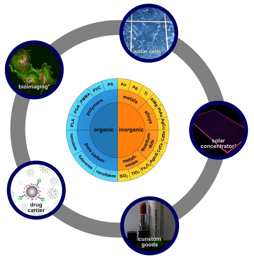
This work was prepared under supervision of
                                   Dr. Ute Resch-Genger
          (Bundesanstalt für Materialforschung und -prüfung Berlin)

                                      in the time frame of
                                  March 2015 to April 2019

                                             at the
               Bundesanstalt für Materialforschung und -prüfung

I hereby declare that this doctoral studies was prepared autonomously and that no illegal
help was used. Contributions of others, e.g., content, quotes, or figures are indicated by
referring to the original work.

Berlin, April 2019                                           (Nithiya Nirmalananthan-Budau)

               1st Reviewer                   Dr. Ute Resch-Genger
               2nd Reviewer                   Prof. Dr. Siegfried Eigler
               Date of Defence                29.07.2019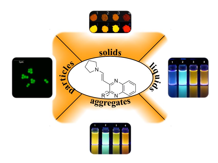
Danksagung

An dieser Stelle möchte ich meinen Dank an diejenigen richten, die mir geholfen haben diese
Arbeit anzufertigen. Beginnen möchte ich hier mit Dr. Ute Resch-Genger für die Vergabe des
spannenden Forschungsthemas, für die vielen Möglichkeiten meine Arbeit auf unterschied-
lichsten Konferenzen als Poster- oder sogar als Vortragsbeiträge zu präsentieren, für ihre
Unterstützung beim Publizieren der Ergebnisse und auch für die vielen spannenden Koope-
rationspartner und –themen. Ebenso danke ich ihr für die Möglichkeit, dass ich während
meiner Doktorarbeit Weiterbildungen in der Qualitätssicherung und in der Nutzung von
Origin machen konnte. Einen sehr großen Dank möchte ich an sie richten für ihren Rat, ihre
konstruktiven Kritiken, ihre unermüdliche Unterstützung bei der Anfertigung dieser Arbeit
und für ihr Vertrauen kleinere organisatorische Aufgaben, sowie Betreuungsaufgaben an mich
zu übergeben.
Herrn Prof. Dr. Siegfried Eigler möchte ich für die schnelle und unkomplizierte Übernahme
der Zweitbegutachtung meiner Arbeit und die fachliche Unterstützung bei der Anfertigung
der Doktorarbeit danken.
Ein besonderer Dank gilt meinen direkten Betreuern Herrn Dr. Thomas Behnke, Frau Dr.
Katrin Hoffmann und Herrn Dr. Daniel Geißler sowie Dipl.-Chem. Marko Moser und Dr.
Bastian Rühle, die mich mit sehr viel Engagement, fachlichen Erklärungen und Diskussionen
in den letzten vier Jahren bei der Anfertigung dieser Arbeit und teilweise auch vor dieser
Zeit während der Masterarbeit schon unterstützten. Bei Rückschlägen ermutigten und un-
terstützten sie mich beim Lösen der Probleme und standen mir tagtäglich mit Rat und Tat
zur Seite. Dr. Daniel Geißler und Dr. Bastian Rühle danke ich zusätzlich für die Korrektur
einzelner Abschnitte dieser Arbeit.
Frau Maria Richter, Frau Dr. Jutta Pauli, Herrn Arne Güttler und Herrn Thomas Schnei-
der danke ich für die Geräte-Einweisungen, Chemikalienbestellungen, Geräte-Kalibrierungen,
Laborsicherheit, Qualitätssicherung, also im Prinzip für die gesamte Laborinfrastruktur, die
ein analytisch akkurates Arbeiten erst ermöglichte.
Ein weiterer großer Dank gilt meinen Kollegen und mittlerweile guten Freunden Lorena D.,
Maysoon S., Sebastian R., Daniel K., Marko M., Norman S., Pablo Q. und Marco K. Euch
danke ich nicht nur für die tollen Frühstücks-, Cafe- und Mittagspausen, sondern auch für
eure Ratschläge, Ideen und Lösungsansätze. Ihr habt es mir ermöglicht mit viel Spaß und
Freude diese Zeit zu erleben. An dieser Stelle möchte ich ebenfalls der gesamten Arbeitsgrup-
pe 1.2 (ehemals 1.10) Biophotonik meinen Dank für die gute Arbeitsatmosphäre aussprechen.
Meinen Auszubildenden/Hilfswissenschaftlern Christopher, Maurice, Martin und Marvin,

                                                                                            i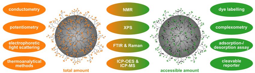
meiner Bachelorstudentin Ella und meiner Praktikantin Shreya möchte ich nicht nur meinen
Dank für die Unterstützung im Labor aussprechen, sondern auch für die Erfahrungenen, die
ich mit euch zusammen sammeln konnte.
Für die vielen tollen und spannenden internen Zusammenarbeiten danke ich weiterhin Dipl.-
Chem. Marko Moser, Dr. Alexander Roloff, Dr. Marc Wegmann und Dr. Harald Tschiche.
Darüber hinaus danke ich Prof. Dr. Thomas J.J. Müller, Dr. Charlotte Gers-Panther, Dr.
Melanie Denißen von der Heinrich-Heine-Universität, Dr. Andreas Schäfer und Prof. Dr. Bea-
te Paulus von der Freien Universität und Prof. Dr. Anke Krüger, Johannes Ackermann MSc.
von der Universität Würzburg für stimulierende Zusammenarbeiten.
Für einen regelkonformen, flüssigen und reibungslosen Ablauf der bürokratischen und tech-
nischen Angelegenheiten während meiner Arbeit danke ich Frau Christin Heinrich, Frau Grit
Ostermann, Frau Julianne Schäfer und Frau Anka Kohl.
Last but not least möchte ich meiner Familie danken. Insbesondere meinen Eltern, die mir
von den ersten Schritten als kleines Kind bis heute den Rücken stärken und mir mit Rat
und Tat bei Tag und Nacht zur Seite stehen, die mich lehrten nie aufzugeben und immer
ein Ziel vor Augen zu haben. Ganz besonders danken möchte ich meinem Freund, meiner
besseren Hälfte, meinem Mann Johannes Horst für seinen unermüdlichen und selbstlosen
Beistand während meiner gesamten bisherigen akademischen Laufbahn, für seine ruhige Art
(mein Ruhepol), für seinen Durchblick, für seine ehrlichen Worte, und natürlich auch für die
Durchsicht meiner gesamten Arbeiten während meiner chemischen Laufbahn.

                    VIELEN HERZLICHEN DANK EUCH ALLEN!!!

ii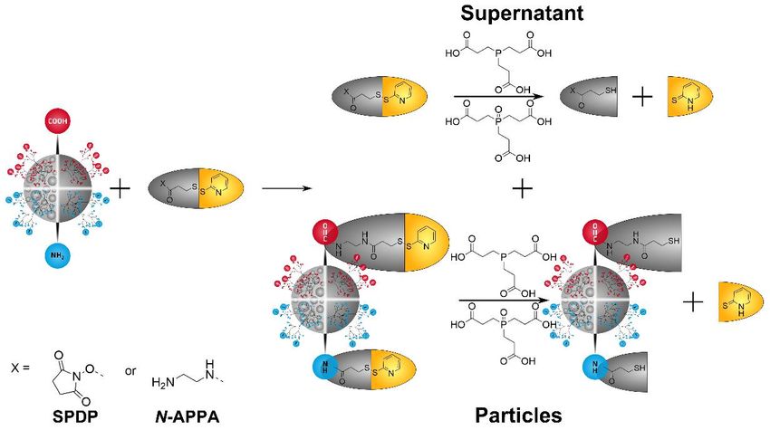
Contents

Abstract                                                                                     vii

Kurzzusammenfassung                                                                          ix

1 Introduction & Motivation                                                                1
  1.1 Introduction to nanoparticles . . . . . . . . . . . . . . . . . . . . . . . . . . .  1
  1.2 Application relevant properties of PNP . . . . . . . . . . . . . . . . . . . . . .   3
       1.2.1 Composition of PNP . . . . . . . . . . . . . . . . . . . . . . . . . . . .    3
       1.2.2 Size and size distribution of PNP . . . . . . . . . . . . . . . . . . . . .   3
       1.2.3 Shape of PNP . . . . . . . . . . . . . . . . . . . . . . . . . . . . . . . .  4
       1.2.4 Optical properties of PNP (fluorescent PNP) . . . . . . . . . . . . . .       4
             1.2.4.1 Aggregation induced property changes and aggregation-induced
                       emission (AIE) PNP . . . . . . . . . . . . . . . . . . . . . . .    6
       1.2.5 Surface functional groups (FGs) of PNP . . . . . . . . . . . . . . . . .      7
             1.2.5.1 Stability of PNP . . . . . . . . . . . . . . . . . . . . . . . . .    7
             1.2.5.2 Functionalization of PNP . . . . . . . . . . . . . . . . . . . .      8
             1.2.5.3 Biocompatibility of PNP . . . . . . . . . . . . . . . . . . . .       8
             1.2.5.4 Toxicity of PNP . . . . . . . . . . . . . . . . . . . . . . . . .     9
  1.3 Motivation and objectives . . . . . . . . . . . . . . . . . . . . . . . . . . . . . 10

2 Synthesis, Characterization and Quantification Methods for PNP                             11
  2.1 Synthesis of PNP . . . . . . . . . . . . . . . . . . . . . . . . . . . . . . . . . .   11
      2.1.1 Preparation of fluorescent PNP and AIE-PNP . . . . . . . . . . . . .             13
  2.2 Characterization of PNP . . . . . . . . . . . . . . . . . . . . . . . . . . . . . .    15
      2.2.1 Characterization of size, size distribution and shape . . . . . . . . . .        15
             2.2.1.1 Scanning electron microscopy (SEM) . . . . . . . . . . . . .            15
             2.2.1.2 Dynamic light scattering (DLS) . . . . . . . . . . . . . . . .          16
      2.2.2 Surface charge . . . . . . . . . . . . . . . . . . . . . . . . . . . . . . .     17
      2.2.3 Optical properties . . . . . . . . . . . . . . . . . . . . . . . . . . . . .     17
             2.2.3.1 Absorption . . . . . . . . . . . . . . . . . . . . . . . . . . . .      18
             2.2.3.2 Emission . . . . . . . . . . . . . . . . . . . . . . . . . . . . .      18
             2.2.3.3 Fluorescence quantum yield (ϕf ) . . . . . . . . . . . . . . . .        19
             2.2.3.4 Fluorescence lifetime (τ ) . . . . . . . . . . . . . . . . . . . .      19
      2.2.4 Surface FGs . . . . . . . . . . . . . . . . . . . . . . . . . . . . . . . . .    20
             2.2.4.1 Conductometric titration . . . . . . . . . . . . . . . . . . . .        21
             2.2.4.2 Optical assays . . . . . . . . . . . . . . . . . . . . . . . . . .      23
             2.2.4.3 BCA assay for protein quantification . . . . . . . . . . . . . .        24
             2.2.4.4 Inductively coupled plasma optical emission spectroscopy (ICP-
                      OES) . . . . . . . . . . . . . . . . . . . . . . . . . . . . . . .     25
             2.2.4.5 Quantitative nuclear magnetic resonance (qNMR) . . . . . .              26

                                                                                             iii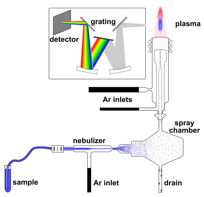
3 Results: Publications and Manuscripts 29
3.1 Major contributions . . . . . . . . . . . . . . . . . . . . . . . . . . . . . . . . 29
3.1.1 Crystallization and Aggregation-Induced Emission in a Series of Pyrro-
lidinylvinylquinoxaline Derivatives . . . . . . . . . . . . . . . . . . . . 29
3.1.2 Multimodal Cleavable Reporters vs Conventional Labels for Optical
Quantification of Accessible Amino and Carboxy Groups on Nano-
and Microparticles . . . . . . . . . . . . . . . . . . . . . . . . . . . . . 41
3.1.3 Multimodal Cleavable Reporters for Quantifying Carboxy and Amino
Groups on on Organic and Inorganic Nanoparticles . . . . . . . . . . . 63
3.2 Minor contributions . . . . . . . . . . . . . . . . . . . . . . . . . . . . . . . . 90
3.2.1 3-Piperazinyl propenylidene indolone merocyanines: consecutive Three-
component Synthesis and Electronic Properties of Solid-state Luminophores
with AIE Properties . . . . . . . . . . . . . . . . . . . . . . . . . . . . 90
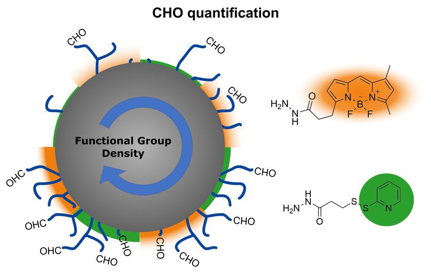
3.2.2 Quantification of Aldehydes on Polymeric Microbead Surfaces via Catch
and Release of Reporter Chromophores . . . . . . . . . . . . . . . . . 149
4 Synopsis of the Results, Conclusion & Outlook 171
5 Reference 177
A Appendix 187
A.1 Publications and planned publications . . . . . . . . . . . . . . . . . . . . . . 187
A.2 Conference contributions . . . . . . . . . . . . . . . . . . . . . . . . . . . . . . 188
iv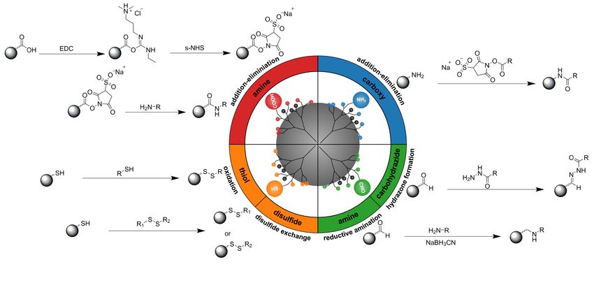
Abbreviations and Glossary
N-APPA N -(aminoethyl)-3-(pyridin-2-yldisulfanyl)propanamide

ACQ aggregation-caused quenching

AFM atomic force microscopy

AIE aggregation-induced emission

APTES (3-aminopropyl)triethoxysilane

BCA bicinchoninic acid

BDP-hzd BODIPY-hydrazide

CLSM confocal laser scanning microscope

DCC dicyclohexylcarbodiimide

DIC diisopropylcarbodiimide

DLS dynamic light scattering

EDC 1-ethyl-3-(3-dimethylaminopropyl)carbodiimide

FDA Food and Drug Administration

FG functional group

FLIM fluorescence lifetime imaging microscopy

FRET fluorescence resonance energy transfer

FTIR Fourier-transform infrared spectroscopy

IC internal conversion

ICP-MS inductively coupled plasma mass spectrometry

ICP-OES inductively coupled plasma optical emission spectroscopy

IS internal standard

ISC intersystem crossing

MSN mesoporous silica nanoparticles

                                                                   v
NHS N -hydroxysuccinimide

NMR nuclear magnetic resonance

NP nanoparticle

NTA nanoparticle tracking analysis

PAA polyacrylic acid

PDPH 3-(2-pyridyldithio)propionyl hydrazide

PGA poly(glycolic acid)

PLA poly(lactic acid)

PMMA poly(methyl methacrylate)

PNP polymer nanoparticles

PSP polystyrene particles

PVC poly(vinyl chloride)

RAFT reversible addition-fragmentation chain transfer

ROS reactive oxygen species

SAXS small-angle X-ray scattering

SEM scanning electron microscopy

SPDP N -succinimidyl-3-(2-pyridyldithio) propionate

sulfo-NHS N -hydroxysulfosuccinimide

TCEP tris(2-carboxyethyl)phosphine hydrochloride

TCSPC time-correlated single-photon counting

TEM transmission electron microscopy

TSEM transmission scanning electron microscopy

XPS X-ray photoelectron spectroscopy

XRD X-ray diffraction

vi
Abstract

Polymer nanoparticles (PNP) are increasingly used as tools in (bio)analytics. Typical appli-
cations of PNP include carriers for drugs, carriers for dye molecules for signal amplification
in optical assays, nanosensors and targeted probes for bioimaging studies. These applications
in life sciences impose stringent requirements on particle size, size distribution, morphology,
colloidal stability, biocompatibility, optical properties, and ease of surface functionalization
with, for example, targeting ligands, sensor molecules, and linkers.
PNP, including the polystyrene particles (PSP) which are in the focus of this work, are
non-fluorescent by nature but can be made fluorescent with the aid of luminophores such
as organic dyes. Fluorescent PNP can be obtained by coupling reactive fluorophores to the
surface groups of the PNP or by encapsulation of fluorophores into the PNP matrix. The
latter approach is particularly attractive due to its versatility since the encapsulation into
preformed PNP does not require luminophores with reactive functional groups (FGs), but
rather only requires hydrophobic luminophores. Moreover, the fluorophores are protected
from the potentially fluorescence quenching environment surrounding the PNP matrix and
the reactive groups on the PNP surface can be exclusively used for the attachment of targeting
ligands. Dye loading of PNP typically requires a dye-specific optimization of the loading
concentration with respect to signal strength as conventional dyes commonly form barely
emissive or even non-fluorescent aggregates at high loading concentrations. Exceptions exist,
which are dyes with propeller-like groups that show aggregation-induced emission (AIE).
These dyes can be loaded with high concentrations into PNP without detrimental fluorescence
quenching effects and can even exhibit fluorescence and hence signal amplification upon
aggregation. Due to these attractive properties, AIE dyes for use in PNP were investigated.
The spectroscopic properties of different AIE dye derivatives were systematically studied
in organic solvents, solvent−water mixtures, and in the solid state. Dyes with optimal
performance were entrapped in PSP. The staining of PSP with these AIE dyes resulted in
a considerable increase in the dye fluorescence quantum yield and lifetime, reflecting the
combined influence of the restricted molecular motion and the reduced polarity of the dye
microenvironment.
Functionalization of undoped and dye loaded PNP with, for example, targeting ligands re-
quires knowledge of the chemical nature and total amount of the surface groups as well as
the amount of surface FGs accessible for coupling reactions such as the conjugation of bio-
molecules. These numbers can differ considerably depending on factors including particle
morphology and sterical constraints. Ideal methods for surface group quantification should

                                                                                             vii
be robust, reliable, and fast. Moreover, they should not require expensive instrumentation and should be versatile to enable the characterization of a broad variety of particle systems independent of their optical properties, including systems that scatter or include systems with encoded dyes. For this respect, we studied a variety of optical assays for the quantification of carboxy and amino surface groups commercial and custom-made particles with varying surface group densities. We performed these studies using both conventional reporters such as fluorescein derivatives, Fluram, or IR 797 as well as synthetically customized cleavable labels. Special emphasis was dedicated to the development of a platform of cleavable and multimodal labels which consist of a suitable reactive group such as NHS-esters or amine, a quantitatively cleavable linker such as disulfide, and an optically active moiety such as 2-thiopyridone for optical assays. Conventional reporters are measured when bound to the particle surface, which renders the resulting optical signals prone to distortions by scattering and interferences from encoding dyes. In contrast, these cleavable labels can be detected pho- tometrically or fluorometrically in the supernatant after quantitative cleavage of the linker. Moreover, the linker unit is designed in such a way that the products of the cleaved linkers remaining at the particle surface can also be detected optically. In addition, the presence of a heteroatom such as sulfur, nitrogen or fluorine in the reporter and/or the linker can be detected by an analytical method relying on a different measurement principle. This allows for straightforward validation by method comparison with, for instance, ICP-OES. Thereby, FGs on a broad variety of different particles such as PNP, silica nanoparticles (NPs), and metal particles can also be quantified. viii
Kurzzusammenfassung

Polymernanopartikel (PNP) werden in der Bioanalytik zunehmend als Trägermaterial für
Medikamente oder Farbstoffmoleküle, die als Reporter zur Signalverstärkung in optischen
Assays, als Nanosensor oder als Zielsonden für die Bildgebung eingesetzt werden, verwendet.
Partikelanwendungen in der Material- und Lebenswissenschaft hängen stark von der Grö-
ße, Größenverteilung, Form, Dispersionsstabilität, Bioverträglichkeit, optischen Eigenschaf-
ten sowie der Oberflächenfunktionalisierung mit beispielsweise Zielliganden, Sensormolekülen
und Linkern, ab.
PNP, im speziellen hier in dieser Arbeit Polystyrolpartikel (PSP), sind von Natur aus nicht
emissiv, durch Anbindung von reaktiven Farbstoffen an den Oberflächenfunktionsgruppen
oder durch Einlagerung von Farbstoffen in die Polymermatrix können dennoch fluoreszieren-
de Partikel realisiert werden. Letztere Methode wird vorallem dadurch attraktiv, dass keine
reaktiven Gruppen an den hydrophoben Farbstoffen notwendig sind. Außerdem können die
Farbstoffe in der Polymermatrix vor der Partikelumgebung und vor Emissionsauslöschungen
geschützt werden und die Oberflächenfunktionsgruppen stehen für anderweitige Anbindun-
gen von Zielliganden zu Verfügung. Die Einlagerung von Farbstoffen benötigt eine farb-
stoffspezifische Optimierung der Beladungskonzentration, um ein optimales Emissionssignal
zu erhalten. Während konventionelle Farbstoffe beim Einlagern von hohen Konzentratio-
nen wenig bis nicht fluoreszierende Aggregate bilden, können verdrillte bzw. Farbstoffe mit
propellerartigen Gruppen (AIE Farbstoffe), in hohen Konzentrationen in PNP eingelagert
werden. Dabei kommt es statt Auslöschungseffekten zu einer Emissionssignalverstärkung.
Aus diesem Grund wurden in dieser Arbeit verschiedene AIE Farbstoffderivate systematisch
spektroskopisch in organischen Lösemitteln, in Lösemittel-Wasser Gemischen und als Fest-
stoff untersucht und Farbstoffe mit optimalen Eigenschaften in PSP eingelagert. Eingelagerte
Farbstoffe in PSP zeigen eine erhöhte Quantenausbeute und eine verlängerte Lebensdauer,
was durch die Kombination der Einschränkung der Molekülbewegung und der Reduzierung
der Polarität in der Farbstoffumgebung erreicht wird.
Die Funktionalisierung von sowohl unbeladenen, als auch mit Farbstoffen beladenen Partikeln
mit zum Beispiel Biomolekülen, benötigt die Kenntnis der chemischen Natur, der Gesamtan-
zahl an Funktionsgruppen (FGs), sowie der tatsächlich zugänglichen FGs, die für anschlie-
ßende Kupplungsreaktionen wichtig sind. Diese Anzahl hängt stark von der Partikelform und
der sterischen Einschränkung ab. Idealerweise sind die Analysemethoden robust, vertrauens-
würdig, schnell, mit kostengünstigen Instrumenten durchführbar und für eine breite Vielfalt
an Partikelsystemen einsetzbar, unabhängig von deren optischen Eigenschaften wie Streu-

                                                                                          ix
ung oder die Anwesenheit von eingelagerten Farbstoffen. In diesem Zusammenhang haben wir verschiedene konventionelle Reporter wie Fluoresceinderivate, Fluram oder IR797 und synthetisch angepasste spaltbare Linker für die Quantifizierung von käuflich erworbenen so- wie selbsthergestellten Partikeln mit unterschiedlichen Carboxy und Amin Funktionsdichten untersucht. Dabei wurde der Fokus auf die Entwicklung spaltbarer multimodaler Linker mit reaktiven Gruppen wie NHS-Ester oder Amin zur Anbindung an Partikeln, quantitativ ab- spaltbaren Linkern wie Disulfidbrücken und optisch auslesbarer Einheiten wie 2-Thiopyridon gesetzt. Im Vergleich zu den konventiellen Reportern, deren direkte Quantifizierung an Parti- keln durch Partikelstreuung und der eingelagerten Farbstoffe gestört wird, können die spalt- baren Reporter nach der quantitativen Abspaltung von der Partikeloberfläche im Überstand ohne jegliche Störung optisch vermessen werden. Außerdem können die optischen Ergebnis- se der spaltbaren Linker, wenn sie Heteroatome wie beispielsweise Schwefel, Stickstoff oder Fluor tragen, mit Hilfe einer etablierten Methode wie ICP-OES validiert werden. Mit der spaltbaren Linker Methode können auch farbstoffbeladene Partikel problemlos quantifiziert werden. Weiterhin ist diese Methode nicht nur auf PNP begrenzt, sondern eignet sich auch für die Funktionsgruppenquantifizierung an Silikapartikeln oder Metallpartikeln. x
1 Introduction & Motivation

1.1 Introduction to nanoparticles
In general, nanoparticles (NPs) are defined as materials with dimensions smaller than 100 nm.1
One nanometer is one billionth of a meter or equal to ten hydrogen atoms or five silicon atoms
lying in a line.2 The history of NPs dates back to thousands of years ago, when Chinese artists
used gold NPs as pigments. The first colloidally stable gold NPs that are stable for almost a
century were prepared and reported by Faraday in 1857.3 Nowadays, there is a vast variety of
NPs. This demands a way of NP classification. Basically, NPs comprise (i) inorganic mate-
rials (e.g. metals, metal chalcogenides and other metal salts, alloys, or semiconductors), (ii)
organic materials (polymers, amphiphilic systems such as micelles or liposomes, or carbon
allotropes),4 and (iii) hybrid materials consisting of both inorganic and organic materials. A
more detailed classification of (i) and (ii) is given in ??.

Figure 1: Classification of organic and inorganic NPs and some famous examples and their appli-
cations;1 poly(vinyl chloride) (PVC), poly(methyl methacrylate) (PMMA), poly(lactic acid) (PLA),
    poly(glycolic acid) (PGA) *Copyright by Bundesanstalt für Materialforschung und -prüfung.

                                                                                              1
1 Introduction & Motivation Advanced syntheses make it possible to prepare not only (quasi) spherical NPs,5 which is the most common shape, but also various other shapes such as rods,6,7 cages,8 cubes,9,10 disks,11 hexagons,12–14 prisms,15,16 wires,17 or tubes,18 etc. The interest in these systems arises from the combination of nanoscale size with the unique diversity of physical properties and chemical functionality. NPs can be described as intermediates between bulk materials and molecules. While micrometer-sized particles show properties that are similar to the re- spective bulk materials, NPs can exhibit unique properties that are not found in their bulk forms.19 This includes high surface-to-volume ratios, enabling surface modification with many different molecules to impart a variety of functionalities and for same materials, also unique mechanical, thermal, electrical, magnetic, and optical properties. This versatility provides the basis for a broad range of applications in different areas, such as material sciences, life sciences, and medicine.1 Depending on the desired application, the NP composition can be chosen to fit its purpose. Particularly, there is a choice between single and multi-composition to build up NPs. The most common multi-composition material is a spherical core/shell sys- tem, which consists of a core (inner material) and a shell (outer layer material). Typically, the core particles are completely coated by a shell of a different material. Different combinations are possible for such systems. The most interesting organic ones are inorganic/organic and organic/organic materials. Inorganic/organic core/shell NPs are made of a metal, a metallic compound, a metal oxide, a semiconductor, or a silica core with a surface stabilizing polymer shell. An organic shell on an inorganic core has a lot of advantages as it can reduce unspecific adsorptions, provide stabilization in the suspension media, and increase the biocompatibility for bioapplications.20–22 Organic/organic core/shell NPs are made of a polymer core and a polymer shell. The majority of organic NPs are made from polymers. Simple non-crosslinked polymer nanoparticles (PNP) are considered as agglomerates of polymer chains with nanoscale dimen- sions and represent the most common organic NP structures used nowadays.1 A growing area of application of PNP is in life sciences. This includes their use as carriers for e.g., analyte- responsive ligands, drugs23–25 as well as dye molecules for their use as multichromophoric reporters for signal enhancement in optical assays26–28 or the fabrication of nanosensors29 and targeted probes in bioimaging studies.30,31 One of the main reasons for such a broad area of application is their design flexibility which results from the polymer diversity and enables a plethora of different sizes, morphologies, and surface functionalities, as well as the synthesis of NPs with structural stability even at dilute concentrations, low production cost, high resistance and of course low/non-toxicity.24,32 Particularly, polystyrene particles (PSP) present an attractive example for an organic/organic core/shell NPs. PSP can be easily synthesized at low costs with high monodispersity and good reproducibility. As PSP are non-functionalized NPs, they need functional groups (FGs) on their surface for their aqueous dispersibility and subsequent functionalization. This can be done easily by grafting with a polymer shell with functional groups.33,34 Nowadays, or- 2
1.2 Application relevant properties of PNP

ganic/organic core/shell PSP are commercially available with various surface chemistries and
narrow size distributions in different sizes from 15 nm up to several micrometers and they are
considered as inert and commonly non-toxic.35–37 Therefore, PSP are attractive materials,
for example, for their use as fluorescent reporters for imaging or bioanalytics.31

1.2 Application relevant properties of PNP
PNP offer a great versatility. Hence, these particles have various applications in different
areas as described in detail in ??. Depending on their specific application, differently designed
PNP are desired. PNP composition, size, shape, surface properties including surface FGs,
and surface charge, as well as optical properties can have a considerable impact on their
performance.38

1.2.1 Composition of PNP
PNP can be classified into natural PNP such as chitosan, alginate, gelatin, and albumin
and synthetic PNP such as poly(lactides), poly(lactide-co-glycoside), poly(methyl methacry-
late) (PMMA), polyacrylamide, PSP. While natural PNP vary in purity and have often
lack of consistence that make them less reproducible, synthetic PNP show good purity and
batch-to-batch reproducibility. This characteristic of synthetic polymers favors them clearly
for different PNP applications.39 Synthetic PNP can be divided again into two chemical
composition types. PNP with heteroatomic (−C−X−) backbones such as poly(lactides),
poly(lactide-co-glycoside) copolymers, poly(�-caprolactones), and poly(amino acids) are eas-
ily hydrolysable, and thus, biodegradable.40,41 Polymers with carbon-only (−C−C−) back-
bones such as PMMA, polyacrylamide, PSP, and polyacrylates are stable in the body and
non-biodegradable.39 Both synthetic PNP compositions are commonly used as carriers for
small molecules, drugs, dyes or biomolecules by incorporating them into the PNP matrix or
attaching them onto the particle surface.42

1.2.2 Size and size distribution of PNP
Two key parameters of PNP are particle size and size distribution, which determine the basic
physiochemical behavior of the particles (e.g. sedimentation rate) and their surface area.43
The size of PNP can be influence by material selection and different synthesis methods.
Different examples show that the size of NPs has also a large impact on biological applications.
One example for this was reported by Kulkarni and Feng.44 They studied the uptake of PSP
in kidney cells and focused on the size by using non-deformable PSP. While the uptake of
100-200 nm-sized PSP has the highest efficiency, 500 nm PSP are poorly internalized and
small particles are cleared rapidly from the blood via extravasation. Thus, the size of the
PSP influences in this example the uptake. This example shows that 50–200 nm-sized PNP
have the highest potential for in vivo applications, due to their ability to circulate in the

                                                                                               3
1 Introduction & Motivation
blood for long periods of time without uptake in the liver while also being small enough
to avoid filtration in the spleen.38,45,46 An ideal NP size for biological applications cannot
be generalized. It is not only size dependent. Additionally, it depends on the cell line, NP
material, surface properties, complexity of the area.38
1.2.3 Shape of PNP
Typically, PNP are synthesized in a spherical shape, due to physical facts such as surface
tension. However, the synthesis of discs, rods, fibers, and elliptical disks were also reported
and studied for different applications.47,48 The uptake of PNP carriers by target cells and
their degradation are also influenced by their shape.47 Champion and Mitragotri showed
that worm-like particles exhibit insignificant phagocytosis compared to traditional spherical
particles. Thus, due to the change of the particle curvature the application of such biological
processes can be inhibited.48
1.2.4 Optical properties of PNP (fluorescent PNP)
Depending on particle size, particle concentration and particle refractive index, all kind of
particles can exhibit Raleigh and Mie scattering by interaction with electromagnetic radia-
tion.49 While for the small particles the Rayleigh scattering dominate, for the bigger ones
the Mie scattering affects more. In ?? absorbance spectra with the specific scattering profile
of 25 nm – 1 µm-sized PSP in water with identical mass concentrations are depicted.
Figure 2: Absorption spectra of differently sized PSP with their typical scattering profiles with
identical 0.17 mg/mL mass concentration.
Fluorescence-based techniques are promising tools for the analysis of complex biological
systems and processes. While commonly used fluorescent organic dye molecules and flu-
41.2 Application relevant properties of PNP
orescent proteins are small and biocompatible, fluorescent NPs have the potential to be
10-1000 times brighter and more photostable than the former. Additionally, they can be
surface-functionalized with biocompatible molecules,50 and stay in the blood circulation for
an extended period of time, which can be important for specific bioimaging and/or drug
delivery applications. The advantages of fluorescent NPs strongly attract scientists from
biological research fields, but not all NPs are suitable for biological applications.51,52 For
example, quantum dots are perfect candidates for multiplex imaging due to their photo-
stability and narrow emission band; their optical properties are tightly linked to their core
composition and size.53,54 Up-conversion NPs can be excited with a low energy, near-infrared
light source and emit higher energy photons in the visible range, which can be beneficial for
deep tissue imaging.55 However, their relatively low brightness in water and possible toxicity
(release of fluoride and lanthanide ions) needs to be considered.55–57 PNP, in turn, exhibit
a remarkable stability in biological environments, a well-controlled surface chemistry, and
biocompatibility. Compared to inherently fluorescent quantum dots and up-conversion NPs,
PNP are non-fluorescent, yet can be made emissive by combining them with, for example,
organic dyes.
Non-fluorescent PNP can be used as nm-sized matrices for organic dyes to generate fluo-
rescent NP-based emitters as well as to stabilize less photochemically and chemically stable
NIR dyes to increase their quantum yield. Such a system provides an enormous flexibility in
synthesis, in matrix-dye combinations, and in optical properties which also allows the con-
trol of brightness, absorption and emission wavelength, size (ranging between 15-500 nm),
surface chemistry, etc.58 Furthermore, it offers the opportunity to synthesize multiple exci-
tation/emission PNP by encapsulating more than one type of dye.29
Fluorescent PNP can be made by attaching reactive fluorophores to the surface FGs of
the PNP or by encapsulation of the fluorophores into the PNP matrix. The advantages,
disadvantages and different synthesis approaches are discribed in ??. For the application of
fluorescent PNP, the brightness which is proportional to the absorption coefficient, quantum
yield, and number of dyes per molecule is a relevant aspect. Important parameters that
influence the brightness are the number of dye molecules, the brightness of the dye itself,
and quenching effects such as reabsorption, dye-dye interactions, and dye-matrix interactions.
Encapsulation of conventional dyes such as rhodamines, cyanines, Nile Red, etc. in high
concentrations leads to self-quenching due to dye-dye interactions and is called aggregation-
caused quenching (ACQ). One example, given by Behnke et al.,27 exhibits the decrease of
quantum yield from 76% to 22% by an increase of the dye loading from 0.05 wt% to 0.8
wt%.27,59,60
51 Introduction & Motivation
1.2.4.1 Aggregation induced property changes and aggregation-induced
emission (AIE) PNP
Conventional hydrophobic dyes are often highly fluorescent as monomeric species in organic
dilute solutions. In cases of dye-dye-interactions, which are encouraged in concentrated
solutions and in hydrophilic surroundings,61,62 through close attachment of several dyes to a
biomolecule63 or to functional groups on the surface of NPs,64,65 or high dye concentrations
in a polymer matrix,27 the dyes can become less or even non-emissive. The main reason
for ACQ is π-π stacking interactions of the chromophores with their extended π-conjugated
systems66 favoring the formation of H-type dimers and aggregates that are barely or non-
emissive. These dimers can then act as energy sinks for fluorescence resonance energy transfer
(FRET) processes between monomeric dyes and aggregated dyes (so called homo-FRET
between chemically identical dyes).67 Additionally, hydrogen-bonding interactions can also
lead to intermolecular fluorescence quenching, particularly in the case of charge transfer
dyes.68–70
π-π stacking of conventional dyes can be prevented or at least reduced by introducing bulky
aromatic substituents such as diarylamin or tetraphenylethylene groups,62 which prevent
strong π-π interactions due to steric hindrance. Tang and coworkers observed a strong
emission enhancement upon dye aggregation behavior for the twisted dye 1-methyl-1,2,3,4,5-
pentaphenylsilole in 2001.71 This opposite phenomenon to ACQ is called aggregation-induced
emission (AIE) and was observed for many dyes with a twisted skeleton conformation and
propeller-like moieties.70,72 In diluted solution, particularly in polar solvents, the monomeric
AIE dyes show very low or even no emission, as non-radiative relaxation processes are fa-
vored due to intramolecular rotations and vibrations of the bulky substituents. Aggregation,
however, results in a rigidization of the dye’s intramolecular conformational flexibility and
the packing structure of the molecule’s propeller blade-like substituents. The completely or
at least partly blocked intramolecular motions of the substituents and the prevented direct
π-π stacking lead to a blocking of the nonradiative decay pathways of the excited singlet
state,73–76 and thus, increase the dye’s emission.
Since the AIE concept was proposed in 2001, these kinds of dyes received great attention.
Consequently, a variety of AIE dyes have been developed in recent years (see some famous
examples in ??). They can be classified in serval categories according to their skeletons:
i) heterocycles such as siloles,76 tetraphenylpyrazines,77 and quinoline-malononitriles;78 ii)
pure carbon aromatics such as distyrylanthracene79 and hexaphenylbenzenes; as well as iii)
ethylene derivatives such as tetraphenylethenes and cyanostilbenes.80
Fluorophores showing AIE are promising candidates for novel signal enhancement strategies
in bioassays or improved NP-based bioimaging approaches as such dyes enable higher dye
loading densities than conventional ACQ dyes.81
AIE dyes already found a wide range of applications, and thus the varied reported AIE NPs
syntheses are summarized in ??. They are used in bioimaging to visualize cells, organelles,
61.2 Application relevant properties of PNP
Figure 3: Recently reported AIE dye skeletons.76–78
tissues, and biological processes in living systems by rapid, noninvasive, sensitive, and in-
expensive fluorescence imaging.82–84 Due to their brightness, biocompatibility, and photo-
stability, AIE-based PNP are also applied as contrast agents for long-term staining and as
reporters for cancer cell tracking.66,85,86 They enable the monitoring of cells, depending on
the chosen dye, for up to 42 days. Some selected AIE PNP are also used in photodynamic
therapy due to their reactive oxygen species (ROS) generation efficiency and high in vitro
photocytotoxicity.87 Various in vivo and in vitro studies reported about the successful use of
AIE NPs as drug delivery systems, for example, for doxorubicin.88
1.2.5 Surface functional groups (FGs) of PNP
One of the key parameters which have to be considered for the synthesis of stable and biocom-
patible core-shell NPs is their surface chemistry.64,89–91 FGs play a pivotal role in colloidal
stability (aggregation tendency), reactivity (special attention biochemical reactivity), bio-
compatibility, pharmacodynamics, biodistribution, etc. They build the interface between
the particle and their environment, and thus, the first point of contact and first interaction
point.92
1.2.5.1 Stability of PNP
The stability of a NP suspension depends on the van der Waals forces, Coulomb forces, and
on the distance between neighboring NPs. If attractive forces dominate, the NPs can aggre-
gate while a dominance of repulsive forces leads to NP stabilization. NPs can be stabilized in
different ways: electrostatic repulsion, steric repulsion, and entropic stabilization. For elec-
trostatic repulsion, the NPs must be charged by surface FGs, which need to be introduced
by covalent attachment or adsorption on the NP surface. Thus, the same surface charge of
neighboring NPs leads to NP repulsion and stabilization. Steric repulsion can be obtained
by covalent attachment or adsorption of long chain polymers to the NPs and increase the
distance between the NPs. Electrosteric stabilization, the combination of both effects, can
also be used for the stabilization of NP dispersions.93,94
71 Introduction & Motivation 1.2.5.2 Functionalization of PNP Many applications of NPs require further surface functionalization, for example, with biomol- ecules such as proteins, DNA, and antibodies or other target-specific molecules or stimuli- responsive dyes such as fluorescein for the preparation of nanosensors. The terminal surface functionalities on the NPs, which can be formed by co-polymerization of a co-monomer, can act as anchoring points for further surface functionalization.1,95 Such molecules can be co- valently attached by traditional organic chemistry reactions or by using small cross-linking molecules.28 The most commonly used FGs are carboxy and amino groups, as they can be easily introduced on polymer core NPs and can be further modified with many established (bio)functionalization reactions known from protein chemistry.28,95 The carboxy groups give the PNP a negative zeta potential at neutral pH. As carboxy groups have only a limited reactivity in amide formation, they need to be activated prior to reaction, for example, using a carbodiimide such as 1-ethyl-3-(3-dimethylaminopropyl)carbodiimide (EDC), dicyclohexylcarbodiimide (DCC), or diisopropylcarbodiimide (DIC). One disadvan- tage of the formed O-acylisourea intermediate is the rapid hydrolysis. In consequence, a high excess is used to achieve a complete activation, but this can cause a loss of colloidal stability due to poor solubility of O-acylisourea. This in turn can be mitigated by converting the O- acylisourea to a more stable intermediate by using N -hydroxysulfosuccinimide (sulfo-NHS) or N -hydroxysuccinimide (NHS). The formed active ester can undergo an addition-elimination reaction with amino groups. Biomolecules, small molecules, or dyes with amino functions can be coupled to the carboxylated PNP surface by formation of an amide bond.96–98 The second common terminal functions are amino groups, which yields PNP with a positive zeta potential at neutral pH. They can undergo an addition-elimination reaction with active esters, as described above. Biomolecules, small molecules, or dyes with active ester groups can be used as reactants for the coupling to amino groups.99,100 Thiol, maleimide, aldehyde, azide, and vinyl groups are also often used for PNP surface modification and extend the possible reactions and reagents on PNP surfaces. Thiol groups can undergo an oxidation reaction with another thiol function and generate a disulfide bound, but it can also perform an exchange reaction with a disulfide and generate a new asymmetric disulfide bond.101–103 Aldehyde PNP are used to attach hydrazides to the surface by the formation of a hydrazone, but also amino groups can undergo a (reductive) amination with carbonyls.104,105 An overview of FGs and their subsequent reaction possibilities that are important for this work are shown in ??. 1.2.5.3 Biocompatibility of PNP A surface modification is a useful strategy to make NPs more biocompatible. Since biomol- ecules often have ϵ-amino groups as found, for example, on the lysine side chains as well 8
1.2 Application relevant properties of PNP

Figure 4: FGs and the further reactions possibilities: carboxy and amino FGs can undergo addition-
elimination reactions (blue and red), thiol functions oxidation or disulfide exchange reactions (yellow),
 and aldehyde functions hydrazine formation or reductive amination reactions (green), respectively.

as N-terminal primary amines of proteins, an addition-elimination reaction is often used to
couple biomolecules to the activated carboxy PNP surface. Additionally, the carboxy groups
in aspartic acid, glutamic acid, or the C-terminus of biomolecules can also be used to couple
proteins, peptide, or antibodies to aminated PNP surfaces.106 Such bioconjugation or also,
for example, PEGylations are used to coat the surface and enhance their hydrophilicity and
biocompatibility. PEG is a hydrophilic, biologically inert polymer that has been approved
by the U.S. Food and Drug Administration (FDA) for internal use. PEG-coated PNP show
significant differences compared to non-coated ones in blood circulation times, it prevents
the aggregation of NPs, and hinders the non-specific interactions with cells by forming a
neutral stabilizing interface.107,108 Such a surface modification can have a large influence on
the biological transport, mediating interactions with cellular membranes and proteins, ki-
netics and mechanism of uptake into cells, diffusion through various biological barriers, and
biodistribution to specific tissues, cells, or organelles.109

1.2.5.4 Toxicity of PNP

The surface chemistry stands out as the main parameter of biological performance as the
NP surface is the first point of exposure, thus, it has also a major impact on the toxicity.
Free reactive groups on NPs can lead to toxicity. NPs with high cationic surface charge
and hydrophobic character can more strongly interact with anionic functions on the cellular
membranes than NPs with anionic or neutral FGs.109–111

                                                                                                       9
1 Introduction & Motivation
1.3 Motivation and objectives
Fluorescent PNP with high brightness and accurately characterized properties, especially a
complete knowledge about the type and amount of the surface chemistry, are prerequisites
for sophisticated and versatile applications in the material and life sciences and in particular
as signal amplification systems in bioanalytics. Knowledge of these fundamental properties
is also necessary for a sustainable and resource efficient application of surface functionalized
fluorescent PNP, which is a major goal in today’s research. The aim of this work is
• (i) development of signal enhancement strategies
• (ii) development of methods for the precise FG quantification on PNP surfaces
The former is addressed by loading preformed PNP with different organic dyes with special
emphasis on AIE emitters. In this respect the understanding of AIE behavior of dye deriva-
tives with different substitution patterns on optical properties in solution and in preformed
PNP is essential. Since no standardized methods for the FG analysis and no reference
materials with known FG densities are currently available, the latter is addressed to the
determination of the accessible amount of FGs on simple and dye-loaded PNP. The main
objectives of this thesis are summarized in ??.
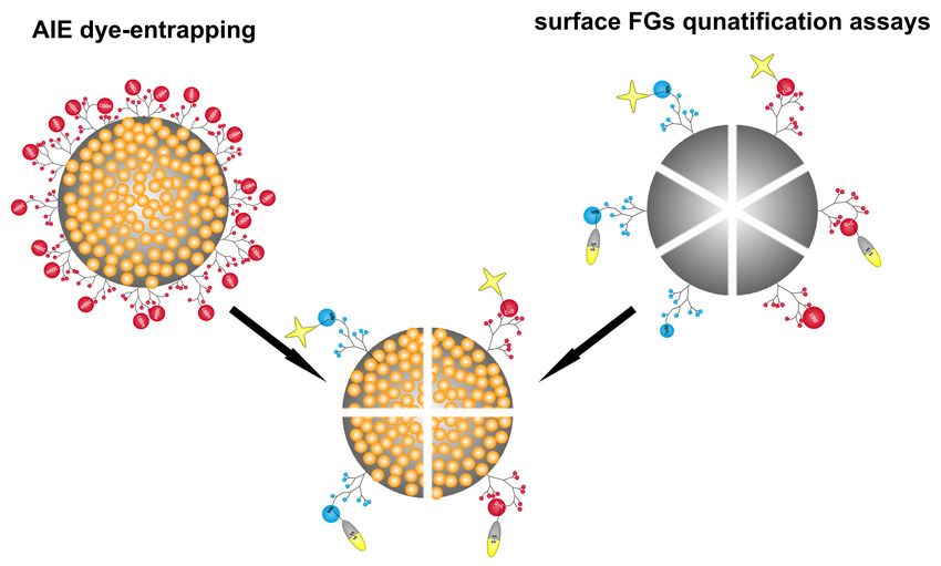
Figure 5: Objectives of the doctoral thesis a) development of signal enhancement strategies by AIE
dye entrapping b) development of surface FG quantification assays.
102 Synthesis, Characterization and Quantification
Methods for PNP
2.1 Synthesis of PNP
The synthesis of monodisperse, uniformly shaped and chemically homogeneous PNP cannot
be simply generalized. PNP can be prepared either by “top-down” synthesis approaches,
starting from a preformed polymer and shape the “bulk material” into to the desired struc-
ture, or via a “bottom-up” synthesis, where the chemical properties of the monomers are
exploited to polymerize monomer-by-monomer and create the desired NPs.2,43 Procedures
belonging to the top-down and bottom-up approaches are summarized in ??. With the above
mentioned techniques not only simple NPs, but also different types of core/shell NPs can be
synthesized.
Figure 6: Various top-down and bottom-up approaches for the preparation of PNP.
The progress in controlled polymerization enables the engineering of multifunctional PNP
with precise control over chemical composition, shape, size, shell thickness, surface charge,
and functionality. The conventional radical emulsion polymerization is the most common way
to synthesize PNP. Herein, the monomer is added stepwise in the presence of a suitable ini-
tiator, which starts the polymerization reaction by forming free radicals and interacting with
the monomers in two immiscible liquids, e.g., water and oil. Hence, the carefully controlled
emulsion polymerization needs additional surface modifiers, emulsifying agents, or surfac-
tants. These molecules can significantly reduce the interfacial tension and form nanometer-
sized micelle droplets of oil-in-water emulsions (nanoreactors/nanocavities), which serve as
centers for nucleation and growth and protect the nonpolar monomers. Additionally, they
also work as a steric stabilizer to prevent the aggregation of the reacting species during the
reaction period. Such an emulsion polymerization can also be done surfactant free, but in
that case the monomer must be soluble in water and stabilized by using ionic co-monomers
112 Synthesis, Characterization and Quantification Methods for PNP
or ionizable initiators. In most cases, a mini-emulsion polymerization with an initiator in
the organic phase or a micro-emulsion polymerization with a water-soluble initiator is per-
formed. Due to the water insolubility of the monomer, a surfactant is necessary in both
polymerizations.33,43,112,113
During the polymerization synthesis, different factors can influence the size and the shape
of formed particle, such as the stirring speed and temperature conditions. Also, the applied
surfactant and the water-to-surfactant ratio are of great importance, as they control the
polymer chain length as well as the size and shape of the final PNP via the formed reaction
cavity that depends additionally on the surfactant’s hydrophilic-lipophilic balance value.
Finally, the reactant concentration is important for the amount of formed nuclei and the
period of particle growth, because only the monomers remaining in the reaction solution are
available for a collision with the formed nuclei and for deposition on the surface of nuclei.114
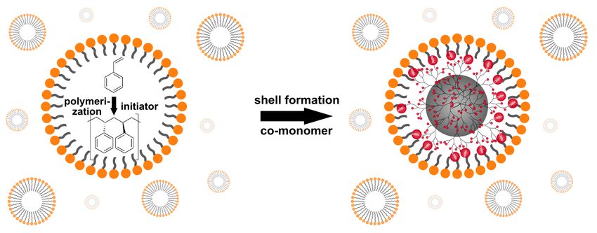
The coating of the synthesized core PNP can be performed by using the in-situ polymerization
method. In this method, after the synthesis of the core particles, a co-monomer is added to
the particle nuclei, which has the desired FG (see ??).115,116
(a)
(b)
Figure 7: Emulsion polymerization of styrene to polystyrene, and in-situ addition of acrylic acid to
achieve the formation of a carboxy shell in micelles.33,34
The shell formation reaction can be challenging as particle agglomeration and formation of
shell particles have to be prevented. Agglomeration of the particles in the reaction media
122.1 Synthesis of PNP
can be controlled via stabilization with surfactants. The separate formation of shell material
particles can be inhibited by adjusting the stirring speed, the co-monomer concentration,
and the co-monomer addition rate. An incomplete coverage of the core can be prevented
by using a sufficient amount of co-monomer molecules to fully cover the surface of the PNP
cores. Some common shell-forming molecules (co-monomers) are listed in ??.
Table 1: Co-monomers with reactive functionalities for the surface functionalization of PNP, espe-
cially interesting for PSP cores.
Co-monomers Functional groups
4-N -Boc-vinylaniline117 amino
aminoethyl methacrylate hydrochloride33 amino
acrylic acid33 carboxy
methyl methacrylate118 carboxy
vinylbenzaldehyde119 aldehyde
vinyl aryl azide120 azide
2.1.1 Preparation of fluorescent PNP and AIE-PNP
Fluorescent PNP can be made via different approaches. One way is attaching reactive flu-
orophores to the surface FGs of the PNP. The disadvantages of this strategy are that only
reactive dyes with suitable functionality can be coupled covalently to the surface, only acces-
sible FGs can be modified, and the dyes are located at the interface between PNP and the
surrounding medium, which makes them accessible for quenchers present in the solution.121
This strategy is often used for the preparation of nanosensors using stimuli-responsive dyes.122
Another elegant way is to encapsulate the dyes into the PNP matrix, either during the poly-
merization reaction or after the synthesis of preformed PNP. The dye can be added during
the synthesis in the reaction mixture as dye monomers, which are then incorporated cova-
lently in the PNP matrix or through physical trapping.123 The incorporation of dyes during
the synthesis can impact particle properties such as size, size distribution, and shape. Addi-
tionally, this requires the synthesis of fluorophore-modified monomers, which can be tedious,
and dyes, which survive the sometimes stringent polymerization conditions. The advantage
of such a dye polymerization into the PNP matrix is that it prevents leaking. The other
approach is the simple swelling procedure studied, for example, by Behnke et al.26,27 using
preformed PSP and conventional water-insoluble dyes without reactive groups (see ??). In
this case, hydrophobic organic dyes are dissolved in an organic solvent, for example, THF or
DMF, and added to the aqueous PNP suspension. The organic solvent leads to a swelling
of the particles. Depending on their log D value (see ??), the hydrophobic organic dyes are
distributed preferentially within the hydrophobic polymer matrix and can then be entrapped
by reducing the organic solvent content. This leads to a deswelling of the PNP back to their
original size and shape. The excess dye is removed by several washing steps.26,27 In this way,
132 Synthesis, Characterization and Quantification Methods for PNP
PNP can act as carriers for water-insoluble materials such as hydrophobic dyes and provide
long-term chemical stability and protection against chemical species which can lead to photo-
or chemical degradation.
Figure 8: Simple encapsulation procedure for producing fluorescent PNP with hydrophobic dyes.
An important aspect for the application of dye-loaded particles is to prevent dye leaking.
Leaking of dyes into the environment leads to a decrease of the NP brightness, increases
the background signal and can have toxic effects. In order to avoid dye release and achieve
high dye loading efficiencies, sufficiently hydrophobic dyes that can be entrapped within the
hydrophobic polymer matrix are required.124,125 For every dye the degree of hydrophobicity
can be calculated. The log D is the logarithm of the ratio of the concentration sums of all i
species in water and in octanol as a measure for the hydrophobicity or hydrophilicity of the
dye:
∑i
·ci
logD = log ∑i 1 i okt . (1)
1 ·cwater
The higher the log D value, the higher the hydrophobicity of the dye.
A special form of fluorescent NPs are AIE PNP. Different synthesis methods for produc-
ing them were reported in the recent years. Examples involve the covalent attachment
of AIE dyes to the polymer or the entrapment of the dyes in PNP. For example, Wei
and co-workers used cross-linkable/polymerizable AIE dyes for the preparation of NPs in
a reversible addition-fragmentation chain transfer (RAFT) polymerization,126,127 emulsion
polymerization,128,129 radical polymerization,130,131 and redox polymerization.132 Another
synthesis of AIE PNP is based on the nanoprecipitation with an amphiphilic polymer such
as 1,2-distearoyl-sn-glycero-3-phosphoethanolamine-PEG (DSPE-PEG) as encapsulating or
coating agent, which additionally enables further functionalization of the PNP.85 Another
study reports the synthesis of AIE PNP by an emulsion solvent evaporation method.133
The amount of emulsifier is one of the important aspects for this kind of synthesis, because
the lower the amount, the higher the aggregation state of the AIE dye. A high amount of
emulsifier encourages a homogeneous dye distribution in the polymer matrix, and thus, no
142.2 Characterization of PNP
aggregation takes place, leading to a reduced quantum yield. The generation of AIE PNP
by a simple swelling procedure followed in this work has not been reported yet.
2.2 Characterization of PNP
To obtain a complete picture about size, size distribution, shape, shell thickness, and surface
charge of the synthesized NPs, suitable analytical methods are required. Serval techniques
are established for the characterization of NPs.
• (i) microscopic analyses134–137 such as atomic force microscopy (AFM), scanning elec-
tron microscopy (SEM), transmission electron microscopy (TEM)
• (ii) spectroscopic analyses136,138–140 such as UV-Vis spectroscopy, fluorescence spec-
troscopy, X-ray photoelectron spectroscopy (XPS), Fourier-transform infrared spec-
troscopy (FTIR)
• (iii) scattering analyses141,142 such as dynamic light scattering (DLS), nanoparticle
tracking analysis (NTA), small-angle X-ray scattering (SAXS)
For the determination of the NP properties, the characterization method must be suitable
for both, the core and the shell material. In the case of core/shell PSP, both materials are
organic polymers. Methods that are suitable for the core are also suitable for the shell but
distinguishing between core and shell could be challenging.
2.2.1 Characterization of size, size distribution and shape
2.2.1.1 Scanning electron microscopy (SEM)
SEM is the most common and reliable technique for visualization of different types of NPs
and for their size and shape analysis (see ??).
Compared to a conventional light microscope, which is typically working with a visible light
source in a wavelength range of 400 to 800 nm, the resolution of an electron microscope is
several orders of magnitude higher, down to the sub-nanometer region. The generated elec-
tron beam at a tungsten cathode is focused by condenser lenses. Subsequently, the focused
electron beam passes through deflector plates and interacts with the sample line by line in
a raster pattern. Different processes can take place when the beam hits the sample. The
collimated electrons can be deflected by elastic scattering or they can ionize the sample and
generate secondary electrons with lower energy, that can be detected. With a CCD detector,
the resulting image is obtained by mapping the intensity of the detected signal.143,144 For
the measurement, the samples are in a dried form, fixed on a substrate, and measured under
high vacuum. Instead of detecting secondary electrons, SEM can also be used in transmis-
sion mode in the so-called transmission scanning electron microscopy (TSEM). In this special
15Sie können auch lesen