Mitteilungen der Deutschen Gesellschaft für Elektronenmikroskopie e.V - Nummer 43
←
→
Transkription von Seiteninhalten
Wenn Ihr Browser die Seite nicht korrekt rendert, bitte, lesen Sie den Inhalt der Seite unten
Nummer 43 · Dezember 2017                                 Nummer   43
                          ISSN 0936-6911

Mitteilungen der
Deutschen Gesellschaft für Elektronenmikroskopie e.V.

                                                                     http://www.dge-homepage.de

                                                                                             43
S. Hirzel Verlag · Wissenschaftliche Verlagsgesellschaft Stuttgart
Postfach 101061 · 70009 Stuttgart
Unlocking
 crystallographic
 information in your lab.
 ZEISS Xradia 520 Versa with LabDCT

// INSPIRATION
 MADE BY ZEISS

                 Your advanced imaging module
                 for diffraction contrast tomography
                 Extend the performance of your Xadia 520 Versa X-ray
                 microscope with LabDCT. Achieve direct visualization
                 of 3D crystallographic grain orientation in your lab.
                 This unique technology enables non-destructive
                 mapping of orientation and microstructure in 3D and 4D.
                 LabDCT opens up a new dimension in the characterization
                 of metal alloys and polycrystalline materials.

                 www.zeiss.com/520-versa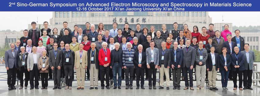
43

Nummer 43 . Dezember 2017
www.dge-homepage.de

Herausgeber:
Deutsche Gesellschaft für Elektronenmikroskopie e.V.
                                                          INHALT
                                                          Deutsche Gesellschaft für Elektronenmikroskopie e.V.:                                  2
Redaktion:
Prof. Dr. Andreas Rosenauer                                Geschäftsführung 2016 – 2017
– Electron Microscopy –
Institute of Solid State Physics                          Aktivitäten                                                                            3
Universität Bremen                                         Wissenschaftliche Tagungen
Otto-Hahn-Allee NW1                                        Offizielle Publikationsorgane
28359 Bremen                                               Arbeitskreise der DGE
Telefon (0421) 218-62270
E-Mail: rosenauer@ifp.uni-bremen.de
                                                          Geschäftsführung                                                                       4
                                                           Technikpreis der DGE an Frau Doris Meertens
Dr. Andreas Graff                                          DGE Förderpreis 2017 an Dr. Andreas Müller
                                                           Wahlen zum DGE Vorstand 2018/19
Fraunhofer Institute for Mechanics of Materials
                                                           Neuer Schwerpunkt zu tomographischen Methoden in der Materialforschung bei der DGM
Walter-Hülse-Straße 1                                      Ausschreibung Konferenz-Reiseförderung
06120 Halle                                                Ausschreibung Mess-Reiseförderung
Telefon (0345) 55 89-113                                   Reisekostenzuschüsse IMC-19
E-Mail: andreas.graff@iwmh.fraunhofer.de
                                                          Ernst-Ruska Preis 2017                                                                10
Prof. Dr. Armin Feldhoff                                   Ernst-Ruska-Prize 2017
Institut für Physikalische Chemie und Elektrochemie der    New Members elected into the Ernst-Ruska-Prize Committee
Leibniz Universität Hannover                               Ausschreibung ERP 2019
Callinstraße 3a
30167 Hannover                                            Personalia                                                                            12
Telefon (0511) 762-29 40                                   Nobelpreis für Chemie 2017
E-Mail: armin.feldhoff@pci.uni-hannover.de                 Anecdotes related to memorable occasions in the academic life of Harald Rose

                                                          Wissenschaftliche Veranstaltungen                                                     17
                                                           Konferenzbericht: MC2017 – Lausanne
                                                           Harald Rose Lecture Award 2017: Laudation for Prof. Hannes Lichte
                                                           Erfahrungsberichte (DGE Reiseförderung)
                                                           Second Sino-German Symposium
                                                           Erfahrungsbericht zum Besuch der
                                                           „21st International Conference on Solid State Ionics“ 2017 in Padua
                                                           Microscopy & Microanalysis (M&M 2017) in St. Louis

                                                          DGE-Arbeitskreise                                                                     32
                                                           AK-EMED Meeting 2017
                                                           Internationaler Workshop zur Elektronenmikroskopie
                                                           von Infektionskrankheiten (Glienicke-Workshop)
                                                           AK-IGEME Meeting 2017
                                                           1. EuFN-Workshop findet in Graz statt

                                                          Laborkurse                                                                            39
                                                           EM-Herbstschule vom 9. – 12. Oktober 2017 in Berlin
Beilagenhinweis:
Diese Ausgabe enthält eine Beilage der Binder
                                                          Aus Forschung und Industrie                                                           40
Labortechnik, 85241 Hebertshausen. Wir bitten
unsere Leser um Beachtung.                                 ZEISS introduces update of LabDCT diffraction contrast tomography
                                                           module for X-ray microscopy
                                                           Sub 30meV energy resolution in a monochromated cubed Themis for
                                                           low voltage STEM and EELS applications

                                                          Buchbesprechung                                                                       42
                                                           Liquid Cell Electron Microscopy

                                                          Nationaler und Internationaler Veranstaltungskalender                                 44

                                                          Mitgliederwerbung                                                                     46

                                                          Impressum                                                                             48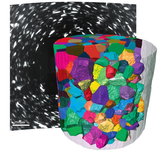
Deutsche Gesellschaft für Elektronenmikroskopie e.V.

    Deutsche Gesellschaft für Elektronenmikroskopie e.V.

    Geschäftsführung 2016 – 2017

Vorsitzender                               Schatzmeister                               Beisitzer

Prof. Dr. Michael Lehmann                  Dr. Wilfried Sigle                          Prof. Dr. Andreas Rosenauer
Institut für Optik und Atomare             Max-Planck-Institut für                     – Electron Microscopy –
Physik (Sekr. ER1-1)                       Festkörperforschung • Stuttgarter           Institute of Solid State Physics
Technische Universität Berlin              Zentrum für Elektronenmikroskopie           Universität Bremen
Straße des 17. Juni 135, 10623 Berlin      Heisenbergstraße 1                          Otto-Hahn-Allee NW1
Tel.: +49 (0)30 / 314-22567                D-70569 Stuttgart                           28359 Bremen
Fax: +49 (0)30 / 314-27850                 Tel.: +49 (0)711 / 689-3525                 Tel.: +49 (0)421 / 218-62270
E-Mail:                                    Fax: +49 (0)711 / 689-3522                  E-Mail: rosenauer@ifp.uni-bremen.de
Michael.Lehmann@tu-berlin.de               E-Mail: w.sigle@fkf.mpg.de
                                                                                       Ehrenbeisitzer
Stellvertretender Vorsitzender             Beisitzer                                   Dr.-Ing. Gerhard Schimmel
                                                                                       Südring 75
                                                                                       69519 Laudenbach
                                                                                       E-Mail: GeSchimmel@aol.com

                                                                                       Von der Mitgliederversammlung
                                                                                       wurden für die Amtsperiode 2018/19
                                                                                       gewählt:
Prof. Dr. Thomas Müller-Reichert           Dr. Christian Kübel                         Rechnungsprüfer
TU Dresden                                 Karlsruhe Institute of Technology           Prof. Dr. Andreas Klingl
Medizinische Fakultät Carl Gustav Carus    Hermann-von-Helmholtz-Platz 1               Pflanzliche Entwicklungsbiologie
Core Facility Cellular Imaging (CFCI)      76344 Eggenstein-Leopoldshafen              Biozentrum der LMU
Fetscherstr. 74                            Tel.: +49 (0)721 / 608-28970                Großhadernerstraße 2-4
01307 Dresden                              E-Mail: christian.kuebel@kit.edu            82152 Planegg-Martinsried
Tel.: +49 (0)351 / 458-6442                                                            Tel.: +49 (0) 89 2180 74416
Fax: +49 (0)351 / 458-6305                 Beisitzerin                                 E-Mail: andreas.klingl@biologie.
E-Mail: mueller-reichert@tu-dresden.de                                                 uni-muenchen.de
Geschäftsführer                                                                        Rechnungsprüfer
                                                                                       Dr. Holm Kirmse
                                                                                       Humboldt-Universität zu Berlin
                                                                                       Institut für Physik
                                                                                       AG Strukturforschung / Elektronen-
                                                                                       mikroskopie
                                                                                       Newtonstraße 15
                                           Dr. Wiebke Möbius                           12489 Berlin, Germany
                                           Max-Planck-Institute of                     Tel.: +49 (0)30 / 2093-7641
                                           Experimental Medicine                       Fax: +49 (0)30 / 2093-7643
Dr. Thomas Gemming                         Department of Neurogenetics,                E-Mail: holm.kirmse@physik.hu-berlin.de
IFW Dresden                                Electron Microscopy
Helmholtzstr. 20                           Hermann-Rein-Str. 3                         Mitglied des Wahlausschusses
01069 Dresden                              37075 Göttingen                             Dr. Ines Häusler
Tel.: +49 (0)351 / 4659-298                Tel.: +49 (0)551 / 3899-786                 Institut für Optik und Atomare Physik
Fax: +49 (0)351 / 4659-9298                Fax: +49 (0)551 / 3899-753                  (Sekr. ER1-1)
E-Mail: T.Gemming@ifw-dresden.de           E-Mail: moebius@em.mpg.de                   Technische Universität Berlin

2                                                                              Elektronenmikroskopie · Nr. 43 · Dezember 2017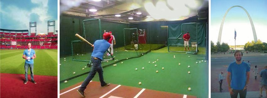
Aktivitäten

Straße des 17. Juni 135                            Mitglied des Wahlausschusses                          Albert-Einstein-Allee 11
10623 Berlin                                       Dr. Johannes Biskupek                                 89081 Ulm
Tel.: +49 (0)30 / 314 29068                        Universität Ulm                                       Tel.: +49 (0)731 / 50 22926
Fax: +49 (0)30 / 314 27850                         Zentrale Einrichtung Elektronen-                      Fax: +49(0)731 / 50 22951
E-Mail: haeusler@tu-berlin.de                      mikroskopie • AG Materialwissenschaft-                E-Mail: johannes.biskupek@uni-ulm.de
                                                   liche Elektronenmikroskopie

Redaktionsschluss für Nummer 44: 31. Oktober 2018

Redaktion:              Prof. Dr. Andreas Rosenauer, U Bremen
                        Dr. Andreas Graff, Fraunhofer IWM, Halle
                        Prof. Dr. Armin Feldhoff, U Hannover                                             Die Zeitschrift ist gegründet worden
                        Prof. Michael Lehmann, TU Berlin                                                 von Dr. Bernd Tesche und Cilly Weichan.

   Aktivitäten
Wissenschaftliche Tagungen                         Aktivitäten: Arbeitskreistreffen, Workshops, Infor-   2. Sprecher:
einschließlich der Mitgliederversammlungen         mationsaustausch, Firmenkontakte                      PD Dr. Thomas Müller-Reichert
DGE-Laborkurse                                                                                           TU Dresden
DGE-Arbeitskreise                                  Energiefilterung und Elektronen-Energieverlust-       Medizin-Theoretisches Zentrum
                                                   spektroskopie (EF & EELS)                             01307 Dresden
Offizielle Publikationsorgane                      Sprecherin:                                           Telefon: (0351) 458-6442
European Journal of Cell Biology                   Dr. Martina Luysberg                                  Telefax: (0351) 458-6305
Ultramicroscopy                                    ER-C und PGI-5                                        E-Mail: mueller-reichert@tu-dresden.de
                                                   FZ Jülich GmbH                                        Aktivitäten: Laborkurse, Symposium
Arbeitskreise der DGE                              Wilhelm-Johnen-Straße
Interessengemeinschaft elektronenmikroskopischer   52428 Jülich
                                                   Germany                                               Elektronenmikroskopische Erregerdiagnostik
Einrichtungen (IGEME)
                                                   E-Mail: m.luysberg (at) fz-juelich.de                 (EMED)
1. Sprecher:
Dr. Dirk Berger                                    Stellvertretender Sprecher:                           1. Sprecher:
TU Berlin                                          Dr. Markus Wollgarten                                 Dr. Bärbel Hauröder
ZE Elektronenmikroskopie                           EE-ANSMA                                              ZInstSanBw Koblenz
Sekr. KWT 2 / Abt. ZELMI                           Helmholtz Zentrum Berlin                              Elektronenmikroskopie
Straße des 17. Juni 135                            Hahn-Meitner-Platz 1                                  Andernacherstr. 100
10623 Berlin                                       14109 Berlin                                          56070 Koblenz
E-Mail: dirk.berger@tu-berlin.de                   Germany                                               Telefon: (0261) 896-7260
                                                   E-Mail: wollgarten (at) helmholtz-berlin.de           Telefax: (0261) 896-7269
2. Sprecher:
                                                                                                         E-Mail: b.hauroeder@zinstkob.de
Dr. Martin Ritter                                  Ländervertreter:
TU Hamburg-Harburg                                 • Österreich:                                         2. Sprecher:
Betriebseinheit Elektronenmikroskopie                PD Dr. M. Stöger                                    Dr. Matthias König
Eißendorfer Straße 42 (M)                            TU Wien, USTEM                                      Institut für Virologie FB10
21073 Hamburg                                        Wiedner Hauptstrasse 8-10/052                       Veterinärmedizin
E-Mail: ritter@tuhh.de                               A-1040 Wien                                         Justus-Liebig-Universität Gießen
                                                     E-Mail: stoeger@ustem.tuwien.ac.at                  Frankfurter Str. 107
Focussed Ion Beam (FIB)                            • Schweiz:                                            35392 Gießen
Dreiländerarbeitskreis FIB                           Prof. Dr. C. Hebert                                 Telefon: (0641) 9938-363
                                                     EPFL Lausanne                                       Telefax: (0641) 9938-379
Hauptsprecher:
                                                     Ch-1015 Lausanne                                    E-Mail: matthias.koenig@vetmed.uni-giessen.de
Marco Cantoni
EPFL SB CIME-GE                                      Switzerland                                         Aktivitäten: jährliches Labormeeting
MXC 133 (Bâtiment MXC)                               E-Mail: cecile.hebert@epfl.ch
Station 12                                         • Deutschland:                                        Hochauflösende Transmissions-Elektronen-
CH-1015 Lausanne                                     Dr. M. Wollgarten                                   mikroskopie (HREM)
Schweiz                                              Institut für Technologie;
Telefon: +41 (21) 69-34816                           Bereich Solarenergieforschung                       1. Sprecher:
E-Mail: marco.cantoni@epfl.ch                        Helmholtz-Zentrum Berlin für Materialien            Dr. Tore Niermann
Stellvertretender Sprecher:                          und Energie GmbH                                    Technische Universität Berlin
Dipl.-Ing. Michael Rogers                            Hahn-Meitner-Platz 1                                Institut für Optik und Atomare Physik
Austriamicrosystems                                  14109 Berlin                                        Arbeitsgruppe M. Lehmann
Schloss Premstätten                                  E-Mail: wollgarten@helmholtz-berlin.de              Straße des 17. Juni 135
A-8141 Unterpremstätten, Österreich                                                                      10623 Berlin
                                                   Aktivitäten: jährliches Kolloquium
E-Mail: michael.rogers@austriamicrosystems.com                                                           Ernst-Ruska Building, Room ER 296
                                                   Präparation und Abbildung Nativer Systeme (PANOS)     Tel.: +49 – (0)30 – 314 21562
Stellvertretender Sprecher:                                                                              Fax.: +49 – (0)30 – 314 27850
Dr. Siegfried Menzel                               1. Sprecherin:                                        E-Mail: Tore.Niermann@tu-berlin.de
IFW Dresden                                        Dr. Wiebke Möbius
Helmholtzstr. 20                                   Department of Neurogenetics, Electron Microscopy      2. Sprecher:
D-01069 Dresden                                    Max-Planck-Institute of Experimental Medicine         Dr. Andriy Lotnyk
Tel.: +49 (351) 4659-214                           Hermann-Rein-Str. 3                                   Leibniz Institute of Surface Modification (IOM)
Fax: +49 (351) 4659-452                            37075 Göttingen                                       Permoserstr. 15
E-Mail: s.menzel@ifw-dresden.de                    Tel.: (0551) 3899 786                                 04318 Leipzig
Gründung: Sept. 2005 in Davos als                  Fax : (0551) 3899 753                                 Tel.: +49 – (0)341 – 235 2840
Dreiländerarbeitskreis D, CH, A                    E-Mail: moebius@em.mpg.de                             E-Mail: andriy.lotnyk@iom-leipzig.de

Elektronenmikroskopie · Nr. 43 · Dezember 2017                                                                                                             3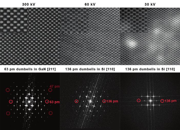
Geschäftsführung

    Geschäftsführung

Technikpreis 2017                      eingeschränkte Anzahl aufgeführt         Förderpreis der DGE an
                                       werden kann (aus der Nominierung
der DGE an Frau Doris                  von Frau Meertens für den Technik-
                                                                                Dr. Andreas Müller
Meertens                               preis 2017):

                                       • Entwicklung technischer Verfahren      Während der diesjährigen Microsco-
                                         zur Probenpräparation für die mo-      py Conference MC2017 wurde auf
                                         derne Elektronenmikroskopie in         der DGE Mitgliederversammlung
                                         der Materialforschung für halblei-     der Förderpreis der DGE an Dr. An-
                                         tende, supraleitende und kerami-       dreas Müller für seine Doktorarbeit
                                         sche Materialien für nachgelagerte     „Ultrastructural Analysis of Insulin
                                         transmissionselektronenmikrosko-       Secretory Granule Ageing“ verlie-
                                         pische Untersuchungen.                 hen. Die Dissertationsschrift wurde
                                       • Entwicklung technischer Verfahren      2016 an der Medizinischen Fakultät
                                         zur TEM-Probenpräparation von          Carl Gustav Carus der Technischen
                                         Materialien mit speziellen Geome-      Universität Dresden angenommen.
                                         trien mit konventionellen und FIB-
                                         Techniken.                             Im Folgenden möchte ich aus der
                                       • Selbständige Entwicklung von Ver-      Nominierung zitieren, die am besten
                                         fahren und Durchführung der Ziel-      den besonderen Stellenwert der Dis-
                                         präparationen elektronischer Bau-      sertation von Dr. Andreas Müller
                                         elementstrukturen im nanoskaligen      darstellen kann: „Die Arbeit zeich-
                                         Bereich.                               net sich durch die gelungene Verbin-
                                       • Eigenverantwortliche Entwicklung       dung von Methodenetablierung und
                                         und Anwendung maßgeschneider-          Beantwortung einer biologischen
Der Technikpreis der DGE ist ein         ter Präparationsverfahren für          Fragestellung aus. Herr Dr. Müller
Preis zur Auszeichnung von techni-       nachfolgende elektronenmikrosko-       hat neue Methoden für Korrelative
schen Mitarbeiterinnen und Mitar-        pische in situ-Experimente z.B. un-    Licht und Elektronenmikroskopie
beitern für herausragende Beiträge       ter thermischer und chemischer         (CLEM) entwickelt. Sein Augen-
zur Entwicklung und Anwendung            Belastung der Präparate.               merk lag dabei auf der Untersuchung
der Mikroskopie. Auf Vorschlag ei-     • Eigenverantwortliche Konzeption        von Insulingranula in primären Be-
ner ganzen Reihe von DGE-Mitglie-        und Durchführung anspruchsvoller       tazellen. Deren Eigenschaften (wie
dern hatte der DGE Vorstand ein-         elektronenmikroskopischer Präpa-       z.B. die Fähigkeit zur Sekretion) hän-
stimmig entschieden, den DGE             rationen und Untersuchungen.           gen stark mit ihrem Alter zusammen.
Technikpreis 2017 an Frau Doris        • Optimierung der Kontrolle von          Herr Dr. Müller nutzte in seiner
Meertens vom ER-C Jülich für ihre        FIB-Experimenten und Analysen          Doktorarbeit ein knock-in Maus-
Entwicklung innovativer Verfahren        möglicher Probleme nachfolgen-         Modell, in dem Insulin mit dem
zur Präparation von elektronen-          der TEM-Experimente zwecks Op-         SNAP-tag fusioniert ist. Der SNAP-
transparenten Proben auf Messchips       timierung der Präparationsverfah-      tag selbst ist nicht fluoreszent, bindet
für in-situ Experimente zu vergeben.     ren, z.B. im Hinblick auf die          aber spezifische fluoreszente Subst-
Der Preis bestehend aus einer Ur-        Reduktion amorpher Probenberei-        rate. Durch die Kombination von Su-
kunde und einem Preisgeld wurden         che und die Präparation hochver-       per Resolution Mikroskopie und
auf der MC2017 im Rahmen der Mit-        spannter Schichtsysteme.               Transmissionselektronenmikrosko-
gliederversammlung verliehen.                                                   pie (TEM) konnte Herr Dr. Müller
                                       Der DGE-Vorstand gratuliert noch         fluoreszent markierte Insulingranula
Elektronenmikroskopie steht und        einmal ganz herzlich Frau Doris          bestimmter Altersgruppen präzise in
fällt mit einer hochqualitativen und   Meertens zum DGE Technikpreis            TEM-Bild lokalisieren. Dies ermög-
verlässlichen     Probenpräparation.   2017!                                    lichte erstmalig quantitative Super
Hier hat Frau Meertens in vielen Be-                                            Resolution CLEM, und erlaubte die
reichen Pionierarbeit geleistet, wo-   Michael Lehmann                          Bestimmung der Halbwertszeit von
von hier aus Platzgründen nur eine     (Vorsitzender)                           Insulin in primären Betazellen. Au-

4                                                                       Elektronenmikroskopie · Nr. 43 · Dezember 2017
Geschäftsführung

ßerdem lassen die Daten den Schluss                                                 Themen der DGE ist in vielen Berei-
zu, dass der intrazelluläre Abbau von
                                          Wahlen zum DGE                            chen gegeben und eine konstruktive
Insulin abhängig vom Alter der Gra-       Vorstand 2018/19                          Zusammenarbeit erwünscht. Weitere
nula und ein regulierter Prozess ist.                                               Details sind unter https://www.dgm.
                                                                                    de/netzwerk/fachausschuesse-ge-
Im Rahmen seiner Doktorarbeit             Aufgrund von Arbeitsüberlastung           samtuebersicht/materialographie/ zu
machte Herr Dr. Müller außerdem           unseres Geschäftsführers sind die         finden.
die Entdeckung, dass die Fluores-         Wahlen zum Vorstand 2018/19 noch
zenzfarbstoffe, die mit dem SNAP          nicht wie sonst üblich im Vorfeld der     Dr. Christian Kübel /
tag verbunden sind, ihre Fluoreszenz      Microscopy Conference als Brief-          Karlsruhe
auch in Epon Epoxykunstharzen er-         wahl durchgeführt worden. Daher ist
halten. Dies ist eine völlig neue Ent-    vorne in dieser Zeitschrift noch der
deckung, die den bisherigen Erfah-        „alte“ Vorstand aufgeführt. Zum
rungen mit fluoreszenten Proteinen        Zeitpunkt des Redaktionsschlusses
widerspricht. Diese verlieren ihre        dieser Ausgabe werden aber gerade
Fluoreszenz durch die denaturieren-       die Briefwahlunterlagen verschickt,
den Bedingungen bei der Kontrastie-       so dass das Ergebnis der Wahl noch
rung und Einbettung in Epon. Des-         vor Weihnachten 2017 über die
halb eignet sich Epon in der Regel        DGE-Homepage und Mailingliste
nicht für CLEM, die direkt auf dem        bekannt gegeben werden soll.
Ultradünnschnitt ausgeführt werden
soll. Herr Dr. Müller konnte hinge-       Michael Lehmann
gen zeigen, dass die Fluoreszenz von      (Vorsitzender)
SNAP-Substraten, welche organische
Fluorophore sind, in Epon erhalten
bleibt. Dies konnte er für verschiede-
ne Proteine und mit verschiedenen
                                          Neuer Schwerpunkt zu
Fixierungsmethoden         nachweisen.
Dieses Ergebnis ermöglicht eine star-     tomographischen Metho-
ke Vereinfachung von CLEM Proto-          den in der Materialfor-
kollen und ist mit fortgeschrittenen      schung bei der DGM
Fixierungsmethoden wie Hochdruck-
gefrieren aber auch mit einfacher
chemischer Fixierung kombinierbar.        Die Deutsche Gesellschaft für Mate-
                                          rialforschung hat am 26. Januar die-
Die Arbeit wurde kürzlich unter dem       ses Jahres innerhalb des Fachaus-
Titel „A Global Approach for Quan-        schusses Materialographie mit dem
titative Super Resolution and Elect-      Fachforum 3D-Gefügeanalyse und
ron Microscopy on Cryo and Epoxy          Tomographie einen neuen Schwer-
Sections Using Self-labeling Protein      punkt zu tomographischen Metho-
Tags“ [Ergänzung der Redaktion:           den in der Materialforschung etab-
Scientific Reports 7, 23 (2017), DOI:     liert. Hier sollen alle Aspekte der
10.1038/s41598-017-00033-x] veröf-        skalenübergreifenden        3D-Abbil-
fentlicht und leistet einen wesentli-     dung, Rekonstruktion, Analyse, Si-
chen Beitrag zu korrelativen Mikros-      mulation und Visualisierung von
kopie biologischer Systeme.“ Der          Werkstoffgefügen bearbeitet werden
Förderpreis der DGE wird in der Re-       und ein regelmäßiger fachlicher Aus-
gel alle zwei Jahre ausgeschrieben und    tausch zwischen Experten die inter-
vergeben für herausragende, bei einer     disziplinäre Zusammenarbeit auf
deutschen Universität oder Hoch-          dem Gebiet erleichtern. Die genau-
schule eingereichte Diplom-, Master-      en Themenschwerpunkte werden
oder Doktorarbeiten vorzugsweise          durch die speziellen Arbeitskreise
mit methodisch-innovativen Charak-        gesetzt, die sich an den unterschiedli-
ters auf dem Gebiet der Mikroskopie.      chen methodischen Schwerpunkten
Die nächste Ausschreibung erfolgt         Atom Probe Tomography, Elektro-
Ende 2018 für das Jahr 2019.              nentomographie, Röntgentomogra-
                                          phie, Serienschnitttomographie und
Michael Lehmann                           3D Data Science orientieren. Ein di-
(Vorsitzender)                            rekter fachlicher Überlapp mit den

Elektronenmikroskopie · Nr. 43 · Dezember 2017                                                                        5
Geschäftsführung

                      Reisekostenzuschüsse
                                 Ausschreibung für den

               International Microscopy Congress 2018 (IMC-19)

                                        in Sydney

    Die Deutsche Gesellschaft für Elektronenmikroskopie e.V. (DGE) fördert die
    Teilnahme junger Wissenschaftler/-innen an der Tagung IMC-19 in Sydney durch die
    Vergabe von Zuschüssen in Höhe von bis zu 1.500,- EUR. Die DGE erwartet dafür
    die Teilnahme an der Tagung mit einem wissenschaftlichen Beitrag und einen
    Erfahrungsbericht zur Veröffentlichung in den Mitteilungen der DGE.

    Mitglieder der DGE oder Personen, die mit dem Förderantrag einen Aufnahmeantrag
    in die DGE stellen, können Anträge stellen an den Geschäftsführer:

    Dr. Thomas Gemming
    IFW Dresden
    Helmholtzstr. 20
    01069 Dresden
    E-Mail: gs (at) dge-homepage.de

    Der Antrag soll enthalten:

    - Anschreiben mit kurzer Darstellung der eigenen beruflichen und wissenschaftlichen
      Situation
    - Kopie des angemeldeten Abstracts
    - Kurze Befürwortung des wissenschaftlichen Betreuers

        Über die Auswahl der Geförderten entscheidet der Vorstand der DGE.
    Gefördert werden können Masterstudierende und Promovierende, die zum Zeitpunkt
           der Antragsstellung ihre Promotion noch nicht abgeschlossen haben.

                Bitte den eigenen Antrag und Abstract elektronisch einreichen,
          die Kurzbefürwortung des wiss. Betreuers elektronisch oder als Papierbrief.

                       Bewerbungsschluss ist der 15. Februar 2018

6                                                            Elektronenmikroskopie · Nr. 43 · Dezember 2017
Geschäftsführung

                    Konferenzreise-Förderung für Studierende und
                                   Promovierende

             der Deutschen Gesellschaft für Elektronenmikroskopie (DGE)

               Studierende, die an deutschen Hochschulen eingeschrieben sind, und
               Doktorandinnen und Doktoranden, die an deutschen Hochschulen oder
         Forschungseinrichtungen promovieren, können bei der Deutschen Gesellschaft für
         Elektronenmikroskopie (DGE) eine Konferenzreiseförderung beantragen. Die DGE
            bietet eine finanzielle Förderung auch für nicht von der DGE mitveranstaltete
         Konferenzen an. Die direkten Tagungsreisekosten werden mit maximal 75% Anteil
          und bis zu maximal 2.500,- Euro gefördert. Fördervoraussetzung ist ein eigener
            Konferenzbeitrag aus dem Bereich der Elektronenmikroskopie. Des Weiteren
              müssen Antragsteller und Befürworter DGE-Mitglieder und an deutschen
         Hochschulen oder Forschungseinrichtungen beschäftigt sein. Der Antrag muss eine
                                     Reisekostenplanung enthalten.

          Anträge können jederzeit an den Geschäftsführer der DGE gerichtet werden:

                                             Dr. Thomas Gemming
                                                  IFW Dresden
                                                 Helmholtzstr. 20
                                                 01069 Dresden
                                     E-Mail: T.Gemming (at) ifw-dresden.de

              Der Vorstand entscheidet über die eingegangenen Anträge spätestens
                             4 Wochen nach folgenden Stichtagen:

                                                   28.02.18
                                                   31.05.18
                                                   31.08.18
                                                   30.11.18

Elektronenmikroskopie · Nr. 43 · Dezember 2017                                                 7
Geschäftsführung

                   „Messreise-Förderung für Studierende und
                        Doktorandinnen/Doktoranden“

           der Deutschen Gesellschaft für Elektronenmikroskopie (DGE)

              Studierende, die an deutschen Hochschulen eingeschrieben sind, und
        Doktorandinnen und Doktoranden, die in Deutschland ihre Promotion vorbereiten,
          können bei der Deutschen Gesellschaft für Elektronenmikroskopie (DGE) eine
       Förderung beantragen, um, im Rahmen von Abschlussarbeiten ihres Studiums, oder
        ihrer Promotion, Elektronenmikroskopie an Instituten durchzuführen, die nicht am
          Studienort lokalisiert sind. Die DGE bietet eine finanzielle Förderung an, die für
           Reise-, Unterbringungskosten und Sachmittel verwendet werden kann (siehe
             separate Hinweise zu den Bedingungen der Förderung und der Form der
                                            Beantragung).

        Anträge können jederzeit an den Geschäftsführer der DGE gerichtet werden:

                                     Dr. Thomas Gemming
                                          IFW Dresden
                                         Helmholtzstr. 20
                                         01069 Dresden
                             E-Mail: T.Gemming (at) ifw-dresden.de

            Der Vorstand entscheidet über die eingegangenen Anträge spätestens
                           4 Wochen nach folgenden Stichtagen:

                                             28.02.18
                                             31.05.18
                                             31.08.18
                                             30.11.18

8                                                            Elektronenmikroskopie · Nr. 43 · Dezember 2017
Geschäftsführung
bei Bewilligung des Antrages Mitglied • Die beantragten Arbeiten müssen
Bedingungen der werden. Die Anträge müssen folgende den Einsatz elektronenmikroskopi-
„Messreise-Förderung“ Informationen enthalten: scher Methoden umfassen, oder ih-
rer Entwicklung dienen (Ausschluss-
• Beschreibung der wissenschaftlichen kriterium)
Gefördert werden Master (Diplom)- Fragestellung (maximal 2 Textseiten • Der Finanzplan muss alle Kosten un-
Studierende deutscher Universitäten/ DIN A4, Schrift 12 pt, Zeilenabstand ter Einbeziehung der beantragten
Fachhochschulen und Doktorandin- 1.5, plus maximal eine Abbildungsseite. Mittel erläutern. Beantragte Mittel
nen und Doktoranden deutscher For- • Arbeitsprogramm (maximal eine können Reise-/Unterbringungskos-
schungsinstitutionen, die für ihre Ab- Seite DIN A4, Format s.o.) ten sowie Sachmittel wie z.B. Nut-
schlussarbeit zu Laboraufenthalten in • Finanzplan (maximal eine Seite DIN zungsentgelte für Mikroskope bein-
externe Labore reisen müssen. Die A4, Format s.o.) halten. (Ausschlusskriterium)
Mittel werden für die Begleichung von • Stellungnahme des wissenschaftli- • Wissenschaftliche Relevanz und Ori-
Reise-/Unterbringungskosten und chen Betreuers (Heimat-Hochschu- ginalität der wissenschaftlichen Fra-
Sachmittel (inkl. Nutzungsentgelte für le) und des Gast-Betreuers gestellung (Ausschlusskriterium)
Mikroskope gemäß Richtlinien http:// • Lebenslauf des Antragstellers • Qualität der Darstellung
w w w. d f g . d e / fo r m u l a r e / 5 5 _ 0 4 / • Durchführbarkeit des Arbeitspro-
55_04_de.pdf der Deutschen For- Alle zum Stichtag eingegangenen grammes während der anvisierten
schungsgemeinschaft) bewilligt (Nach- vollständigen Anträge werden von Zeit
weis erforderlich). Die Maximalsum- zwei Gutachtern begutachtet. Der
me der Förderung beträgt 2500 Euro Vorstand entscheidet spätestens 4
pro Antragsteller. Die Auszahlung er- Wochen nach Abgabefrist.
folgt nach Beleg und nach Eingang ei-
nes Berichts, der vom Antragsteller Kriterien für die Begutachtung:
und dem Betreuer unterzeichnet sein • Vollständigkeit der Unterlagen und
muss. Der/die geförderte Antragstel- Einhaltung der Vorgaben (Aus-
ler/in muss DGE-Mitglied sein oder schlusskriterium)
Von Dr. Christian Colliex
Aus dem Französischen übersetzt und bearbeitet von Prof. Dr. Helmut Kohl
2008. X, 125 Seiten. 52 Abbildungen. Kartoniert. € 21,90 [D]
ISBN 978-3-8047-2399-3
Dieses leicht verständliche Buch führt den Leser in die wichtigsten Prinzipien
dieser mehr denn je modernen Untersuchungsmethode ein und spannt den
Bogen zwischen
■ zugrunde liegenden physikalischen Gesetzmäßigkeiten
■ Geräteaufbau
■ praktischer Anwendung
■ Probenvorbereitung
■ Bildinterpretation
■ analytischem Einsatz.
Eine kompakte Einführung für alle, die sich mit Elektronenmikroskopie
beschäftigen.
„... exzellente Übersicht über die Möglichkeiten der Elektronenmikroskopie...“
Annals of Anatomy 190/4 2008
Wissenschaftliche Verlagsgesellschaft Stuttgart
Birkenwaldstraße 44 | 70191 Stuttgart
Telefon 0711 2582 -341 | Telefax 0711 2582 -390
www.wissenschaftliche-verlagsgesellschaft.de
Alle Preise inklusive MwSt. [D], sofern nicht anders angegeben. Lieferung erfolgt versandkostenfrei innerhalb Deutschlands.
Lieferung ins Ausland zuzüglich Versandkostenpauschale von € 7,95 pro Versandstück.
Anz_Colliex_1-2_quer_4c_DEZ 2017.indd 1 06.12.17 12:20
Elektronenmikroskopie · Nr. 43 · Dezember 2017 9Ernst-Ruska-Preis 2017
Ernst-Ruska-Preis 2017
The Ernst-Ruska Prize is awarded by in a thin carbon film, could by itself be based on statistical parameter esti-
the German Society for Electron Mi- used to produce a 90-degree phase mation theory. This has enabled atom
croscopy every two years for out- shift of the unscattered electrons. counting with single atom sensitivity.
standing achievements in the field of Danev’s development uses a conti- More recently, Sandra van Aert has
electron microscopy. The prize is gi- nuous (hole-free) carbon film with the also shown that this method can be
ven for work carried out by younger amount of charging controlled by hea- extended to use only a single image
scientists pioneering new capabilities ting the carbon film to ~250°C. This enabling a potential, significant re-
of electron microscopy as a scientific effect allows the “Volta” phase plate, duction in the required electron
technique through innovative instru- which has been commercialized to be beam dose. The method has also
mentation or novel methods of basic reused an indefinite number of times. been extended to mixed element sys-
and general interest. The decision is Stunning results have been achieved tems and has been disseminated to
made by the independent Ernst Rus- in the context of electron cryo-tomo- the wider community through the
ka Prize committee. graphy using this device, including one open source STATATEM software.
describing the structure of the 20 S
As current president of the German proteasome at a resolution of 3.2 Å Michael Lehmann
Society for Electron Microscopy, it is and one describing the nucleosome at (DGE President)
an honor for me to present this year’s a resolution of 3.9 Å.
Ernst-Ruska-Prize 2017, which is
shared between two outstanding sci- Sandra Van Aert is awarded the
New Members elected into
entists: Prof. Sandra van Aert (Uni- other half of the Ernst Ruska Prize
versity of Antwerp, Belgium) and Dr. 2017 for her work in the develop- the Ernst-Ruska-Prize
Radostin Danev (Max-Planck-Insti- ment and application of new tech- Committee
tute for Biochemistry, Martinsried, niques for optimum quantitative ana-
Germany). The prize is handed over lysis of electron microscopy data.
at this year’s electron microscopy With the end of this year, Paul Midgley
conference MC2017 in Lausanne. She has pioneered the development and Andreas Engel will leave the com-
and application of statistical, model mittee after four years of service. In
Radostin Danev is awarded the one- based methods for atom counting in the name of the German Society for
half of the Ernst Ruska Prize 2017 for STEM data in both two and three di- Electron Microscopy (DGE), I would
his work in developing a new type of mensions, which led to the publica- like to thank you for all your efforts!
hole-free phase plate with applica- tion of a full 3D structure of a nano-
tions in cryo-EM. In Martinsried, Ra- particle at atomic resolution. The At the general assembly of the Ger-
dostin Danev made the key discovery method developed is based on an ac- man Society for Electron Microscopy
that the charging that caused unac- curate measurement of column-by held at MC2017 in Lausanne, two new
ceptable effects at the edge of the hole column scattering cross sections members for the Ernst-Ruska-Prize
committee 2018-2021 have been elec-
ted: Bram Koster and Jo Verbeeck. I
would like to welcome you in the
ERP-committee! For the coming two
years, we have the following six mem-
bers in the committee:
Angus Kirkland, Oxford (chair); Hel-
mut Kohl, Münster; Bram Koster, Lei-
den; Daniel Studer, Bern; Jo Verbeeck,
Antwerp; Roger Wepf, Brisbane
The next Ernst-Ruska-Prize will be
(Bild: Conventus)
given in 2019. Correspondingly, the
deadline for nominations for the pri-
ze will be November 30, 2018.
The Ernst-Ruska-Prize 2017 Awardees Sandra Van Aert (middle) and Radostin
Danev (right) together with Michael Lehmann (left) as current DGE president. Michael Lehmann / President
10 Elektronenmikroskopie · Nr. 43 · Dezember 2017Ernst-Ruska-Preis 2017
Deutsche Gesellschaft für Elektronenmikroskopie e.V.
(German Society for Electron Microscopy)
announces the
ERNST RUSKA PRIZE 2019
for outstanding achievements in the field of electron microscopy.
The Deutsche Gesellschaft für Elektronenmikroskopie invites to propose candidates
for the Ernst-Ruska-Prize. The prize is awarded for work carried out by younger
scientists pioneering new capabilities of electron microscopy as a scientific technique
through innovative instrumentation or novel methods of basic and general interest.
Work carried out by pure application of existing techniques will not be considered.
The eligible work should not date back more than 7 years. It must be published or it
must be accepted for publication at the time of submission of the proposal.
The decision will be made by an independent committee. The Ernst-Ruska-Prize
consists of a certificate, a financial award, as well as the honor of giving an Ernst-
Ruska Distinguished Lecture at the Ceremony of Award. If a group of authors
receives the award, they will be awarded jointly. The ceremony will take place at the
Microscopy Conference 2019 in Berlin, Germany, Sept. 1st- 5th, 2019.
Proposals including appraisal of the achievement, reprints or preprints, and short CV
including list of publications of the authors should be received (on paper and CD) not
later than November 30th, 2018, addressed to
President of DGE
Prof. Dr. Thomas Müller-Reichert
Core Facility Cellular Imaging (CFCI)
Medizinische Fakultät Carl Gustav Carus
Fetscherstr. 74
01307 Dresden
GERMANY
E-Mail: mueller-reichert (at) tu-dresden.de
Elektronenmikroskopie · Nr. 43 · Dezember 2017 11Personalien
Personalien
ration müssen Proben fixiert, entwäs- Im Namen des Vorstands gratuliert die
„And the winner is … “ sert, ‘angefärbt’ und meist in Kunstharz Deutsche Gesellschaft für Elektronen-
eingebettet werden, um die Ultrastruk- mikroskopie den Trägern des Nobel-
tur abbilden zu können. Besonders die preises für Chemie 2017 sehr herzlich
Entwässerung bei Raumtemperatur und wünscht Ihnen für Ihre weiteren
zerstört die Struktur der Proteine. Die Arbeiten viel Erfolg und vor allem Ge-
‘Anfärbung’ mit Schwermetallen führt sundheit!
zudem zu einer indirekten Abbildung,
indem die bevorzugte Ablagerung der Thomas Müller-Reichert,
Schwermetallatome die Lipid- und Dresden
Proteinzusammensetzung repräsen-
tiert. Zur Abbildung der Proteine
selbst muss die Wasserumgebung er-
Anecdotes related to
halten bleiben. Der Schlüssel zur hoch-
aufgelösten Abbildung nativer Protei- memorable occasions
(Bild: Conventus)
ne war hier die Kryoimmobilisierung in the academic life of
mittels Vitrifizierung, wodurch eine Harald Rose
direkte elektronenmikroskopische
Jacque Dubochet während seines Untersuchung von ‘frozen-hydrated
Plenarvortrages @ MC2017 specimens’ im Vakuum unter Kryobe-
dingungen möglich wurde. Hydratisier-
Es war eine positive Überraschung, am te, ungefärbte Proben werden inzwi-
Mittag des 4. Oktober dieses Jahres von schen weltweit und routinemäßig in
der Verleihung des Nobelpreises für Kryo-TEMs untersucht.
Chemie an drei Kollegen im Feld der
Elektronenmikroskopie zu erfahren. Es war Joachim Frank, der wesentlich
Mit der Verleihung des Preises an zu einer Entwicklung einer dreidimen-
Jacques Dubochet, Joachim Frank und sionalen Abbildung mit dem Elektro-
Richard Henderson werden bahn- nenmikroskop beigetragen hat. In Ver-
brechende Methodenentwicklungen bindung mit gefroren-hydratisierten
ausgezeichnet und auch die herausra- Proben hat sich Joachim Frank für die
gende Bedeutung der Kryo-Elektro- funktionellen Stadien der Proteinbio-
nenmikroskopie in der Strukturaufklä- synthese an den Ribosomen interes-
rung von Proteinen gewürdigt. Es siert. Diese molekularen Maschinen
werden so wegweisende Arbeiten um übersetzen die Information der Mes-
die Präparation, Abbildung und Bild- senger-RNA in die Aminosäureabfol- Den von Herrn Prof. Harald Rose
auswertung biologischer Präparate ex- ge der Proteine. Die Entwicklung der zusammengetragenen Anekdoten
poniert. Die Nobelpreisträger haben dreidimensionalen Abbildung und die möchten wir gerne wichtige Ereig-
neue Wege in der Elektronenmikros- Anwendung einer Strukturanalyse der nisse aus seinem Lebenslauf voran-
kopie beschritten, und letztendlich pro- Ribosomenstruktur hat schließlich un- stellen, damit der Leser / die Leserin
fitieren wir in unserer täglichen Arbeit ser Bild von der Proteinbiosynthese die zum Teil sehr persönlichen Ge-
immer wieder von den Erkenntnissen wesentlich geprägt. schichten im Gesamtkontext besser
dieser ausgezeichneten Arbeiten. Hier einordnen kann:
nur kurz: Was wurde für nobelpreis- Richard Henderson war es schließlich,
würdig angesehen? der mit der Kryo-Elektronemikrosko- Prof. Harald Rose wurde im Februar
pie erste atomare Auflösungen von 1935 in Bremen geboren.
Es waren Jacque Dubochet und Kolle- Membranproteinen und von Memb-
gen seinerzeit am EMBL in Heidel- ran-Protein-Komplexen erreichen Nach dem Abitur 1955 studierte Ha-
berg, die den Weg für die Kryopräpara- konnte. Dies ist vor dem Hintergrund rald Rose Physik an der TH Darm-
tion biologischer Proben öffneten. zu sehen, dass sich viele Proteine einer stadt. Schon in seiner Diplomarbeit
Biologische Proben sind von Wasser Kristallisierung entziehen und somit beschäftigte er sich mit Fragen der
umgeben, und diese Wasserumgebung nicht mit Röntgenstrahlkristallogra- Elektronenmikroskopie. 1961 schloss
ist für die Struktur der Proteine uner- phie oder Elektronenkristallographie Harald Rose sein Physikstudium ab.
lässlich. In der gängigen Probenpräpa- untersucht werden können. Anschließend war er Mitarbeiter bei
12 Elektronenmikroskopie · Nr. 43 · Dezember 2017Personalien
Otto Scherzer. Von diesem wurde er • 2003 Distinguished Scientist Award pact on Ernst Ruska. Because he was
Anfang 1965 mit einem Thema in der of the Microscopy Society of Ame- not aware of the wave nature of ele-
theoretischen Elektronenoptik pro- rica mentary particles, he concluded that
moviert. 1967 erhielt er eine Assis- • 2003 Ehrenmitglied der Deutschen it should be possible to construct an
tentenstelle in der Fakultät für Ma- Gesellschaft für Elektronenmikro- electron microscope consisting of so-
thematik und Physik. Im Februar skopie. lenoids thus avoiding the limitation
1970 wurde er habilitiert und erhielt • 2005 Award of the 141 Committee of the resolution by diffraction. On
die venia legendi für das Fach Physik. of the Japanese Society for the Pro- the occasion of a dinner at his home,
motion of Sciences he told me that he was very disap-
Der Titel seiner Habilitationsschrift • 2006 Karl Heinz Beckurts-Preis pointed and very frustrated when his
lautete: Korrektur elektronenmikro- (zusammen mit Maximilian Haider brother in law, Bodo von Borries,
skopischer Objektive. und Knut Urban) made him aware of de Broglie’s hy-
• 2008 Honda Preis (zusammen mit pothesis which attributes a wave to
Zusammen mit Otto Scherzer leitete Maximilian Haider und Knut Ur- the electron. About a week later he
er die Gruppe Theorie im Institut für ban) was very relieved after he had recei-
Angewandte Physik. Diese bearbei- • 2008 Honorary fellowship of the ved a phone call from von Borries
tete Probleme der elektronenopti- Royal Microscopical Society telling him that he must not worry
schen Abbildung und der Schädigung • 2009 Robert-Wichard-Pohl-Preis because the wavelength of the elect-
elektronenmikroskopischer Objekte der Deutschen Physikalischen Ge- rons (at 60 kV) was about 5 orders of
durch die abbildende Strahlung. sellschaft magnitude smaller than that of visib-
• 2011 Wolf-Preis für Physik (zusam- le light. Nevertheless, owing to the
Von 1976-1980 arbeitete Harald men mit Maximilian Haider und large spherical aberration of round
Rose als Principal Research Scientist Knut Urban) electron lenses, the achievable instru-
am New York State Department of • 2011 Ehrenprofessur der Jiaotong mental resolution is limited by both
Health University, Xi’an, China the wavelength of the electrons and
• 2013 Frontiers of Knowledge the size of the usable aperture angle.
Von 1980 bis zu seiner Emeritierung Award (zusammen mit Maximilian Hence the Abbe-Rayleigh criterion
2000 war er als Professor an der TU Haider und Knut Urban) der Stif- turned out to be also valid for the re-
Darmstadt im Fachbereich Physik tätig. tung der Banco Bilbao Vizcaya Ar- solution of the electron microscope.
gentaria SA
Ende der 1980er Jahre berechnete • 2015 NIMS Award of the National 2) After the “Black Friday”, in 1929, it
Rose ein Korrektursystem für die Institute of Materials Science of Ja- was almost impossible to obtain fi-
sphärische Aberration elektromag- pan (zusammen mit M. Haider und nancial support for research. At the
netischer Linsen. Das Bahnbrechen- K. Urban) end of 1931 Sommerfeld ran out of
de daran ist, dass die klassische run- • 2017 Lee Hsun Lecture Award of funds because the German Research
de Elektronenlinse mit unrunden the Chinese Academy of Sciences Foundation went bankrupt. In order
Elementen kombiniert wird, so dass to find some support for his post-doc
ein weitgehend fehlerfreies optisches Die Redaktion der Zeitschrift Elekt- Otto Scherzer, Sommerfeld recom-
Gesamtsystem entsteht. ronenmikroskopie ist Herrn Prof. mended him to Professor Ernst Brue-
Rose sehr dankbar, dass er seine An- che who was director of research at
Im Jahr 2000 ging er für ein Jahr an ekdoten einen breiteren Kreis an Le- the company AEG in Berlin at that
das Department of Materials Sci- serinnen und Lesern zugänglich time. His main research activities
ence, Oak Ridge National Laborato- macht. concerned the development of elect-
ry. Anschließend hatte er einen Auf- ron optical components for novel ins-
enthalt als Research Fellow am truments and electronic devices. At
Anecdotes related to
Department of Materials Science, the beginning of 1932 Scherzer joined
Argonne National Laboratory. memorable occasions Brueche’s group. The results of his
in the academic life of two year investigations at AEG cul-
Von 2003 bis 2005 war er an der Ad- Harald Rose minated in the book “Geometrische
vanced Light Source, Lawrence Ber- Elektronenoptik”, which he published
keley National Laboratory tätig. together with Ernst Brueche in 1934.
Collected by H. Rose This first comprehensive treatise on
Seit 2010 ist er Inhaber einer von Ulm University geometrical electron optics was the
Carl Zeiss gestifteten Senior-Profes- standard book on the subject for
sur an der Universität Ulm. 1) The discovery of Hans Busch, in many years. After the recovery of the
1926, showing that an axially symme- funding situation, Scherzer went back
Nach der Realisierung seines Kor- tric magnetic field acts on electrons to the Sommerfeld institute in Mu-
rektors hat er viele Ehrungen und in the same way as a focusing glass nich where he was working again in
Auszeichnungen erhalten: lens on the light rays, had a large im- the field of quantum mechanics. After
Elektronenmikroskopie · Nr. 43 · Dezember 2017 13Personalien
he had received his “Habilitation”, he in vain to find such a system. When I on was wishful thinking for me be-
got a professorship at the Technical told Scherzer about my fruitless ef- cause due to our experience the
University in Darmstadt. At the age forts he answered: “There should parasitic aberrations resulting from
of 28 he was the youngest Professor come a genius who will clarify this the magnetic inhomogeneities of the
in Germany at that time. In Darm- problem or I should have an hour of pole pieces turned out to be at least
stadt he changed his field of research free time available“. His statement as large as those caused by mechani-
by investigating again electron opti- gave the impetus for me to prove cal inaccuracies. Therefore, I told him
cal problems, in particular aberra- that his assumption was wrong. It that I am convinced that his efforts
tions of electron lenses although the took my some time before I found will fail as they did in Darmstadt. Be-
resolution of electron microscopes the mathematical prove that the cause Albert did not believe my ar-
was limited by mechanical and elec- equations were not separable, thus guments, he offered me a bet that he
tric instabilities at that time rather refuting Scherzer’s presumption. will succeed with his corrector within
than by aberrations. During a conver- When I showed him my result he about two years. I accepted the bet.
sation with Professor Scherzer at accepted it without any comment. The looser had to pay 6 bottles of
some occasion he told me that Som- champagne. However, even after he
merfeld was not very happy about 4) About 1970, when the resolution had incorporated additional stigma-
the kind of research activities of his of electron microscopes was limited tor coils compensating for the se-
former student because to in his opi- by misalignment and insufficient sta- cond-order aberrations he was unab-
nion quantum mechanics was the bility of the lenses rather than by le to surmount all obstacles which
prime scientific topic of physicists. their spherical aberration, Scherzer prevented an improvement in resolu-
Therefore, he said to Scherzer regar- had a discussion with A. Seeger of tion due the insufficient technology
ding the merits of the electron micro- the Max Planck Institute in Stuttgart available at that time. Although we
scope: “All you can obtain with the regarding his efforts to correct this met each other several times later, he
electron microscope are pictures wit- aberration. Owing to the dominant neither paid for the lost bet nor men-
hout any relevant physical informati- effect of the incoherent aberrations tioned it ever.
on. However, we are physicists who on the information limit at that time,
want to get quantitative information Seeger was very skeptical about the 6) The secretary of Albert Crewe was
about the atomic structure and the benefits of correcting the spherical Judith Reiffel who had an amazing
chemical and electronic properties aberration. Therefore, he said to poetic gift. At the beginning of my
of the object”. Because Scherzer was Scherzer: “You act like a horseman sabbatical, in 1972, my English was
convinced that this quantitative who let his horse jump before it sufficient but not very good. Therefo-
information could be obtained with reaches the fence”. Scherzer respon- re, I asked Judith for help in writing
an appropriate electron microscope ded by saying: “Indeed you are right, the two-page abstract of my presen-
sometime in the future, he devoted however, because the fence is very tation for the proceedings of the
all of his following research activities high, the horseman must practice in EMSA meeting 1973 in New Orle-
to reach this goal. advance in order to take the fence ans. According to the instructions,
when he reaches it”. the abstract had to be written in good
3) Within the frame of my Ph.D. English and to be of high scientific
thesis, I was confronted with the 5) During my sabbatical stay from quality in order to be accepted. When
problem to determine the imaging September 1972 until October 1973 I told her these requirements Judith
properties of arbitrary static electro- at the Enrico Fermi Institute of the was laughing and saying: “Your abs-
magnetic fields with a straight optic University of Chicago, Albert Crewe tract will be accepted regardless of
axis which is generally chosen as the said to me: “We will achieve 0.5 Å its quality because nobody will ever
z-axis of the Cartesian coordinate with our quadrupole-octopole cor- read it”. Nevertheless, she helped me
system. My investigations showed rector in the near future”. I doubted to write in good English a reasonable
that in paraxial approximation the that he would reach this goal because abstract which was accepted. Be-
off-axial x (z) and y (z) components his corrector did not have appropria- cause I doubted her assertion that
of the electron trajectories are solu- te stigmators compensating for the nobody will read the abstract she of-
tions of two coupled linear second- second-order misalignment aberra- fered me a bet at the farewell party
order differential equations. Scherz- tions which were necessary accor- before I moved back to Darmstadt.
er supposed that the equations could ding to our experiences with the The bet was that she could prove her
be decoupled by introducing a non- Darmstadt corrector. He responded presumption by submitting a hoax
orthogonal rotating coordinate sys- to my doubts by telling me that there for the next EMSA meeting in 1974. I
tem similar to the orthogonal rota- would be no need for these stigma- agreed because I was convinced that
ting coordinate system applied to tors because the people of his machi- a hoax abstract would never be ac-
separate the differential equations ne shop were able to achieve the cepted. Three months after I had left
for the ray components in the case of required mechanical accuracy and Chicago, she wrote me a letter indi-
rotationally symmetric magnetic the necessary electrical and mecha- cating that she will submit an abs-
fields. According to his advice, I tried nical stability. I replied that his opini- tract for the next EMSA meeting in
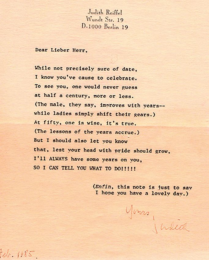
14 Elektronenmikroskopie · Nr. 43 · Dezember 2017Personalien 1974 in order to prove that her asser- lowing Limerick showing her great the neighbor table, I was able to hear tions concerning the reviewing poetic gift: their conversation. The organizer practice of EMSA are realistic. Inde- told him that I was involved in char- ed, she submitted to EMSA, in Feb- 7) In 1980, at a time when I had just ged-particle optics trying to find a ruary 1974, together with her fantasy found the basic design of the hexa- suitable corrector for improving the coauthor L. Gandolfi the two-page pole corrector, I used to attend regu- performance of magnetic solenoids abstract of their paper entitled: “En- larly the physics colloquium at the using the terminology of the accele- hancement of contrast in CEM and Technical University of Darmstadt. rator community. However, my colle- STEM using bumpy specimens and At the occasion of the installation of ague considered my investigations to employing the dappled field mode of the linear accelerator DALINAC in be fruitless because I did neither operation”. Although her abstract the Institute of Nuclear Physics the have the expertise nor the sophisti- was a hoax and the content was com- guest speaker addressed problems of cated computer programs of his insti- pletely unphysical, it was accepted particle motion in beam-guiding sys- tute for determining the path of and published in the EMSA procee- tems. Within the frame of the discus- charged particles. Moreover, if such dings 1974. For example, a specific sions after his talk, I asked the spea- systems would be of importance, detail of one of the pictures was de- ker for his opinion on the effect of they would have already done the scribed as “moon over Lake Michi- aberrations regarding the stability of calculations for their design. gan”. Nobody had looked at the abs- the particle trajectories in accelera- About twenty years later, I was asked tract and the organizers even gave tors. Obviously, my question must in Berkeley by a member of the Ad- her an oral presentation owing to the have been of interest for him because vanced Light Source at LBNL how I interesting title of her contribution. he asked the organizer at the fol- was able to find the design of the he- Judith even wanted to present her lowing dinner what kind of research xapole corrector. He told me that he hoax contribution at the conference. I was doing. Although I was sitting at first doubted the outstanding optical However, Albert Crewe did not al- low her to go to the conference after she had made him aware of the situa- tion. At the conference the chairman told the audience that the author was prevented to present her contributi- on because her coauthor had passed away and she had to attend his fune- ral. He even asked the audience to stand up for a minute’s silence in commemoration of the coauthor. Obviously, also the chairman had not read the abstract. It was later that the congress participants were informed of the hoax by somebody who had read the abstract. As a result of this embarrassing event, thorough revie- wing of the submitted abstracts was made thereafter. The hoax gave rise to tensions between Albert Crewe and Judith Reiffel. As a consequence she left Chicago some time later and moved together with the editor of Ultramicroscopy, Professor Elmar Zeitler, to Berlin where she found a new job at the Fritz Haber Institute connected with the editing of articles to be published in Ultramicroscopy. Owing to my close cooperation with Dr. Dieter Krahl, who realized suc- cessfully the imaging Omega filter at the Fritz-Haber Institute, I stayed in permanent contact with Judith until she moved back to the US after her retirement. On the occasion of my fiftieth birthday she sent me the fol- Elektronenmikroskopie · Nr. 43 · Dezember 2017 15
Personalien

performance of this corrector. There-        after the end of Dr Phillipp’s presen-     rather non-physical picture is the one
fore, he recalculated the optical pro-       tation: “Instead of spending all this      of all of my drawings which is most
perties of my design by means of the         money for producing primarily radia-       often shown.
well-established computer program            tion damage, it would be better to
used by the accelerator community.           provide money for the realization of       9) Before I published the hexapole
The calculations confirmed to his sur-       the correction of aberration by me-        semi-aplanat in 1990, I approached
prise the correctness of my calculati-       ans of the novel hexapole corrector.       Dr. Kurz who was the head of the
ons which I performed mainly ana-            The necessary funds will be signifi-       electron microscope division of the
lytically. He hardly could believe this      cantly lower than those for the Stutt-     company Zeiss at that time. He was
fact and asked me how long it took           gart project”. The answer of the chair-    interested in aberration correction de-
me to have the idea. My answer was:          man was: “Dr. Rose show that your          spite the fact that the BMBF (German
“I had the idea within a short moment        corrector will work, then you will get     Ministry of Education and Research)
however it took me almost twenty             the money”. In response I said: “We        had denied our earlier proposal to
years for giving birth to this idea”.        need the money to build the correc-        support the development of a correc-
                                             tor and to prove that it will work with    tor previously with the argument:
8) During the eighties, the perfor-          present technology. After achieving        “Our expert reviewers have told us
mance of the electron microscopes            this goal, I have finished my job”. For-   that aberration correction by means
had improved to such an extent that          tunately, this statement convinced         of correctors will never work in practi-
the resolution of medium-voltage             Knut Urban. Only a short time later        ce. If this would be possible the Japa-
electron microscopes was limited to          he initiated together with Maximilian      nese manufacturers would already
about 2 Å by the unavoidable spheri-         Haider and me a proposal to the            have done so because they are 10 ye-
cal aberration of the objective lens. In     Volkswagen Foundation for realizing        ars ahead in the development of high-
order to lower this limit, high-voltage      a spherically corrected 200 kV trans-      performance electron microscopes”.
electron microscopes seemed to be            mission electron microscope. The           This argument was the principle rea-
the way to go. This route was pionee-        Volkswagen Stiftung was the only           son for me to prove that the “experts”
red commercially primarily by the            German funding agency who was wil-         were wrong and ignorant. In order to
manufacturer       JEOL.       Professor     ling to finance this high-risk project     realize the hexapole corrector, I orga-
Gareth Thomas at Berkeley was the            which is known today as the “Volks-        nized a meeting in Darmstadt with
first to roughly reach 1 Å in his 1 MV       wagen Project”. All other agencies re-     participants from EMBL (Dr. Haider)
TEM. At about the same time A. See-          fused to support such an ill-posed         and industry (Zeiss and Philips). Be-
ger started an initiative to install a 1.2   project which had no chances of suc-       cause I did not want to show the de-
MV TEM at the Max Planck Institute           cess according to the opinion of the       sign, I only outlined the feasibility and
in Stuttgart. His coworker Dr. Phil-         “experts”. In particular, one of the re-   simplicity of the novel corrector. In
lipp reported about the project in a         viewers wrote in his review of a pre-      order to obtain a real improvement in
session chaired by Professor Knut            vious BMBF proposal by Zeiss and           resolution by means of this corrector,
Urban at the Dreiländer-Tagung 1989          me: “Japan is about ten years ahead        it was necessary that the basic instru-
in Salzburg. During his talk I was sit-      of Germany in the development of           ment had an information limit of
ting next to Dr. Oppolzer who was            high-resolution electron microscopes.      about 1 Å down from about 2.1 Å
investigating the structure of semi-         If correction would work, they would       which was the information limit of the
conductors at the Siemens Research           already have achieved it”.                 best standard electron microscopes at
Laboratories in Munich. Due to my            Despite of my critics concerning the       that time. To achieve this improve-
experience with the problem of radi-         usefulness of high-voltage TEMs for        ment it was necessary to equip the ba-
ation damage acquired during my              obtaining atomic resolution, Profes-       sic instrument with a field emission
time at the New York State Depart-           sor Seeger invited me to give a talk       gun and to use accelerating voltages
ment of Health in Albany, I said in a        within the symposium held at the in-       higher than about 200 kV. Dr. Kurz
low voice to Oppolzer: “All what they        auguration of the Stuttgart 1.2 MV         showed interest in the project and told
will obtain with this brute-force me-        TEM. My talk, published in Ultrami-        me when he left that he would inform
thod is radiation damage”. He agreed         croscopy, was entitled “Correction of      me within a week if Zeiss would consi-
by stating: “You are absolutely right,       Aberrations – the Promising Route          der developing such an instrument.
I am working with the JEOL 400 kV            to Sub-Angstrom Electron Micro-            Indeed he gave me a phone call about
TEM. However, for investigating se-          scopy”. In this talk, I presented for      one week later. His answer was: “the
miconductors I must operate the mi-          the first time the nowadays often          studies of our sales experts have
croscope below about 180 kV in or-           used picture showing the increase of       shown that 12 corrected electron mi-
der that the sample is not destroyed         resolution in microscopy as a func-        croscopes will be sold worldwide at
immediately. What I need is an aber-         tion of time including the assumed         most. However, we must sell many
ration corrected 150 kV TEM”. Owing          improvement resulting from aberra-         more microscopes in order to com-
to his remarks and the high costs of         tion correction. This prediction has       pensate for our development costs.
high-voltage TEMs, I made the fol-           come true due to the success of the        Therefore, Zeiss will not participate in
lowing statement in the discussion           Volkswagen project. As a result, my        the project”.

16                                                                              Elektronenmikroskopie · Nr. 43 · Dezember 2017
Sie können auch lesen